There are contraindications. Specialist consultation is required.
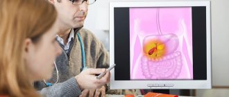
Manifestations of diseases Diagnostic methods Treatment methods
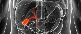
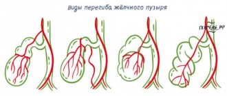
Bile, which is produced in the liver, enters the gallbladder and bile ducts through the hepatic duct. Part of the bile is immediately sent through the common channel to the duodenum. Sometimes there are malfunctions in this system, which can be immediately determined by the characteristic pain in the right hypochondrium. The nature of this pain is related to the type of disease. For example, if there are large stones in the excretory ducts, a person experiences stabbing pain.
Clinical manifestations and symptoms
Manifestations of the disease depend on the type of damage.
1. If bile flows freely into the abdominal cavity through the damaged bile duct
- Pain appears in the right hypochondrium and in the upper abdomen. The pain is constant, dull.
- There is a feeling of heaviness in the stomach.
- Then an infection sets in and the body temperature rises.
- Intoxication and bile peritonitis occur, requiring emergency surgical treatment.
2. If there is no leakage of bile into the abdominal cavity, then as a result of an obstruction (stricture), bile does not enter the duodenum
The pressure in the bile duct itself increases, as a result of which bile enters the blood through the vascular system of the liver and obstructive jaundice develops.
- Patients develop pain and increased body temperature.
- Yellowness of the skin, sclera and visible mucous membranes.
- If treatment for this complication is not started on time, the patient becomes lethargic and apathetic.
- General weakness, confusion and other severe disorders of the basic functions and systems of the body develop.
Complications of strictures and tumors of the bile ducts
- Mechanical jaundice.
- Biliary peritonitis.
- Abdominal abscesses are an accumulation of bile in certain areas of the abdominal cavity accompanied by infection.
- Cholangitis is inflammation of the bile ducts as a result of the action of complex negative factors.
Clinical forms of obstructive jaundice in choledocholithiasis
- Jaundice-pain: the main symptoms are pain, nausea, vomiting, jaundice, fever.
- Icteric-pancreatic: develops when a stone is strangulated in the large duodenal papilla or cicatricial stenosis of the large duodenal papilla. There are symptoms of obstructive jaundice and acute pancreatitis.
- Jaundice-cholecystitis: jaundice develops against the background of an attack of acute cholecystitis.
- Icteric-septic: this form is based on the development of obstructive purulent cholangitis, sometimes complicated by cholangiogenic liver abscesses and sepsis.
- Jaundice-painless: jaundice increases gradually, the patient’s condition suffers little, there is no indication in the anamnesis of a painful attack. Requires differential diagnosis with jaundice of tumor etiology.
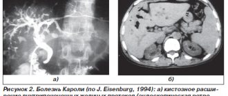
Causes of hardening of the gallbladder walls
Thickening of the gallbladder walls during ultrasound may be a reflection of the following problems:
- Cholesterosis. When the lipid balance in the body is disturbed, cholesterol is deposited on the wall of the bladder.
- Gallstone disease and chronic cholecystitis (chronic inflammation in the gallbladder) against this background. In this case, the stones can injure the walls of the bladder, provoking subsequent inflammation.
- “Porcelain” gallbladder with calcification (calcification) of the bladder as a result of chronic inflammation.
A case from the practice of gastroenterologist Daniela Purgina, an expert on the website Pokhmelye.rf.
Once, a patient came to see me; several years ago, during treatment at a sanatorium, he had undergone an ultrasound of the abdominal cavity and was diagnosed with a “porcelain gallbladder.” Disabled gallbladder." For several years he lived with the idea that his gallbladder was not working and needed to be removed. And so he finally decided to have the operation and came to me for a referral to the hospital. As expected, I sent him for an ultrasound before this to make sure the diagnosis was correct and to see if any changes had occurred in the bladder during this time. The ultrasound was planned to be done with a choleretic breakfast: in order to make sure that it was really “disabled”, because the patient had not experienced any discomfort in the right hypochondrium over the several years that he had not consulted a doctor. The study showed the presence of cholesterol deposits in the walls of the organ, and the contractile function of the gallbladder was reduced, that is, the bladder was functioning, but it was just working slowly. Next, the patient underwent a computed tomography scan, the results of which confirmed the deposition of cholesterol in the walls of the organ. Thus. the patient did not require surgical treatment or organ removal.
Causes of strictures
Most often, there are three groups of causes of the disease:
- traumatic - develop after surgical interventions: cholecystectomy, endoscopic manipulation, resection of the stomach or liver;
- inflammatory - they are caused by cicatricial changes in the walls of the ducts in various diseases (chronic pancreatitis, low duodenal ulcers, parasitic liver diseases, etc.);
- tumor - strictures that occur with cancer of the gallbladder or extrahepatic bile ducts.
Physical examination
Physical examination is an opportunity to diagnose pathology without the use of equipment, using external stimuli: palpation, tapping, auscultation.
| Palpation | A slight swelling, rising in the liver area, sharply thins towards the hips and navel; pain radiates to the navel and kidneys; concentrated below the liver. |
| Percussion (tapping) | Tap the front and back of the right side of the ribs; pain radiates to the kidneys - high acidity of urine; liver, lower abdomen - multiple fusion of folds. |
| Listening | Dull, inhomogeneous sound. Swelling of the liver from the back; gurgling – compression of the gallbladder with fluid; hissing air movements - injury, muscle swelling - antibiotics, kidneys, colds. |
Classification of narrowing of the bile ducts
By etiology
- Post-traumatic scar strictures;
- Strictures of biliary anastomoses;
- Primary sclerosing cholangitis;
- Secondary inflammatory strictures;
- Tumor stenoses of the bile ducts 5.1. Bismuth-Corlette classification (for proximal bile duct cancer)
- Type I. Damage to the common hepatic duct
- Type II. Damage to the confluence of the hepatic ducts.
- Type IIIA. Damage to the right hepatic duct.
- Type IIIB. Lesion of the left hepatic duct.
- Type IV. Damage to both hepatic ducts.
- 5.2. Extrahepatic bile duct cancer
- Cancer of the distal common bile duct.
- Cancer of the large duodenal nipple.
According to the degree of narrowing of the ducts
- Complete strictures.
- Incomplete strictures.
- Limited strictures (up to 1 cm).
- Widespread strictures (1 - 3 cm).
- Subtotal strictures (more than 3 cm).
- Total damage to the extrahepatic bile ducts
According to the clinical course
- With external biliary fistula.
- With jaundice.
- With cholangitis.
- With liver and kidney failure.
- With biliary cirrhosis and portal hypertension.
Forecast
In general, the prognosis is favorable. Exceptions: cirrhosis, cancer, acute cholecystitis, polyposis, hepatitis.
Cancer survival prognosis:
| 1-2 stages | 90% |
| 3 stages | 60% |
| 4 stages | no more than 15% |
Survival prognosis for cirrhosis:
| Stage 1-2 | 85% |
| Stage 3 | 40% |
| Stage 4 | 15-20% |
In other cases - 90% if you consult a doctor in a timely manner.
Treatment of strictures and tumors of the bile ducts
Currently, various types of treatment are used: from drainage to reconstructive operations. However, there is no systematic approach that takes into account both the main characteristics of ductal stenosis and the advantages and disadvantages of a particular surgical treatment technique for a particular patient. Today, an individual approach to each clinical situation is required, allowing the use of both modern minimally invasive techniques and time-tested reconstructive operations.
For non-tumor injuries of the bile ducts, the following types of surgical interventions are distinguished:
- drainage operations (bile diversion to the outside);
- reconstructive operations;
- reconstructive operations.
Drainage operations include: drainage of the common bile duct according to Vishnevsky, Kerta, Halsted, drainage of the proximal end of the crossed duct, percutaneous - transhepatic drainage.
In case of marginal injuries, preference is given to T-shaped drainage compared to other methods, since it has been proven that in choledochotomies completed with T-shaped drainage, neither gross deformations of the hepaticocholedochus nor its stricture at the site of the drainage were detected in the long term.
When the tumor affects the distal part of the common bile duct, preference is given to gastropancreatoduodenal resection.
Treatment
Treatment cannot be 100% effective without the right diet
The main role in treatment is given to medications and diet. Folk remedies as an auxiliary method.
The main objectives of drug therapy; relieving inflammation and spasms; relief of pain for a while; normalization of bile acidity to reduce the load on the mucous membrane; normalization of phospholipid metabolism if there are fat deposits. Concomitant diseases are treated after the walls are thinned.
Exceptions:
| Cancer | Treatment is carried out despite the thickness of the walls. |
| Cirrhosis | At stages 1-2 - therapy for cirrhosis - the walls of the gallbladder return to normal as a result of its use. |
| Infectious | Antibiotics and hepatoprotectors in combination. |
| Hepatitis | In chronic cases, if there is cirrhosis, complex treatment; in other cases - therapy for hepatitis after inflammation is relieved. |
To normalize wall thickness:
| Antispasmodics | No-shpa; for mild spasms - Drotaverine. |
| Relieving inflammation | Glycyrrhizic acid. |
| Normalization of phospholipid metabolism | Phosphogliv, Forsliv. |
| Choleretic | Allohol, Holenzim. |
| Normalization of acidity | Preparations with artichoke, and for strong ones - Vikalin, sodium sulfate. |
Traditional methods
Before taking herbal formulations, consultation with a doctor is required.
Folk remedies are not able to resolve scars, adhesions, or remove fat deposits. They partially relieve the inflammatory process of a non-infectious nature. Before taking herbal formulations, consultation with a doctor is required.
For 0.5 liters of cold water, 1 teaspoon each of bearberry, nettle, and corn silk. Boil for 5 minutes. Leave for 12 hours. Drink 50 ml over 6 hours. Softens fatty deposits.
For 200 ml of boiling water, 0.5 tsp of immortelle, corn silk, mint, lemon balm, 1 tsp of chamomile, 1 tbsp. l. plantain. Boil in a water bath for 1 minute. Don't take it off. Let cool. Strain. Drink in 2 doses over 2 hours. The decoction is effective for injuries and inflammatory processes.
Grate 0.5 kg of peeled apples on a beet grater. Grind the pulp of the baked pumpkin in a mixer. Mix with 0.5 liters of liquid honey. Leave for 12 hours. In the future, store in the refrigerator. Use within 3-5 days. Helps with obesity caused by an unbalanced diet; inflammation due to helminthiasis.
Diet
Eliminate junk food from your diet
Exclude fatty and smoked meat from the diet; snacks, legumes; strong tea and coffee; fried and cooked dishes. Minimize baked goods, dairy products, eggs.
Give preference to soft casseroles, first courses, meat, fish - low-fat varieties, cooked in the oven, steamed. Add well-cooked or baked vegetables to vegetable salads. For fruit - boil or bake fruit until half cooked. First courses are based on lean fillet. If there is a lot of meat, drain the first broth.
Treatment in the department of thoracoabdominal surgery and oncology of the Russian Scientific Center for Surgery
Treatment of tumors and strictures of the bile ducts in our department is carried out within the framework of the compulsory medical insurance , voluntary medical insurance, VMP , as well as on a commercial basis . Find out how to get treatment at the department of thoracoabdominal surgery and oncology of the Russian Scientific Center for Surgery.
We provide surgical treatment with highly qualified doctors , the best modern equipment and attentive staff. Read patient reviews here .
To schedule a consultation, call:
+7 (499) 248 13 91 +7 (903) 728 24 52 +7 (499) 248 15 55
Submit a request for a consultation by filling out the form on our website and attaching the necessary documents.
Complications
The following complications may occur.
- Organ rupture due to contraction by adhesions.
- Malignant tumors are degeneration of benign tumors or constant inflammation.
- Reactive inflammations carried by blood - immune diseases of bone tissue, meningitis, inflammation of the heart muscle; severe leukocytosis - blood sepsis.
- Due to stagnation of bile - suppuration in the gastrointestinal tract, dysbiosis, susceptibility to infectious diseases.
- Uneven thickening-thinning – peritonitis, perforation, rupture.
- High probability of cirrhosis and hepatitis - rapid development.