If distant metastases are detected in the liver, this always indicates a widespread tumor process, that is, it means the fourth stage of cancer. Therefore, in such a situation you cannot hesitate; the main thing is to start treatment immediately.
What treatment is possible for stage IV cancer with liver metastases?
Metastatic foci in the liver have a histological structure and phenotype similar to the primary tumor, and therefore metastases are treated, as a rule, according to the same scheme as the underlying disease.
When choosing a treatment method for liver metastases
The oncologist takes into account many factors, including:
- Primary focus of the disease (localization and biological type of tumor, degree of malignancy);
- Single or multiple liver lesions;
- Size of metastatic foci in the liver;
- The presence or absence of metastases in lymph nodes and other organs;
- History of the disease (antitumor treatment methods completed);
Symptoms of liver metastases
Symptoms of metastases begin to appear when they disrupt the functioning of the liver and gallbladder. With single metastases, they appear when the tumor reaches a size of 5-7 cm.
Signs of pathology:
- heaviness and dull pain in the right hypochondrium;
- cardiopalmus;
- constant slightly elevated temperature;
- gynecomastia – enlargement of the mammary glands in men;
- yellowness of the skin and whites of the eyes, dark urine and light feces.
You should notify your doctor if the patient simultaneously exhibits several symptoms from the list. If this is not done and measures are not taken, the symptoms increase and the patient’s condition worsens. This shows up:
- symptoms of intoxication - constant nausea and weakness;
- sudden weight loss;
- anemia;
- swelling in the legs and accumulation of fluid in the abdomen - ascites;
- a venous network on the abdomen, radiating from the navel;
- vomiting "coffee grounds" and tarry stools.
As the process progresses, the liver loses its functions. To identify metastases at an asymptomatic stage, it is advisable for patients with cancer to undergo an ultrasound of the liver once a month and an MRI with contrast once every 3 months.
Forecast
The five-year survival rate for colorectal cancer with metastases is 12%. Treatment methods are being improved, and perhaps this figure will be increased in the future.
| More information about the treatment of rectal cancer at Euroonko: | |
| Treatment of rectal cancer | |
| Proctologist-oncologist | RUB 5,100 |
| Chemotherapy appointment | RUB 6,900 |
| Emergency oncology care | from 12,100 rub. |
| Radiologist consultation | RUB 11,500 |
Book a consultation 24 hours a day
+7+7+78
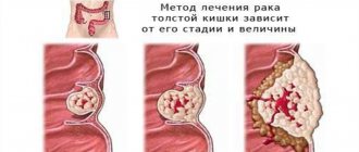
Classification
Depending on the number, metastases are:
- sole, or solitary;
- single (up to 10);
- multiple (more than 10).
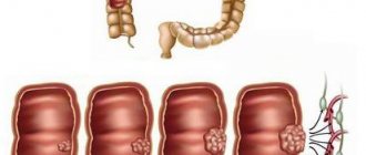
The sizes are small (up to 1 cm in diameter), medium (1-3 cm) and large (more than 3 cm). The speed of their appearance depends on the degree of aggressiveness of the primary tumor.
Metastatic tumors are not classified according to the TNM system. The presence of metastases in the liver itself indicates stage 4 of the primary tumor.
How is colon cancer with metastases diagnosed?
Typically, the primary tumor is detected using colonoscopy - an endoscopic examination during which a special instrument - a colonoscope . If a pathological formation is detected during a colonoscopy, the doctor obtains a piece of tissue from it and sends it to the laboratory for study under a microscope and molecular genetic analysis. This diagnostic method is called a biopsy, it helps to definitively confirm the diagnosis.
When colon cancer is confirmed, various studies are used to look for metastases in the abdominal cavity and beyond:
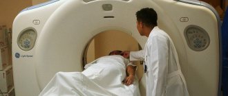
- CT, MRI
- Ultrasound of the abdominal organs
- PET scan
- Chest X-ray
- Bronchoscopy
- X-ray of bones
- Angiography of the liver
- If necessary, diagnostic laparoscopy is performed
Diagnosis of metastatic liver cancer
The diagnosis is made on the basis of laboratory and instrumental studies:
- Ultrasound of the liver
- allows you to detect signs of metastases from 0.4-0.5 cm; - CT and MRI with contrast
, as well as
radioisotope PET scanning
- allow you to reliably identify metastases, their size and location, growth pattern and the degree of involvement of surrounding tissues in the process; - portography
- a type of CT with the introduction of contrast into the portal vein - allows you to detect micrometastases; - biopsy of metastatic nodes
under ultrasound control followed by histological analysis of the biopsy - allows you to determine the histological type of tumor and prescribe effective treatment; - biochemical blood test and liver tests
- help to identify functional disorders in the liver; - tumor marker AFP
is a marker of a malignant tumor in the liver.
Often, liver metastases are detected before the underlying cancer is diagnosed. And it is the biopsy that makes it possible to determine from which organ the cancer cells entered the liver.
Why and how do metastases occur in colon cancer?
Metastasis of malignant tumors is a complex process, its causes and mechanisms are not fully understood. There are several stages in it:
- The cancer cell breaks away from the primary site and penetrates the wall of a blood or lymph vessel.
- The cancer cell migrates with the lymph or blood flow.
- Then it “gets stuck” in vessels of small diameter. Some studies have shown that not only the diameter of the vessel plays an important role in this process, but also the speed of blood flow in it.
- The tumor cell penetrates through the wall of the blood vessel into the surrounding tissues - in the future they will become its new “place of residence”.
- The cancer cell may “sleep” for some time. When a favorable moment comes, it begins to multiply quickly and gives rise to a secondary outbreak.
Tumor cells can spread not only through blood and lymphatic vessels, but also “spreading” throughout the peritoneum.
The biggest problem in diagnosing and treating metastatic cancer of the intestine and other organs is that there are usually many metastases, many of them are microscopic in size and are not visible on photographs, they cannot be detected and removed during surgery.
Treatment methods for cancer metastases in the liver
The treatment regimen for metastatic lesions is developed taking into account a number of factors:
- single or multiple metastases have been identified;
- are there metastases in the lymph nodes;
- they are located on the periphery or near large blood vessels and bile ducts;
- histological type of tumor;
- general condition of the patient.
If the metastatic foci are histologically the same as the primary tumor, then the pathology is treated according to the same scheme as the underlying disease. Single and several small metastases with a capsular location are removed surgically.
This is possible in 5-20% of all cases. If the clinical situation allows, the operation is performed not laparotomically through an incision, but laparoscopically through punctures.
Together with the metastases, a small part of the liver is removed (segmental or lobar resection). In this case, the organ’s ability to self-regenerate plays a major positive role. Therefore, even with severe metastatic damage to the liver by cancer cells, it is possible to remove tumor foci in several stages. For metastases up to 1 mm in size, a laser cyber knife is used. In other cases, radiation and chemotherapy are indicated.
Modern methods of radiation therapy
are highly accurate – this increases the effectiveness of treatment and minimizes the risk of relapse. Their goal is to destroy cancer cells without affecting healthy ones. For liver metastases, strong focused radiation, isotope radiotherapy with hepatic vein bypass, or radiofrequency hyperthermia are used. Radiation therapy is often used at the palliative stage to reduce pain.
Chemotherapy
Cytostatic drugs for liver metastases block the division and growth of cancer cells, thereby slowing down or completely stopping the growth of a metastatic tumor. Often, the method makes it possible to reduce the volume of a tumor and make an inoperable metastatic lesion operable.
In clinical practice, the liver often deactivates many chemotherapy drugs. Then chemoablation is performed instead - a chemotherapy drug is injected into the metastasis vessel along with a drug to block the vessel.
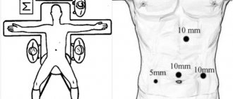
If the clinical situation allows, radiofrequency ablation
– malignant structures are destroyed by high-frequency current supplied from a needle electrode through a puncture in the skin. In addition, immunotherapy with targeted drugs, for example, Sorafenib, approved in Russia, is effective. The drug acts on the target molecule, the death of which causes the death of the entire tumor.
A promising experimental treatment method is the destruction of liver metastases using ultrasonic waves
.
In approximately 50% of cases, palliative therapy
.
In addition to specific anticancer therapy for liver metastases, hepatoprotective treatment to prevent toxic damage to the organ. It is aimed at normalizing liver parameters - ALT and AST, GGTP, bilirubin and alkaline phosphatase. detoxification is also carried out
. If liver metastases disrupt protein synthesis, the body is supported by the administration of protein drugs.
Peritoneal carcinomatosis in colon cancer
If cancer cells spread into the peritoneum, the prognosis is greatly worsened. Ascites occurs - fluid accumulates in the abdominal cavity. To combat this condition, they resort to laparocentesis - a puncture is made in the abdominal wall and the fluid is removed. A peritoneal catheter is installed to ensure constant drainage of fluid . Intracavitary chemotherapy is carried out, and according to indications, surgical interventions are performed aimed at ensuring the outflow of fluid from the abdominal cavity.
The average life expectancy of such patients when using classical treatment methods is 2–6 months. But over the past 20 years, a new treatment method has been actively developed: hyperthermic intraperitoneal chemotherapy (HIPEC). The procedure consists of two stages:
- A surgical procedure is performed during which all visible lesions are removed from the abdominal cavity. This is a long (on average 6–9 hours) and complex operation.
- Then the abdominal cavity is washed with a chemotherapy drug heated to 42–43 °C.
The HIPEC method has helped to greatly increase the survival rate of patients with peritoneal carcinomatosis.
Probability of relapse
In the first 2 years after achieving remission, 40-60% of patients experience relapses of the oncological process. By the end of the 3rd year, the number of patients with relapses reaches 70%. The prognosis for treatment of liver metastases and the likelihood of relapse are influenced by the malignancy of the primary tumor and metastases, their location, size and effectiveness of the treatment regimen.
The presence of metastases significantly worsens the course of the oncological process, but modern medicine allows us to achieve positive results in such difficult cases. For us at SM-Clinic there are no hopeless patients. We always fight for the patient - to either completely defeat the disease, or to prolong the patient’s life and improve its quality.
We treat liver metastases with a multidisciplinary team of specialists. Every year new technologies and drugs appear, which we are intensively mastering, that is, the capabilities of our doctors are constantly growing. To consult a specialist, please contact us.
Information verified by an expert
Mikhailov Alexey Gennadievich operating oncologist, doctor of the highest qualification category, Ph.D. experience: 21 years
The information in this article is provided for reference purposes and does not replace advice from a qualified professional. Don't self-medicate! At the first signs of illness, you should consult a doctor.
Chemotherapy for colon cancer with metastases
For unresectable colon cancer with metastases, one of three chemotherapy regimens is usually used as first-line treatment:
- FOLFOX : leucovorin, 5-fluorouracil and oxaliplatin;
- FOLFIRI : leucovorin, 5-fluorouracil and irinotecan;
- CAPEOX or CAPOX : capecitabine (Xeloda) and oxaliplatin.
These regimens can be used in combination with targeted drugs.
How long does treatment continue ? Until it stops helping (the tumor begins to progress again), or the patient experiences serious side effects. The main problem with regimens that include oxaliplatin is that the drug often leads to the side effect of neuropathy , a condition that causes burning and numbness in the arms and legs. To prevent this complication, some experts recommend treating with first-line drugs for some time and then switching to maintenance chemotherapy with other regimens.
The patient should talk to his doctor in advance to find out what medications are planned to be prescribed, what side effects they may cause, and what the plan of action will be if they occur.
Rectal cancer - symptoms and treatment
Rectal cancer is treated with various methods, most often several at once. Some of them are recognized and considered standards of treatment, some are undergoing clinical trials. Once improved or new therapies surpass the effectiveness of established standard treatments, they may also become part of everyday practice.
There are currently six treatment options for colorectal cancer:
- surgical method;
- radiation therapy;
- chemotherapy;
- active surveillance;
- targeted therapy;
- immunotherapy.
Surgical methods of treatment
Surgery is the main treatment for rectal cancer, but after or before surgery, radiation, chemotherapy, or a combination of both - chemoradiotherapy - may be required.
The type of surgical treatment depends on the stage of the cancer, the location of the tumor and the purpose of the operation. For example, depending on the goal, it can be radical or palliative. Radical surgery is performed in the initial stages of cancer, when the tumor can still be completely removed, and palliative surgery is performed in the final stages of the disease, when the tumor is no longer operable: it helps to improve the patient’s well-being, relieve him of tumor symptoms and prevent complications.
Before surgery, it is important to determine how close the tumor is to the anal sphincter. The type of surgical intervention and its result depend on this [4][5][10][11][12]. The further it is located from the sphincter, the more likely it is that the sphincter will not be damaged during treatment and the patient will not have problems with fecal retention.
The tumor is removed using one of the following methods:
- Polypectomy is the removal of a polyp in which tumor cells were found. The operation is performed during a colonoscopy. Unlike a biopsy, which only removes a small piece of tissue, a polypectomy removes the entire polyp.
- Local excision is a more complex procedure. Instruments to remove small tumors are also inserted through the colonoscope. The formation is removed along with a small part of the healthy rectal mucosa. This method of treatment is possible for non-invasive tumors that do not spread throughout the entire thickness of the intestinal wall and affect only the mucous (most superficial) layer. During the operation, there is no need to make an incision to get to the abdominal cavity. The purpose of this operation is to remove the entire tumor or polyp. If there is a possibility that tumor cells remain (that is, the tumor is not removed within healthy tissue), then the formation may spread. Then the next step may be a more complex type of rectal surgery.
- Transanal excision (TAI) is a local excision of the rectum. This method is used only in cases where the tumor does not spread to the entire depth of the rectal wall and affects only the innermost (mucous) layer. During the operation, the surgeon cuts through all layers of the rectal wall to remove the tumor. Some healthy surrounding tissue is also removed. The resulting defect in the rectal wall is then sutured. During the intervention, the lymph nodes are not removed, therefore, if the tumor could not be completely removed or its cells appeared in the lymph nodes, the issue of prescribing chemoradiotherapy is decided.
- Low anterior resection (LAR) - removal of the part of the intestine containing the tumor and all surrounding lymph nodes. More often, such an operation is performed at stages II or III of rectal cancer, but sometimes at stage I. After the tumor is removed, the free ends of the intestine are connected to restore natural intestinal patency. But they cannot always be connected immediately after removal: it all depends on the patient’s condition and chemoradiotherapy in the postoperative period.
After LAR, most patients spend several days in the hospital. They then recover at home for approximately 3–6 weeks. During this period, it is recommended to follow a gentle diet with sufficient calories, not smoke or drink alcohol, engage in moderate physical activity for 30 minutes every day and take medications that prevent the formation of blood clots.
- Resection of the rectum with colo-anal anastomosis - removal of the entire rectum and surrounding lymph nodes. Basically, the operation is performed on stages II and III of rectal cancer, if the tumor is located in the middle and lower third of the rectum. The free end of the colon is sutured to the anus so that the patient can go to the toilet in his usual way. This connection is called colo-anal anastomosis.
- Abdominoperineal rectal extirpation (APR) is a complete removal of the rectum, more complex than low anterior resection. The operation is performed mainly on stage II or III rectal cancer, if the tumor is located close to the anal sphincter or has grown into the muscles that lift the pelvic floor and the anal sphincter. During the operation, the surgeon makes one incision in the skin of the abdomen and another in the skin of the perineum around the anus. This technique removes the rectum, anus, and tissue around it, including the sphincter muscles. To remove feces after such an operation, a permanent colostomy is formed - the end of the large intestine is brought out to the anterior abdominal wall and sutured to it.
- Creation of a diverting stoma is the elimination of intestinal obstruction without removing the tumor. It is carried out when the tumor closes the lumen of the rectum and interferes with the passage of feces. During this operation, the colon is cut above the tumor and its free upper end is sutured to the skin of the abdomen. This opening is called a diverting colostomy. Often this surgery helps the patient recover before the next stage of treatment, such as chemotherapy. When treatment is completed and the patient feels better, the natural flow of the intestines is restored.
Pain may occur after waking up from anesthesia. To muffle it, you will need to take analgesics for several days.
For two days after the operation, the patient should not drink or eat so that the intestines can recover. All nutrients are administered intravenously at this time. Then the patient is allowed to eat small portions of chemically and thermally gentle food.
Complications after surgery
The possible risks and side effects of surgery depend on several factors, including the complexity of the procedure and the general condition of the patient before surgery.
Complications during or shortly after treatment include postoperative bleeding, infection at the incision sites, and blood clots in the blood vessels of the legs. With the development of such complications, body temperature rises to 38–39 °C, the heartbeat increases, blood pressure decreases, shortness of breath and abdominal pain occur, which is not typical for a normal postoperative period, and pus is released from the wound.
In rare cases, the suture between the two ends of the intestine may come apart, allowing the contents of the intestine to leak into the abdominal cavity. This can cause severe abdominal pain, fever, and very tight abdominal muscles. A slight leakage of feces can cause stool retention, despite a good appetite.
A leaky connection between parts of the intestine can also lead to the development of infectious complications. In this case, additional surgery will be required. And an open wound that may form at the site of the incision in the anterior abdominal wall will require special care.
Excess scar tissue in the abdomen that forms after surgery can cause organs and other tissues to stick together. Such scars are called adhesions. In some cases, they can cause the intestines to twist and block peristalsis. In this case, there may be pain and swelling in the abdomen, which often intensifies after eating. Further surgery may be required to remove scar tissue.
Colostomy or ileostomy
Some patients with rectal cancer require a temporary or permanent stoma after surgery. A stoma formed from a section of the small intestine is called an ileostomy, and one from a section of the large intestine is called a colostomy. They are brought out through an opening in the wall of the abdomen.
Getting used to an ostomy is not easy: it takes time to change your usual lifestyle. But colostomy bags, which are attached to the stoma, and special care products help patients to almost completely rehabilitate and carry out their usual activities without withdrawing from society.
The patient needs to monitor the filling of the colostomy bag and empty it in a timely manner. It usually fills with feces at the same time of day. It is also important to treat the skin around the stoma and choose the right clothing: it should not tighten or squeeze the colostomy bag too much. Excessive physical activity is contraindicated.
Those who are planning to have an ostomy need to prepare: learn how and where to order the necessary supplies and how to use them. You can consult with specially trained nurses, a therapist and your doctor about these issues. There are also online ostomy support groups and chats that you can join. With their help, people share experiences and useful tips on stoma care and lifestyle, as well as provide psychological support.
Sex life and fertility
Complications are always individual, so before the operation you should talk with your doctor about what will change after the intervention and how it will affect your sex life. The patient and his partner must be prepared for possible changes:
- In men, damage to the nerve plexuses during abdominoperineal extirpation (APE) or after radiation therapy may temporarily or completely result in loss of erection or the ability to achieve orgasm. Sometimes the pleasure of orgasm becomes less intense. These changes are usually associated with natural aging, but surgery can make them worse.
- In women, operations on the rectum (with the exception of removal of the pelvic organs - exenteration) usually do not lead to sexual problems. Abdominal adhesions can sometimes cause pain or discomfort during sex. If during the operation it was necessary to remove the uterus affected by metastases, then the woman will lose the opportunity to give birth to a child.
Your lifestyle and level of sexual comfort may also be affected by your stoma, but it should not interfere with your ability to have an active sex life.
Radiation therapy
Radiation therapy is a type of cancer treatment that uses high-energy X-rays or other types of radiation. They destroy tumor cells or prevent them from growing.
There are two types of radiation therapy:
- External radiation therapy - carried out using a special device that irradiates a specific area of the body with a tumor;
- internal radiation therapy - the introduction of a radioactive substance through a catheter, needles, special conductors or in the form of grains directly into the tumor or into the tissue next to it.
Rectal cancer is treated only with external beam radiation therapy. It is carried out as indicated in a short or long course to reduce the tumor before surgery.
Radiation doses are lower than with standard treatment. The operation is performed several days or weeks after the last procedure. There are also postoperative radiation modes.
Radiation therapy may also be the only treatment for colorectal cancer. It is performed when the tumor cannot be completely removed, secondary tumor foci (metastases) need to be irradiated, or there is a severe concomitant disease that does not allow surgery.
In general, radiation therapy is painless. Sometimes patients may experience temporary symptoms of illness.
Chemotherapy
Chemotherapy is a way to treat cancer using drugs that suppress the growth of tumor cells, destroy them, or prevent them from dividing. Due to this, the tumor stops growing and gradually decreases.
There are two types of chemotherapy:
- With systemic chemotherapy, drugs are taken by mouth or injected into a vein or muscle, then the substances enter the bloodstream, reaching tumor cells throughout the body;
- In regional chemotherapy, drugs are injected into the cerebrospinal fluid, the organ itself, a body cavity (for example, the abdominal cavity), or a feeding artery [8].
Chemotherapy, like radiation therapy, can be an independent method of treating cancer, for example, if it is impossible to completely remove the tumor or the patient has a severe concomitant disease that does not allow surgery.
Active Surveillance
If the disease does not progress, the doctor closely monitors the patient's condition without any treatment. This tactic allows you to detect the first signs of worsening the disease in time.
During active observation, patients undergo certain examinations and tests:
- digital rectal examination;
- MRI of the pelvis;
- endoscopic examination (rectoscopy or colonoscopy);
- CT scan of the abdominal cavity and chest with contrast;
- blood test for cancer embryonic antigen (CEA).
Active surveillance must last for at least five years. During the first three years, you need to undergo a colonoscopy and MRI at least once every six months, and then once a year.
After five years of observation, the patient is considered cured of cancer.
Targeted therapy
Targeted therapy uses drugs or other substances that recognize and attack specific tumor cells without harming healthy tissue. These drugs include monoclonal antibodies . They are obtained in the laboratory from one type of immune system cell. These antibodies are administered intravenously. They find altered proteins on the surface of tumor cells or other substances that promote tumor growth. Antibodies attach to these proteins and kill tumor cells, block their growth, or prevent them from spreading.
This method of treatment is used only in isolated cases when surgery and chemoradiotherapy are ineffective.
Immunotherapy (targeted therapy option)
This type of treatment stimulates the immune system to fight tumor cells. Substances that are produced in the laboratory or produced by the body itself can strengthen, direct or restore the body's natural defenses.
This type of cancer treatment is otherwise called biological therapy, or biotherapy. It is prescribed for multiple metastases in other organs, resistance to chemoradiotherapy and ineffectiveness of other treatment methods.
One type of immunotherapy is treatment with immune checkpoint inhibitors . These drugs target the PD-1 protein, which is found on the surface of T cells ("immune cells") and helps control the body's immune responses. When PD-1 attaches to another protein on a tumor cell (PDL-1), it stops the T cell from killing the mutated cell. To help the body fight tumors, PD-1 inhibitors attach to PDL-1 and allow T cells to destroy tumor cells.
Immune checkpoint inhibitors include pembrolizumab. But this drug should be taken with caution and only as directed by a doctor, as it can lead to side effects such as pneumonitis, colitis, diarrhea, fever, adrenal insufficiency, autoimmune hepatitis and damage to the thyroid gland.
Cancer Treatment Options Based on Stage
At stage 0 (carcinoma in situ), the following treatment options may be considered:
- polypectomy;
- local excision, including through the anus (transanal excision);
- resection - indicated when the tumor is large and it is impossible to completely remove it using local excision, for example, if the tumor is inconveniently located or the necessary equipment is not available.
At stage I the following may be considered:
- local excision;
- resection, sometimes followed by radiation or chemotherapy.
At stages II and III you may need:
- surgery;
- chemotherapy in combination with radiation therapy and subsequent surgery;
- a short course of radiation therapy followed by surgery and chemotherapy;
- resection followed by chemoradiotherapy;
- chemoradiotherapy with further active observation (if chemoradiotherapy is ineffective, surgery is performed).
At stage IV the following may be considered:
- surgery, sometimes with chemoradiotherapy;
- systemic chemotherapy, sometimes in combination with targeted therapy (angiogenesis inhibitors);
- systemic chemotherapy, sometimes together with immunotherapy (immune checkpoint inhibitors);
- chemotherapy to control tumor growth;
- radiation therapy, chemotherapy, or a combination of both as palliative therapy to relieve symptoms of the disease and improve the patient's living conditions;
- installation of a stent to maintain patency of the rectum when the lumen is blocked by a tumor - also helps alleviate the symptoms of the disease and improve the quality of life;
- Immunotherapy is carried out when all of the above treatment methods are ineffective and there is resistance to chemoradiotherapy.
Treatment of metastases
The choice of treatment method depends on the affected organ. For example, treatment for colorectal cancer metastases to the liver includes:
- surgical resection - removal of tumor metastases;
- cryosurgery or radiofrequency ablation - influencing abnormal cells using low temperature or electric current;
- chemoembolization or systemic chemotherapy - blockage of the vessels feeding the tumor using a chemotherapy drug or its introduction into the systemic circulation.
Also, the choice of treatment method depends on the resectability of metastases, i.e. the ability to remove them. If metastases are unresectable, treatment begins with chemotherapy to reduce their size and number, thus preparing them for removal.