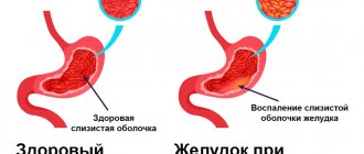
Gastric cancer is one of the most common types of cancer. An effective, informative and accurate means of diagnosing this and a number of other diseases is histological examination of stomach tissue. Histological analysis involves taking samples of stomach tissue through a biopsy and examining them microscopically. This procedure gives the doctor the opportunity to make an accurate diagnosis and prescribe the optimal treatment. First of all, a biopsy of stomach tissue and its histological examination must be performed if the doctor suspects a tumor in the patient. Histological analysis provides information about the type of neoplasm and the cellular composition of the tumor. If decoding the results of gastric histology gives a positive result (the presence of a malignant tumor), it is regarded as the final diagnosis. It is important to understand that a poor histology analysis is not a death sentence, since many types of tumors detected in a timely manner are successfully cured. If the result is negative, an error cannot be ruled out, which may be caused by a mismatch in the location of the cancer cells and the place where the biopsy was taken. If the result of the histological examination is negative, but symptoms of the tumor process are present, the procedure is repeated.
Technical aspects
Numerous methods have been described to obtain an adequate amount of biopsy material. The most commonly used is a pincer biopsy performed using biopsy forceps. Multiple biopsies increase the diagnostic value of the study. Aspects such as the size of the pieces, the location of collection, orientation, fixation and staining of the preparations are also important. With the pinch method, only the mucous membrane is usually included in the biopsy sample. Sometimes large biopsy forceps also capture the submucosal layer. However, such forceps require a biopsy channel of at least 3.6 mm in diameter, and the material they usually remove is 2-3 times larger in surface, but not in depth. Cytological examination of brush biopsy specimens can be a useful adjunct to punch biopsy in the diagnosis of a number of malignant and infectious processes. Loop excision is used to remove large polyps. A combination of techniques can improve diagnostic performance. Aspiration biopsy using a thin needle under endoscopic ultrasound control allows you to take a biopsy from subepithelial lesions, as well as objects located outside the gastrointestinal tract (lymph nodes, pancreatic tumors).
Cost and duration of the study
| Name | Term | Cost, rub. |
| Histology without immunohistochemistry | from 3 days | 12 500 |
| Histology with immunohistochemistry | from 3 days | 28 300 |
* Arrangement and payment for delivery of raw material (not in blocks) is carried out by the client.
For any questions you may have, you can consult our medical administrator by phone: 8-800-555-92-67 or write to us on WhatsApp: +7 925 740 05 87
Esophagus
Esophageal malignancies can be diagnosed by biopsy in 95% of cases, except in situations where obstruction prevents adequate imaging and biopsy from the lesion. 8 to 10 biopsies should be taken. Additional brush cytology can improve diagnostic capabilities.
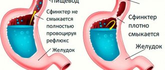
The most common inflammatory changes in the esophagus occur with reflux esophagitis, which develops with gastroesophageal reflux disease (GERD). Endoscopic examination with biopsy is indicated in the diagnosis of Barrett's esophagus, or to exclude infectious or malignant lesions of the esophagus masquerading as gastroesophageal reflux disease. Erosive changes detected during endoscopic examination correlate well with the histological picture, but single erythema is an unreliable criterion for diagnosing esophagitis. On the contrary, histological abnormalities (inflammatory cell infiltration, including neutrophilic and eosinophilic leukocytes) can be detected in biopsies from patients with GERD with a normal endoscopic appearance of the mucous membrane. A biopsy and collection of material for cytological examination from an abnormal-looking mucous membrane are necessary to exclude malignant, infectious processes, some autoimmune diseases and Barrett's esophagus.
Barrett's esophagus is a condition in which the normal lining squamous epithelium is replaced by metaplastic, specialized intestinal epithelium. Its diagnosis requires a biopsy during endoscopic examination. Based on the detection of metaplasia of the esophageal mucosa, patients are included in national cancer programs (registered and periodically examined). Histological examination reveals the lining of the mucous membrane with columnar epithelium devoid of a brush border. The latter is distinguished from the gastric epithelium by the presence of goblet cells, which can be recognized by additional Alcian blue staining.
Destruction of the mucous membrane in the area of Barrett's esophagus is often accompanied by the formation of large ulcerations in the esophagus, and inflammation-induced atypia of epithelial cells at the edges of the defect can be mistakenly regarded as dysplasia. In such cases, intensive drug therapy leads to healing of the mucous membrane and correct subsequent histopathological interpretation of biopsy specimens.
A biopsy is also performed to detect dysplasia or adenocarcinoma. If dysplasia is established or its presence is suspected, a 4-quadrant biopsy should be performed at 1-2 centimeter intervals, as well as additional pieces should be taken from any pathologically changed areas of the mucosa. A 2-cm biopsy misses 50% of cancers in patients with severe dysplasia compared with a 1-cm biopsy. Although large forecept biopsies have previously been recommended, a retrospective analysis found that 4-quadrant biopsies missed the same number of cancers at 2 cm. crayfish when collected with both large forceps (4/12, 33%) and standard size forceps (6/16, 38%). The collection of bipsy material from the mucous membrane of the esophagus should be carried out using the method of rotational aspiration, in which the open forecept is brought close to the end of the endoscope, the endoscope is turned to the wall, aspiration is performed, the forecept is extended, closed and the piece is removed. It is reported that in patients with severe dysplasia who refused surgery, this technique (carried out at 3-6 month intervals) made it possible to effectively diagnose cancer, which at the time of detection in 96% of cases was located within the mucous membrane.
High-resolution endoscopic examination and methylene blue chromoendoscopy increase the detection rate of short segment Barrett's esophagus through targeted biopsy. Chromoendoscopy with Lugol's solution and methylene blue increase the detection rate of squamous cell carcinoma and neoplastic changes in the Barrett's esophagus, respectively, although the value of using methylene blue in monitoring patients with Barrett's esophagus remains controversial.
The study of biopsy samples using flow cytometry with DNA analysis makes it possible to identify patients with aneuploidy, polyploidy, and, using p-53, loss of heterozygosity, which indicates an increased risk of developing cancer.
Local lesions in the esophagus can be removed by endoscopic mucosal resection. In this case, a physiological solution is injected into the submucosal layer in order to elevate the pathologically changed area, and then it is removed using loop electroexcision. This technique has been successfully used to remove neoplastic lesions in areas of Barrett's esophagus and to remove benign tumors of the esophagus.
Infectious esophagitis develops in patients with immunodeficiency conditions caused by systemic antiimmune therapy, inhalation of steroid drugs, malignant tumors (after chemotherapy), diabetes, and AIDS. The most common causative agents of infectious esophagitis are fungi of the genus Candida, herpes simplex virus, and cytomegalovirus. Fungal esophagitis is recognized by the presence of a white coating against the background of inflamed mucosa. Brush biopsy and sampling are performed, but cytological examination of smears after brush biopsy is a more sensitive method. Viral esophagitis is manifested by the formation of ulcerations. A biopsy should be taken from both the edge and the center of ulcerative defects. Histological examination is usually informative, but in case of AIDS, the collection of a large number of pieces (up to 10) is required. Isolation of the virus in culture facilitates diagnosis, but this method is less sensitive than histological examination aimed at diagnosing cytomegalovirus infection.
Stomach
Stomach tumors may appear as ulcers, polyps, submucosal formations, or thickened folds of the mucous membrane. Adequate sampling of material sometimes requires the use of combined techniques. Punch biopsy gives the best results for ulcerative or polypoid formations. Several pieces should be taken from the edge of each quadrant of the ulcer and its bottom. The combination with brush cytology increases the possibilities of morphological diagnosis. Biopsies must be taken from all small polyp-like formations; polyps larger than 2 cm, if technically possible, must be removed entirely. Removal of gastric polyps is associated with a higher risk of bleeding than removal of intestinal polyps, so the possibility of prescribing antisecretory therapy in the postoperative period should be considered.
Endoscopic mucosal resection is used to remove material from thickened folds of the gastric mucosa in order to exclude gastric cancer and to treat its early forms. This method removes foci of early gastric cancer less than 20 mm in size that do not extend beyond the mucous membrane, which is confirmed by endoscopic ultrasound or based on endoscopic criteria. The pathological focus is raised above the submucosal layer by endoscopic injection of fluid, and then resected using one of the technical techniques.
In patients with peptic ulcer disease, maltoma and in persons with an increased risk of developing stomach cancer (gastric cancer in relatives according to anamnestic data), the presence of Hp infection should be excluded or confirmed. For this purpose, methods are used based on the study of a biopsy obtained during endoscopy. This includes testing for urease activity (rapid urease test), identification of typical convoluted bacteria by histological examination, and isolation of bacteria in culture. In untreated patients, material should be collected from the lesser curvature of the antrum near the angle of the stomach. The rapid urease test is inexpensive, highly specific and can be performed in an endoscopy department, giving results within 1 hour. If the urease test is negative, other methods for detecting HP can be used.
Histological examination of a gastric biopsy should include assessment of inflammatory cell infiltration and the presence of typical tortuous bacteria, which may require additional staining techniques. The presence of pronounced inflammatory cell infiltration in the absence of bacteria requires a serological test (determination of Hp-specific antibodies), a urease breath test, or detection of antigen in feces. Culture testing is designed to detect antibiotic resistance, but has low sensitivity and is difficult to perform.
The sensitivity of methods for detecting Hp in tissue samples may be reduced in patients receiving proton pump inhibitors or antibiotics, in those who have recently received anti-Helicobacter therapy (but the infection persists), or in the presence of gastrointestinal bleeding. In such patients, material should be collected from many areas of the mucous membrane of the antrum and body of the stomach, and a negative rapid urease test should be supplemented by other diagnostic methods. If possible, patients should be asked to stop taking proton pump inhibitors one week before testing for Hp.
Pap test and liquid cytology
The material is collected in the same way (standardized sampling): with a combined brush or two cytological brushes (Figure 1), since the epithelium must be taken from both the outer vaginal surface of the cervix (ectocervix) and the inner one - from the cervical canal (endocervix). The need to collect cellular material from the cervical canal is due to the fact that the junction zone of epithelium (cylindrical and stratified squamous non-keratinizing - the place where “bad” processes most often begin (90-96% of cases)) with age moves closer to the center and inside the cervical canal.
Figure 1 – Cytological brushes (on the left – combined, on the right – 2 cytological brushes)
It is recommended to collect cytological material before bimanual (two-handed) vaginal examination, colposcopy and ultrasound examination. You should not take smears if you have vaginitis (inflammatory process in the vagina), during its treatment, or during menstruation. Also, sexual abstinence is necessary for two days.
Technique for collecting biomaterial:
- the patient lies on a gynecological chair;
- a speculum is inserted into the vagina, visualizing the cervix;
- the area of the external pharynx is carefully blotted with a cotton swab to remove mucus;
- when using two cytobrushes: the first brush is placed on the vaginal surface of the cervix and in the exocervix and rotated 360⁰ clockwise 5 times, and the second brush is placed in the cervical canal at a depth of about 2 cm and rotated at least 3 times counterclockwise;
- when using a combined cytobrush: the central part of the brush, which has short bristles arranged horizontally, is inserted into the cervical canal, with long bristles located on the vaginal part of the cervix, the brush is turned clockwise 3 to 5 times.
Differences between Pap test and liquid cytology
- When performing a traditional cytological examination (PAP test), the resulting material is distributed on a pre-defatted glass slide in a uniform thin layer, which is not always possible due to the presence of the human factor (the microslide is prepared directly by a specialist), as well as in the presence of an inflammatory process or bleeding (epithelial cells can often be obscured by accumulations of white blood cells and red blood cells and are not visible under a microscope). In view of the above, 10% of smears will be uninformative, which will require re-analysis. In addition, most of the collected cells remain on the cytobrushes; for additional studies (if a questionable result is obtained), repeated sampling will be necessary.
- The sensitivity of the Pap test (the probability of reliably detecting “unhealthy” cells) is 55-74%, and the specificity (the guarantee that “unhealthy” cells will be detected if they are present in the smear) is 63.2 – 99.4%. The liquid cytology method has a number of advantages over traditional research.
- When performing liquid oncocytology, material is always collected using a combined cytological brush; the collected material, together with a removable brush, is placed in a special container (vial) filled with a stabilizing solution, which prevents the loss of biomaterial and ensures its long-term storage and additional studies if necessary.
- The information content of liquid cytology is higher, which is ensured by an automatic system for preparing and staining microslides, which makes it possible to arrange epithelial cells in one layer, separating them from other cellular elements. Drug evaluation is also performed automatically using the CytoScreen system.
- The number of inadequate smears when using the liquid technique is 10 times lower than when using the traditional one and does not exceed 1%.
- The biological material remaining as a result of liquid oncocytology can subsequently be used for additional studies, for example, immunocytochemical determination of the p16(INK4α) protein or determination of highly oncogenic types of human papillomaviruses.
In 99% of cases, the result obtained using liquid cytology coincides with the results of histological examination.
The only drawback of the method is that it is not included in the compulsory health insurance system, i.e. the analysis is paid.
Small intestine
Biopsy is important in diagnosing diseases of the small intestine. Oral biopsy material is traditionally taken from the trigeminal ligament. Endoscopic biopsy is currently used most often, as it allows several targeted biopsies to be performed in a short time in more comfortable conditions for the patient. The volume of pieces obtained from a punch biopsy is usually sufficient to make a diagnosis in diffuse lesions of the small intestinal mucosa, if at least 3 biopsies are taken from areas distal to the duodenal bulb (to avoid misinterpretation of the morphological picture associated with Brunner's glands). In diseases with a segmental nature of the lesion, several biopsies are taken from more distant parts of the small intestine, which requires a longer endoscope of smaller diameter. Histological examination can be useful for making a diagnosis even with a normal macroscopic picture.
A small bowel biopsy is the standard test to confirm the diagnosis of malabsorption syndrome. When diagnosing celiac disease, histological examination of biopsies of the small intestine is extremely necessary, even with a positive test for the presence of endomysial antibodies or tissue transglutaminase in the blood. A biopsy should be performed before treatment is given as the above tests may be false positive
Infectious lesions of the small intestine can also be determined by histological examination. Giardia and a number of other protozoal pathogens can cause inflammatory changes in the mucous membrane of the small intestine. Typing of mature adult pathogens, their trophozoids, or intermediate life cycle forms within the epithelium or on its surface can help establish an accurate diagnosis. In some patients, morphological manifestations are similar to eosinophilic gastroenteritis; the diagnosis of the latter can only be established after parasitic infestation has been excluded.
In patients with immunodeficiency conditions (post-transplant disease, HIV infection), pathogens such as Isospora belli, Cryptosporidia, Cyclospora and Microsporidia can be detected in biopsies of the small intestine. Other pathogens detected in the small intestine in immunodeficiency states are cytomegalovirus, Candida fungi, histoplasma and Mycobacterium avium-intracellulare complex. When collecting material from such patients, it may be useful to use large biopsy forceps. It is also recommended to use forceps without a fixing needle to avoid mechanical damage to the mucous membrane and loss of exudate on its surface.
Tumors of the 12th colon are detected by endoscopic examination with biopsy. The technique for collecting material depends on the location and size of the tumor. Duodenal and jejunal polyps can occur in 33-100% of patients with a history of familial adenomatous polyposis (FAP). Gastric polyps in patients with FAP most often look like fundic gland polyps. They do not undergo malignant transformation, but require biopsy verification to exclude adenoma. Duodenal polyps are usually adenomatous and are found mainly in the ampulla or periampullary zone. Upper gastrointestinal polyps can develop synchronously or metachronously with respect to detected polyps in the colon. Adenocarcinoma arising from periampullary adenoma is an easily recognized condition and is the most common cause of death in patients with FAP after colon cancer. Patients with FAP should be included in a surveillance program, although the effectiveness of this strategy remains to be confirmed.
Several literature sources describe the occurrence of pancreatitis after biopsy of the major duodenal papilla, but complications after endoscopic examinations with biopsy of the small intestinal mucosa or removal of duodenal adenomas from areas adjacent to the major duodenal papilla are rare.
Differences between the studies
If we talk about the named scientific directions, the difference between them will be immediately clear:
- Histology is a scientific field that deals with the analysis of tissue structure;
- Cytology is a scientific field whose subject of study is cells.
Cytology and histology have certain differences in terms of analysis.
The first is a method for assessing the characteristics of the cell elements of the biomaterial under study. The method is applicable to detect tumors and non-tumor pathologies.
The second is a laboratory diagnostic method, the subject of research of which is biopsy samples, that is, tissue elements taken during manipulation.
Both directions are important from a purely practical point of view: they give the doctor a picture for making a diagnosis, preliminary or final, for various pathologies. They are also widely used in oncology.
Colon
Visualization of a pathological focus in the large intestine necessitates its pathohistological evaluation. If the number of polyps is too large for simultaneous removal, a representative number of pieces should be taken. The smallest polyps detected during screening sigmoidoscopy should be biopsied; larger polyps should be removed entirely during a subsequent colonoscopy. Morphological confirmation of the presence of an adenoma or adenocarcinoma should serve as a reason for examining the entire colon. Published data on the value of identifying hyperplastic polyps during sigmoidoscopy remain controversial. Many US gastroenterologists do not believe that these polyps carry an increased risk of severe proximal neoplasia.
For colitis, endoscopic examination with biopsy helps in establishing the extent of the process, differential diagnosis and treatment planning. An acute biopsy in a patient with bloody diarrhea can distinguish spontaneously resolving acute colitis from a primary or recurrent attack of chronic ulcerative colitis or ischemic colitis. Terminal ileal biopsy may be helpful in the diagnosis of Crohn's disease, infectious ileitis, and lymphoid nodular hyperplasia. Both Crohn's disease and ulcerative colitis are associated with an increased risk of colorectal cancer. For these diseases, in order to identify dysplasia, it is recommended to conduct an endoscopic examination 8 years after the initial diagnosis, if the process was right-sided. In patients with left-sided localization of pathological changes, the risk of developing cancer increases by the 15th year from the onset of the disease. In patients with pancolitis, the most commonly used approach is 4-quadrant sampling every 10 cm (every 5 cm over the distal 25 cm). In left-sided colitis, a biopsy from the proximal colon should also be performed to determine the extent of the process.
Tactical approaches to dysplasia lesions in patients with chronic colitis continue to evolve. If the area of the mucous membrane with dysplastic changes is large, has an uneven surface, or is combined with a stricture, surgical treatment is required. However, a typical type of adenoma that has developed in an area of the colon with signs of chronic colitis should be removed with the collection of biopsy material from adjacent areas of the mucous membrane. If the adenomatous polyp is removed entirely and there are no signs of dysplasia in the surrounding mucous membrane, we can assume that adequate treatment has been carried out for a benign sporadic adenoma and continue to monitor the patient with repeated endoscopic examinations.
It remains unclear how many pieces and in what location should be harvested in chronic diarrhea and endoscopically normal colon. The diagnosis of microscopic colitis is established after identifying the corresponding histological signs in patients with chronic watery diarrhea, in the presence of a normal endoscopic picture and the absence of dysbiosis. Biopsy fragments obtained by flexible fiber optic sigmoidoscopy may be adequate to diagnose this condition.


