How is food digested in the body?
Digestion
is a chemical process during which food is first mixed with gastric juice and then passes through the gastrointestinal tract, gradually breaking down into its components. Digestion begins not even in the stomach, but in the mouth, because in the process of chewing a person already grinds food and partially mixes it with saliva to make swallowing easier. From this moment we can already talk about the beginning of the digestion process, which will end only in the small intestine. It's not such a short journey.
The gastrointestinal tract consists of several sections. Stomach and intestines
are large, hollow organs with a layer of muscle that allows their walls to move so that food and liquid can move through the digestive system.
Without such help, the digestion process would be impossible, food would simply stagnate in the stomach. The process of contraction of the gastrointestinal tract is called peristalsis
and is compared to a wave that passes along the digestive tract and helps food and liquid slowly move forward. If the food was thoroughly chewed beforehand and there was enough liquid, then it will be easier to move it through the digestive tract.
About the diagnosis of intestinal absorption disorders
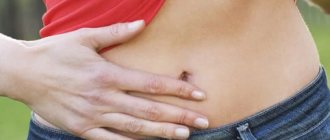
Increased gas formation is a symptom of impaired intestinal absorption.
Like many other diseases, malabsorption syndrome can be diagnosed by some characteristic symptoms:
- frequent diarrhea, accompanied by copious mucus and stench;
- increased gas formation;
- sensations of discomfort, heaviness or even cramps in the stomach, which intensifies many times after each meal;
- fast fatiguability;
- obvious exhaustion, usually accompanied by noticeable weight loss;
- unhealthy pallor and other clinical signs of anemia;
- night blindness (as a rule, such a disorder develops when the body lacks vitamins);
- increased sensitivity of the skin to damage, expressed in the immediate appearance of bruises from almost any mechanical impact, which is a natural result of a lack of vitamin K in the body;
- characteristic bone aches and muscle pain, indicating calcium deficiency.
If you detect several of the above warning signs, the patient should immediately contact a good gastroenterologist to clarify the diagnosis.
The specialist will collect an anamnesis of the disease and prescribe all necessary additional examinations. Thus, today the following research methods are widely used to diagnose intestinal malabsorption syndrome:
- a blood test that determines the deficiency of certain useful substances in the body, as well as confirming (or refuting) the presence of anemia in the patient;
- stool analysis, which reveals the degree of digestibility of healthy fats obtained from food by the body;
- analysis of the current state of the intestinal microflora obtained through a smear;
- a sample of the patient’s exhaled air, confirming or refuting the presence of lactose intolerance in the patient, and also allowing one to determine the approximate number of bacteria in the patient’s intestines;
- endoscopy, used primarily to obtain biological material for another study - biopsy of intestinal tissue;
- X-ray of the intestines (usually using a barium solution, which is necessary to obtain higher-definition images).
How does the digestive system work?
As we said, digestion begins in the mouth.
, where food is crushed during chewing and mixed with saliva.
Saliva is not as aggressive as gastric juice, but it also contains certain enzymes
that start the digestion process and are capable of breaking down starch.
When a person swallows food, it enters the esophagus
, this area located between the pharynx and stomach.
To swallow food, a person must make some effort, because at the junction of the esophagus and stomach there are ring muscles, a kind of valve, the role of which is played by the lower esophageal sphincter
. It opens under pressure from incoming food and allows it to enter the stomach.
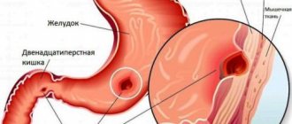
Three important processes occur in the stomach at once
:
- food storage;
- mixing food with gastric juice;
- transporting food to the small intestine.
When a person eats, the stomach works primarily as a “bag” or storage
, where all the food eaten and liquids drunk ends up. To accept all this load, the stomach must be able to increase in size, with the help of the muscles that are located in the upper part of the stomach. As a person consumes food, they relax, which allows the walls of the stomach to stretch.
Mixing of food with gastric juice occurs in the lower part of the stomach
.
A small amount of gastric juice is always present there, but to break down large amounts of food, the body produces additional gastric juice. Gastric juice has a complex chemical composition, based on hydrochloric acid
and digestive enzymes that break down protein. Hydrochloric acid itself is dangerous for the walls of the stomach, but they are covered with a large amount of mucus, which prevents the acid from affecting the walls.
Food mixed with gastric juice passes into the duodenum
, where proteins, fats and carbohydrates are digested under the influence of enzymes of the small intestine and pancreatic juice.
Additional processing occurs with bile, which the rest of the time accumulates in the gallbladder
, and during meals is injected in portions into the duodenum. Bile acids primarily act on fat, breaking it down into small particles that are easily broken down by enzymes.
Substances obtained during the breakdown of food are absorbed through the walls of the small intestine into the blood
and are distributed throughout the body.
Particles that cannot be digested move to the large intestine. In the large intestine, water and remaining vitamins are absorbed from undigested particles, which can be beneficial to the body. The body considers waste to be useless and unsuitable for further use, so it forms feces
, which enter the rectum and are naturally excreted. It is believed that the removal of accumulated feces should occur daily, thus the body cleanses itself.
Library
S.T.
Metelsky Doctor of Biological Sciences, Chief Researcher of the State Research Institute of General Pathology and Pathophysiology of the Russian Academy of Medical Sciences; contact information for correspondence; Moscow, 125315, Baltiyskaya 8. Purpose of the lecture . Consider the physiological mechanisms of absorption in the gastrointestinal tract (GIT). Basic provisions . In the literature, these issues are covered from three sides: 1) topography of absorption of substances in various parts of the gastrointestinal tract - stomach, duodenum, jejunum, ileum and colon; 2) the main functions of enterocytes; 3) the main mechanisms of absorption in the intestine. 7 main mechanisms of absorption of substances in the intestine are considered. Conclusion. Of the entire gastrointestinal tract, the jejunum and ileum are characterized by the widest spectrum of absorption of various compounds.
Understanding the physiological mechanisms of absorption in the small intestine is of great importance in practical gastroenterology. Keywords:
Absorption, ions, sodium, nutrients, gastrointestinal tract, simple diffusion, facilitated diffusion, osmosis, filtration, pericellular transport, active transport, coupled transport, secondary energized transport, endocytosis, transcytosis, P-glycoprotein.
Basic mechanisms of absorption
The wall of the small intestine, where the most intensive absorption of essential nutrients, or nutrients, occurs, consists of the mucosa (villi and intestinal glands), submucosa (where the blood and lymphatic vessels are located), the muscular layer (where the nerve fibers are located) and the serosa. The mucous membrane is formed by villi, covered with single-layer epithelium interspersed with goblet cells; Lymphatic vessels, a capillary network, and nerve fibers pass inside the villi. A characteristic feature of the transport of substances in the epithelium of the small intestine is that it occurs through a monolayer of cells. The absorption surface of such a monolayer is significantly increased due to microvilli. Enterocytes of the small intestine, where the absorption of nutrients (nutrients) mainly occurs, are asymmetrical, or polarized: the apical and basement membranes differ from each other in permeability, a set of enzymes, the magnitude of the electrical potential difference and perform unequal transport functions. Ions enter cells using ion channels or special molecular machines - pumps. Energy for the entry of ions into the cell is usually provided through the plasma membrane by an electrochemical sodium gradient generated and maintained by the functioning of the Na+, K+-ATPase pump. This pump is localized on the basolateral membrane facing the blood (Fig. 1). The energy that can be obtained from the electrochemical potential of Na+ (ion concentration difference + electrical potential difference across the membrane) and which is released when incoming sodium crosses the plasma membrane can be used by other transport systems. Consequently, the Na+, K+-ATPase pump performs two important functions: it pumps Na+ out of cells and generates an electrochemical gradient that provides energy to the mechanisms of solute entry. The term “absorption” refers to a set of processes that ensure the transfer of substances from the intestinal lumen through the epithelial layer into the blood and lymph; secretion is a movement in the opposite direction.
Absorption in various parts of the gastrointestinal tract
20% of alcohol consumed is absorbed in the stomach, as well as short-chain fatty acids. In the duodenum - vitamins A and B1, iron, calcium, glycerol, fatty acids, monoglycerides, amino acids, mono- and disaccharides. In the jejunum - glucose, galactose, amino acids and dipeptides, glycerol and fatty acids, mono- and diglycerides, copper, zinc, potassium, calcium, magnesium, phosphorus, iodine, iron, fat-soluble vitamins D, E and K, a significant part of the vitamin complex B, vitamin C and alcohol residues. The ileum contains disaccharides, sodium, potassium, chloride, calcium, magnesium, phosphorus, iodine, vitamins C, D, E, K, B1, B2, B6, B12 and most of the water. In the large intestine - sodium, potassium, water, gases, some fatty acids formed during the metabolism of plant fibers and undigested starch, vitamins synthesized by bacteria - biotin (vitamin H) and vitamin K.
Main functions of enterocytes
The main functions of enterocytes include the following. Absorption of ions, including sodium, calcium, magnesium and iron, by the mechanism of their active transport. Water absorption (transcellular or pericellular) occurs due to the osmotic gradient formed and maintained by ion pumps, in particular Na+, K+-ATPase. Absorption of sugars. Enzymes (polysaccharidases and disaccharidases) located in the glycocalyx break down large sugar molecules into smaller ones, which are then absorbed. Glucose is transported across the apical membrane of the enterocyte using the Na+-dependent glucose transporter. Glucose moves through the cytosol (cytoplasm) and leaves the enterocyte through the basolateral membrane (into the capillary system) using the GLUT-2 transporter. Galactose is transported using the same transport system. Fructose crosses the apical membrane of the enterocyte using the GLUT-5 transporter. Absorption of peptides and amino acids. In the glycocalyx, peptidase enzymes break down proteins into amino acids and small peptides. Enteropeptidases activate the conversion of pancreatic trypsinogen to trypsin, which in turn activates other pancreatic zymogens. Lipid absorption. Lipids - triglycerides and phospholipids - are broken down and passively diffuse into enterocytes, and free and esterified sterols are absorbed in mixed micelles (see below). Small lipid molecules are transported into the intestinal capillaries through tight junctions. Sterols that enter the enterocyte, including cholesterol, are esterified by the enzyme acyl-CoA: cholesterol acyltransferase (ACAT), together with resynthesized triglycerides, phospholipids and apolipoproteins, is included in chylomicrons, which are secreted into the lymph and then into the bloodstream. Resorption of unconjugated bile salts. Bile that enters the intestinal lumen and is not used in the process of lipid emulsification is reabsorbed in the ileum. The process is known as enterohepatic circulation. Absorption of vitamins. For the absorption of vitamins, as a rule, the absorption mechanisms of other substances are used. A special mechanism exists for the absorption of vitamin B12 (see below). Secretion of immunoglobulins. IgA from mucosal plasma cells is absorbed through the basolateral surface through the mechanism of receptor-mediated endocytosis and released into the intestinal lumen as a receptor-IgA complex. The presence of a receptor gives the molecule additional stability.
Basic mechanisms of absorption of compounds in the intestine
In Fig. 2 presents the main mechanisms of absorption of substances. Let us consider these mechanisms in more detail. Presystemic metabolism, or metabolism (effect) of the first passage of the intestinal wall. A phenomenon in which the concentration of a substance decreases sharply before entering the bloodstream. However, if the administered substance is a substrate of P-glycoprotein (see below), its molecules can be repeatedly transferred into and out of enterocytes, as a result of which the likelihood of metabolism of this compound in enterocytes increases. P-glycoprotein is highly expressed in normal cells lining the intestine, renal proximal tubules, blood-brain barrier capillaries, and liver cells. P-glycoprotein transporters are members of the largest and most ancient transporter superfamily, present in organisms from prokaryotes to humans. These are transmembrane proteins whose function is to transport a wide range of
substances through extra- and intracellular membranes, including metabolic products, lipids and drugs. Such proteins are classified as ATP-binding cassette transporters (ABC transporters) based on their sequence and the arrangement of the ATP-binding domain. ABC transporters influence the drug resistance of tumors, cystic fibrosis, bacterial resistance to many drugs, and some other phenomena. Passive transport of substances through the epithelial layer. Passive transport of substances through a monolayer of enterocytes occurs without the expenditure of free energy and can be carried out either transcellular or pericellular. This type of transport includes simple diffusion (Fig. 3), osmosis (Fig. 4) and filtration (Fig. 5). The driving force for the diffusion of solute molecules is its concentration gradient. The dependence of the rate of diffusion of a substance on its concentration is linear. Diffusion is the least specific and, apparently, the slowest transport process. With osmosis, which is a type of diffusion transfer, movement occurs in accordance with the concentration gradient of free (not associated with the substance) solvent (water) molecules.
The filtration process involves the transfer of a solution through a porous membrane. The passive transfer of substances through membranes also includes facilitated diffusion - the transfer of substances using transporters, i.e., special channels or pores (Fig. 6). Loose diffusion is substrate specific. The dependence of the rate of the process at sufficiently high concentrations of the transported substance reaches saturation, since the transfer of the next molecule is inhibited by waiting for the transporter to be free from transfer of the previous one. Pericellular transport is the transport of connections between cells through the area of tight junctions (Fig. 7); it does not require energy expenditure. The structure and permeability of tight junctions of the small intestine are currently being actively studied and debated. For example, it is known that claudin-2 is responsible for the selectivity of tight junctions for sodium. Another possibility is that intercellular transfer occurs due to some defect in the epithelial layer. Such movement can occur along intercellular areas in those places where desquamation of individual cells occurs. This path may be a gateway for the penetration of foreign macromolecules directly into the blood or tissue fluids. Endocytosis, exocytosis, receptor-mediated transport (Fig. and transcytosis. Endocytosis is the vesicular uptake of fluid, macromolecules or small particles into a cell. There are three mechanisms of endocytosis: pinocytosis (from the Greek words “drink” and “cell”), phagocytosis (from the Greek words “eat” and “cell”) and receptor-mediated endocytosis or clathrin-dependent endocytosis. Violations of this mechanism lead to the development of certain diseases. Many intestinal toxins, in particular cholera, enter enterocytes precisely through this mechanism. During pinocytosis, the flexible plasma membrane forms an invagination ( invagination) in the form of a pit. Such a pit is filled with liquid from the external environment. Then it is detached from the membrane and, in the form of a vesicle, moves into the cytoplasm, where its membrane walls are digested and the contents are released. Thanks to this process, cells can absorb both large molecules and various ions that are unable to cross the membrane on their own.Pinocytosis is often observed in cells whose function is related to absorption. This is an extremely intensive process: in some cells, 100% of the plasma membrane is absorbed and restored in just an hour. During phagocytosis (a phenomenon discovered by the Russian scientist I.I. Mechnikov in 1882), the outgrowths of the cytoplasm capture droplets of liquid containing any dense (living or nonliving) particles (up to 0.5 microns), and draw them into the thickness of the cytoplasm, where hydrolyzing enzymes digest the absorbed material, breaking it down into fragments that can be absorbed by the cell. Phagocytosis occurs via a clathrin-independent actin-dependent mechanism; This is the main mechanism of defense of the host body against microorganisms. Phagocytosis of damaged or aged cells is necessary for tissue renewal and wound healing. In receptor-mediated endocytosis (see Fig., specific surface receptors are used for the transfer of molecules. This mechanism has the following properties - specificity, the ability to concentrate the ligand on the cell surface, refractoriness. If a specific receptor, after binding the ligand and its absorption, does not return to the membrane, the cell becomes refractory to this ligand. Using the endocytotic vesicular mechanism, both high molecular weight compounds such as vitamin B12, ferritin and hemoglobin, and low molecular weight ones - calcium, iron, etc. are absorbed. The role of endocytosis is especially great in the early postnatal period. In an adult, the pinocytotic type of absorption is of significant importance in provision of the body with nutrients apparently does not.Transcytosis is a mechanism by which molecules that enter the cell from outside can be delivered to various compartments within the cell or even move from one layer of cells to another.
One well-studied example of transcytosis is the penetration of some maternal immunoglobulins through the intestinal epithelial cells of the newborn. Maternal antibodies enter the baby's body with milk. Antibodies bound to the appropriate receptors are sorted into early endosomes of digestive tract cells, then, with the help of other vesicles, pass through the epithelial cell and fuse with the plasma membrane at the basolateral surface. Here the ligands are released from the receptors. The immunoglobulins then collect in the lymphatic vessels and enter the newborn's bloodstream. Consideration of absorption mechanisms from the point of view of individual groups of substances and compounds will be presented in one of the following issues of the journal. The work was supported by the Russian Foundation for Basic Research grant 09-04-01698 References:
1. Metelsky S.T. Transport processes and membrane digestion in the mucous membrane of the small intestine. Electrophysiological model. – M.: Anacharsis, 2007. – 272 p. 2. General course of human and animal physiology. - Book 2. Physiology of visceral systems / Ed. HELL. Nozdracheva. – M.: Higher School, 1991. – P. 356–404. 3. Membrane digestion. New facts and concepts / Ed. AM Ugolev. – M.: MIR Publishers, 1989. – 288 p. 4. Tansey T., Christie DA, Tansey EM Intestinal absorption. – London: Wellcome Trust, 2000. – 81 p
article taken from the website of the Russian Journal of Gastroenterology, Hepatology, Coloproctology
The article is posted at:
https://www.gastro-j.ru/article/33-fiziologicheskie-mehanizmyi-vsasyivaniya-v-nbsp-kishechnike/show/full/
What can cause disruptions in the gastrointestinal tract?
A person’s general well-being largely depends on the functioning of his gastrointestinal tract
, various diseases of the stomach and gallbladder can disrupt this work, but the person himself can harm himself.
Frequent “man-made” causes of disturbances in the functioning of the stomach
are:
- smoking and alcohol abuse;
- unbalanced diets;
- food poisoning caused by poor quality food.
It must be remembered that digestion
is a complex multi-step process, so it is important that it is not disrupted at any stage, from the oral cavity to the rectum. The gastrointestinal tract ensures the breakdown of food into the simplest compounds, which the body subsequently uses to build new tissues and to obtain energy; without this, the development and functioning of the body is impossible.
Carbohydrates
present in plant foods mainly in the form of starch. During digestion, it is converted into glucose, which can be stored in the form of a polymer - glycogen - and used by the body. The starch molecule is a very large polymer formed by many glucose molecules. In its raw form, starch is enclosed in granules that must be broken down before it can be converted into glucose. Processing and cooking lead to the destruction of some of the starch granules.
Also on topic:
APPETITE
Some foods contain carbohydrates in the form of disaccharides. These relatively simple sugars, particularly sucrose (cane sugar) and lactose (milk sugar), are converted into even simpler compounds called monosaccharides during digestion. The latter do not need to be digested.