general description
Feces are formed in the large intestine. It consists of water, remnants of food taken and secretions of the gastrointestinal tract, products of the transformation of bile pigments, bacteria, etc. For the diagnosis of diseases associated with the digestive organs, stool examination in some cases can be crucial. A general stool analysis (coprogram) includes macroscopic, chemical and microscopic examination.
Macroscopic examination
Quantity
In pathology, the amount of feces decreases with prolonged constipation caused by chronic colitis, peptic ulcers and other conditions associated with increased absorption of fluid in the intestines. With inflammatory processes in the intestines, colitis with diarrhea, and accelerated evacuation from the intestines, the amount of feces increases.
Consistency
Thick consistency - with constant constipation due to excessive absorption of water. Liquid or mushy consistency of stool - with increased peristalsis (due to insufficient absorption of water) or with abundant secretion of inflammatory exudate and mucus by the intestinal wall. Ointment-like consistency - in chronic pancreatitis with exocrine insufficiency. Foamy consistency - with enhanced fermentation processes in the large intestine and the formation of a large amount of carbon dioxide.
Form
The form of feces in the form of “large lumps” - when feces remain in the colon for a long time (hypomotor dysfunction of the colon in people with a sedentary lifestyle or who do not eat rough food, as well as in cases of colon cancer, diverticular disease). The form in the form of small lumps - “sheep feces” indicates a spastic state of the intestines, during fasting, stomach and duodenal ulcers, a reflex nature after appendectomy, with hemorrhoids, anal fissure. Ribbon or “pencil” shape - for diseases accompanied by stenosis or severe and prolonged spasm of the rectum, for rectal tumors. Unformed feces are a sign of maldigestion and malabsorption syndrome.
Color
If staining of stool by food or drugs is excluded, then color changes are most likely due to pathological changes. Grayish-white, clayey (acholic feces) occurs with obstruction of the biliary tract (stone, tumor, spasm or stenosis of the sphincter of Oddi) or with liver failure (acute hepatitis, cirrhosis of the liver). Black feces (tarry) - bleeding from the stomach, esophagus and small intestine. Pronounced red color - with bleeding from the distal parts of the colon and rectum (tumor, ulcers, hemorrhoids). Gray inflammatory exudate with fibrin flakes and pieces of the colon mucosa (“rice water”) - with cholera. The jelly-like character is deep pink or red in amoebiasis. In typhoid fever, the stool looks like “pea soup.” With putrefactive processes in the intestines, the feces are dark in color, with fermentative dyspepsia - light yellow.
Slime
When the distal colon (especially the rectum) is affected, the mucus occurs in the form of lumps, strands, ribbons, or a glassy mass. With enteritis, the mucus is soft, viscous, mixing with feces, giving it a jelly-like appearance. Mucus, covering the outside of formed feces in the form of thin lumps, occurs with constipation and inflammation of the large intestine (colitis).
Blood
When bleeding from the distal parts of the colon, the blood is located in the form of streaks, shreds and clots on formed stool. Scarlet blood occurs when bleeding from the lower parts of the sigmoid and rectum (hemorrhoids, fissures, ulcers, tumors). Black feces (melena) occur when there is bleeding from the upper digestive system (esophagus, stomach, duodenum). Blood in the stool can be found in infectious diseases (dysentery), ulcerative colitis, Crohn's disease, disintegrating colon tumors.
Pus
Pus on the surface of the stool occurs with severe inflammation and ulceration of the mucous membrane of the colon (ulcerative colitis, dysentery, disintegration of an intestinal tumor, intestinal tuberculosis), often together with blood and mucus. Large amounts of pus without mucus are observed when opening paraintestinal abscesses.
Leftover undigested food (lientorrhea)
The release of undigested food residues occurs with severe insufficiency of gastric and pancreatic digestion.
What can the results of the analysis indicate?
- Color. Normally, it ranges from light brown or olive to dark brown. Black stools are observed with gastrointestinal bleeding, light yellow stools are observed with insufficient pancreatic function.
- Consistency. Loose stools may indicate colitis, impaired motor function of the intestine, or disorders of its innervation. Ointment-like feces are characteristic of diseases of the pancreas, and sheep feces are characteristic of inflammatory bowel diseases.
- Reaction. Normal stool has a pH of 7-8. An increase in indicators is observed with disorders of the stomach functions, colitis, a decrease is observed with increased fermentation processes in the intestines.
- Slime. Determined in inflammatory bowel diseases (colitis, enterocolitis).
- Leftover undigested food. Indicate insufficiency of gastric or intestinal digestion, accelerated evacuation of food through the intestines.
- Muscle fibers. Present in case of insufficiency of gastric juice or digestive enzymes, disturbances of absorption processes in the intestines.
- Fat. The presence of a small amount of fat in the stool is normal in children; in adults, it indicates diseases of the pancreas and liver.
- Leukocytes. Appear against the background of infections and inflammatory diseases of the gastrointestinal tract.
- Red blood cells. A sign of ulcerative or malignant diseases of the gastrointestinal tract, polyps, hemorrhoids, rectal fissures.
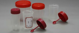
You can submit stool for general clinical examination at the Spectra clinic. Material for stool analysis is accepted on weekdays and weekends. The conclusion is ready within 4-5 working days. To interpret the results, you should contact a gastroenterologist or therapist.
Chemical research
Fecal reaction
An acidic reaction (pH 5.0-6.5) is observed when iodophilic flora is activated, producing carbon dioxide and organic acids (fermentative dyspepsia). An alkaline reaction (pH 8.0-10.0) occurs with insufficient digestion of food, with colitis with constipation, sharply alkaline with putrefactive and fermentative dyspepsia.
Reaction to blood (Gregersen reaction)
A positive reaction to blood indicates bleeding in any part of the gastrointestinal tract (bleeding from the gums, rupture of varicose veins of the esophagus, erosive and ulcerative lesions of the gastrointestinal tract, tumors of any part of the gastrointestinal tract in the stage of decay).
Reaction to stercobilin
The absence or sharp decrease in the amount of stercobilin in the feces (the reaction to stercobilin is negative) indicates obstruction of the common bile duct with a stone, compression by a tumor, stricture, stenosis of the common bile duct, or a sharp decrease in liver function (for example, in acute viral hepatitis). An increase in the amount of stercobilin in feces occurs with massive hemolysis of red blood cells (hemolytic jaundice) or increased bile secretion.
Reaction to bilirubin
The detection of unchanged bilirubin in the stool of an adult indicates a disruption in the process of bilirubin recovery in the intestine under the influence of microbial flora. Bilirubin can appear during rapid evacuation of food (sharp increase in intestinal motility), severe dysbiosis (syndrome of bacterial overgrowth in the colon) after taking antibacterial drugs.
Vishnyakov-Triboulet reaction (for soluble protein)
The Vishnyakov-Triboulet reaction is used to identify a hidden inflammatory process. The detection of soluble protein in stool indicates inflammation of the intestinal mucosa (ulcerative colitis, Crohn's disease).
Why does large stool appear?
Large and thick poop that is quite difficult to move through the intestines is a clear sign of constipation.
The Bristol Stool Form Scale distinguishes 2 types of thick feces:
- Large and thick poop is sausage-shaped and has a lumpy structure. The diameter of such feces reaches 3-4 cm. Defecation can cause severe pain due to the fact that the diameter of the anus is less than 5 cm and is greatly stretched during defecation. Such feces belong to the second type on the scale of fecal forms and indicate fairly rare bowel movements.
- The second type of large poop is similar to the previous one, but has a more modest diameter from 2 to 4 cm and has cracks on the surface. Defecation is also accompanied by pain, the anus is greatly stretched until cracks appear. Based on the scale data, poop of these sizes is classified as the third type. Such fecal masses indicate that bowel movements occur more often than in the second type of feces, but there is hidden constipation.
Microscopic examination
Muscle fibers - with striations (unchanged, undigested) and without striations (changed, digested). A large number of changed and unchanged muscle fibers in the feces (creatorrhoea) indicates a violation of proteolysis (protein digestion):
- in conditions accompanied by achlorhydria (lack of free HCl in gastric juice) and achylia (complete absence of secretion of HCl, pepsin and other components of gastric juice): atrophic pangastritis, a condition after gastric resection;
- with accelerated evacuation of food chyme from the intestines;
- in case of violation of the exocrine function of the pancreas;
- with putrefactive dyspepsia.
Connective tissue (remnants of undigested vessels, ligaments, fascia, cartilage). The presence of connective tissue in the feces indicates a deficiency of proteolytic enzymes of the stomach and is observed with hypo- and achlorhydria, achylia.
Fat is neutral. Fatty acid. Salts of fatty acids (soaps)
The appearance of large amounts of neutral fat, fatty acids and soaps in the stool is called steatorrhea. This happens:
- with exocrine pancreatic insufficiency, a mechanical obstruction to the outflow of pancreatic juice, when steatorrhea is represented by neutral fat;
- if the flow of bile into the duodenum is impaired and if the absorption of fatty acids in the small intestine is impaired, fatty acids or salts of fatty acids (soaps) are found in the feces.
Plant fiber
Digestible - found in the pulp of vegetables, fruits, legumes and grains. Indigestible fiber (the skin of fruits and vegetables, plant hairs, the epidermis of cereals) has no diagnostic value, since there are no enzymes in the human digestive system that break it down. It is found in large quantities during rapid evacuation of food from the stomach, achlorhydria, achylia, and the syndrome of bacterial overgrowth in the colon.
Starch
The presence of a large amount of starch in the feces is called amilorrhea and is observed more often with increased intestinal motility, fermentative dyspepsia, and less often with exocrine insufficiency of pancreatic digestion.
Iodophilic microflora (clostridia)
With a large amount of carbohydrates, clostridia multiply intensively. A large number of clostridia is regarded as fermentative dysbiosis.
Epithelium
A large amount of columnar epithelium in the feces is observed in acute and chronic colitis of various etiologies.
Leukocytes
A large number of leukocytes (usually neutrophils) are observed in acute and chronic enteritis and colitis of various etiologies, ulcerative necrotic lesions of the intestinal mucosa, intestinal tuberculosis, and dysentery.
Red blood cells
The appearance of slightly changed red blood cells in the stool indicates the presence of bleeding from the colon, mainly from its distal parts (ulceration of the mucous membrane, disintegrating tumor of the rectum and sigmoid colon, anal fissures, hemorrhoids). A large number of red blood cells in combination with leukocytes and columnar epithelium is characteristic of ulcerative colitis, Crohn's disease with damage to the colon, polyposis and malignant neoplasms of the colon.
Worm eggs
Eggs of roundworms, tapeworms, etc. indicate a corresponding helminthic infestation.
Pathogenic protozoa
Cysts of dysenteric amoeba, Giardia, etc. indicate a corresponding invasion by protozoa.
Yeast cells
Found in feces during treatment with antibiotics and corticosteroids. Identification of the fungus Candida albicans is carried out by culturing on special media (Sabouraud's medium, Microstix Candida) and indicates a fungal infection of the intestine.
Calcium oxalate (oxalic lime crystals)
Detection of crystals is a sign of achlorhydria.
Triple phosphate crystals (ammonium phosphate-magnesia)
Triple phosphate crystals found in feces (pH 8.5-10.0) immediately after defecation indicate increased protein putrefaction in the colon.
Norms
Macroscopic examination
| Parameter | Norm |
| Quantity | A healthy person produces an average of 100-200 g of feces per day. Normally, feces contain about 80% water and 20% dry matter. With a vegetarian diet, the amount of feces can reach 400-500 g per day; when using easily digestible food, the amount of feces decreases. |
| Consistency | Normally, formed feces have a dense consistency. Pasty feces can occur normally and are caused by the intake of predominantly plant foods. |
| Form | Normally cylindrical. |
| Smell | Normally, stool has a mild odor, which is called fecal (ordinary). It may intensify with the predominance of meat products in the diet, with putrefactive dyspepsia and weaken with a dairy-vegetable diet, constipation. |
| Color | Normally, stool is brown in color. When eating dairy foods, stool turns yellowish-brown, and meat stool turns dark brown. Ingestion of plant foods and some medications can change the color of stool (beets - reddish; blueberries, blackcurrants, blackberries, coffee, cocoa - dark brown; bismuth, iron color stool black). |
| Slime | Normally absent (or in scanty quantities). |
| Blood | Normally absent. |
| Pus | Normally absent. |
| Leftover undigested food (lientorrhea) | Normally none. |
Chemical research
| Parameter | Norm |
| Fecal reaction | Normally neutral, less often slightly alkaline or slightly acidic. Protein nutrition causes a shift in the reaction towards the alkaline side, while carbohydrate nutrition causes the reaction to shift towards the acidic side. |
| Reaction to blood (Gregersen reaction) | Normally negative |
| Reaction to stercobilin | Normally positive. |
| Reaction to bilirubin | Normally negative. |
| Vishnyakov-Triboulet reaction (for soluble protein) | Normally negative. |
Microscopic examination
| Parameter | Norm |
| Muscle fibers | Normally absent or single in the field of view. |
| Connective tissue (remnants of undigested vessels, ligaments, fascia, cartilage) | Normally absent. |
| Fat is neutral. Fatty acid. Salts of fatty acids (soaps). | Normally there are no or scanty amounts of fatty acid salts. |
| Plant fiber | Normally, there are single cells in the p/z. |
| Starch | Normally absent (or single starch cells). |
| Iodophilic microflora (clostridia) | Normally, single in rare areas (normally, iodophilic flora lives in the ileocecal region of the large intestine). |
| Epithelium | Normally, there are no or single columnar epithelial cells in the p/z. |
| Leukocytes | Normally, there are no or single neutrophils in the p/z. |
| Red blood cells | Normally none. |
| Worm eggs | Normally none. |
| Pathogenic protozoa | Normally none. |
| Yeast cells | Normally none. |
| Calcium oxalate (oxalic lime crystals) | Normally none. |
| Triple phosphate crystals (ammonium phosphate-magnesia) | Normally none. |
Diseases for which a doctor may prescribe a general stool test (coprogram)
Anal fissure
With an anal fissure, feces may take the form of small lumps (“sheep feces” indicates a spastic condition of the intestines). Due to bleeding, scarlet blood and slightly changed red blood cells may be present in the stool.
Crohn's disease
With Crohn's disease, you may find blood in your stool. The Vishnyakov-Triboulet reaction reveals soluble protein in it. Crohn's disease affecting the colon is characterized by the presence in the feces of a large number of red blood cells in combination with leukocytes and columnar epithelium.
Colon diverticulosis
With diverticular disease, due to the long stay of stool in the colon, it takes the form of “large lumps”.
Duodenal ulcer
With a duodenal ulcer, the feces have the form of small lumps (“sheep feces” indicates a spastic condition of the intestines).
Stomach ulcer
With a stomach ulcer, the feces have the form of small lumps (“sheep feces” indicates a spastic condition of the intestines).
Chronic pancreatitis
In chronic pancreatitis with exocrine insufficiency, stool may have a pasty consistency.
Hemolytic anemia
With hemolytic jaundice (anemia), due to massive hemolysis of red blood cells, the amount of stercobilin in the feces increases.
Benign neoplasms of the colon
With a tumor accompanied by bleeding from the distal colon, the stool may have a pronounced red color. With disintegrating colon tumors, blood may be found in the stool. Pus on the surface of the stool occurs when there is severe inflammation and ulceration of the colon mucosa (disintegration of an intestinal tumor), often along with blood and mucus. When a colon tumor is in the stage of decay due to bleeding, the reaction to blood (Gregersen reaction) is positive.
Intestinal helminthiases
With helminthic infestation, the feces contain eggs of roundworms, tapeworms, etc.
Cirrhosis of the liver
In liver failure, including liver cirrhosis, the stool is grayish-white, clayey (acholic).
Ulcerative colitis
With colitis, there is mucus covering the outside of the stool in the form of thin lumps. With ulcerative colitis, blood may be found in the stool; pus on the surface of the stool, often along with blood and mucus; soluble protein in the Vishnyakov-Triboulet reaction; a large number of leukocytes (usually neutrophils); a large number of red blood cells in combination with leukocytes and columnar epithelium.
Constipation
With prolonged constipation caused by chronic colitis, peptic ulcers and other conditions associated with increased absorption of fluid in the intestine, the amount of feces decreases. With constant constipation due to excessive absorption of water, the consistency of stool is dense. With constipation, there may be mucus covering the outside of the stool in the form of thin lumps.
Malignant neoplasm of the colon
The form of feces in the form of “large lumps” - when feces remain in the colon for a long time - is noted in colon cancer. Pronounced red color of stool - with a tumor accompanied by bleeding from the distal parts of the colon and rectum. Blood in the stool can be found in disintegrating colon tumors. Pus on the surface of the stool occurs when there is severe inflammation and ulceration of the colon mucosa (disintegration of an intestinal tumor), often along with blood and mucus. A positive reaction to blood (Gregersen reaction) indicates bleeding with a tumor of the colon in the stage of decay. A large number of red blood cells in combination with leukocytes and columnar epithelium is characteristic of malignant neoplasms of the colon.
Irritable bowel syndrome, chronic colitis
With colitis with diarrhea, the amount of feces increases. The amount of feces decreases with prolonged constipation caused by chronic colitis. Mucus covering the outside of formed feces in the form of thin lumps is found in colitis. An alkaline reaction (pH 8.0-10.0) occurs in colitis with constipation. A large number of leukocytes (usually neutrophils) are observed in colitis of various etiologies.
Cholera
In cholera, stool looks like a gray inflammatory exudate with fibrin flakes and pieces of the colon mucosa (“rice water”).
Amoebiasis
With amebiasis, the stool is jelly-like, deep pink or red.
Typhoid fever
In typhoid fever, the stool looks like “pea soup.”
Clinical stool analysis
When studying the physical properties, the quantity, consistency, shape, color and smell of feces are assessed, and macroscopically visible impurities are examined.
The amount of feces excreted per day depends on the composition and amount of food taken the day before and can fluctuate within significant limits.
With a normal diet, the daily amount
feces is 120–200 g. The amount of feces is reduced when animal proteins predominate in the diet and increases with a plant-based diet. An increase in the daily amount of feces (polyfecation) occurs when there are disturbances in the functional state of the gastrointestinal tract: impaired absorption, bile secretion (achilya), damage to the pancreas, and enteritis. A decrease in the daily amount of feces develops with chronic constipation.
Consistency and shape
depend on the percentage of water. Normally, feces are shaped, sausage-shaped, and contain 75–80% water. With an increase in the percentage of water due to increased intestinal motility (insufficient absorption of water), abundant secretion of inflammatory exudate and mucus by the intestinal wall, the stool becomes unformed, mushy or liquid. With constant constipation, due to excessive absorption of water, the feces become dense and may have the appearance of small balls - “sheep feces”. With stenosis or spastic narrowing of the lower part of the sigmoid or rectum, special forms of feces may be observed: “ribbon-shaped”, “pencil-shaped”.
Stool color
in a healthy person is due to the presence of stercobilin and mesobilifuscin, which are formed from bilirubin in bile under the influence of intestinal microflora and give it various shades of brown color.
| Color | Probable Causes |
| Dark brown | Normal feces on a mixed diet |
| Black-brown | Meat diet |
| Light brown | Plant based diet |
| Brown-red | Bleeding in the lower parts of the digestive tract, taking purgen, cocoa |
| Black | Bleeding in the upper digestive tract, taking bismuth |
| Greenish black | Taking iron supplements |
| Green | Presence of bilirubin and biliverdin, increased peristalsis, vegetable diet |
| Greenish yellow | Carbohydrate fermentation |
| Golden yellow | Presence of unchanged bilirubin (in infants) |
| Orange-light yellow | Dairy diet |
| White or grayish-white (acholic stool) | Stopping the flow of bile into the intestines |
Smell
Normally unpleasant, but not harsh and is caused mainly by skatole, indole and, to a lesser extent, by phenol, ortho- and para-cresols. These organic aromatic compounds are formed during the breakdown of proteins. The smell intensifies with the predominance of protein-rich foods in the diet, with diarrhea, with putrefactive dyspepsia. The smell weakens with a predominantly plant-based and dairy diet, with constipation, and with fasting. With fermentative dyspepsia, stool acquires a sour odor. In the analysis, the smell of feces is indicated if it differs sharply from usual.
Macroscopically
visible impurities in the stool can be represented by undigested food debris, mucus, blood, pus, stones, and parasites. Normally, undigested food remains in the stool are not macroscopically detectable. Severe insufficiency of gastric and pancreatic digestion is accompanied by the release of lumps of undigested food - lientorrhea. Excessive mucus is detected macroscopically in the form of strands, flakes, and dense formations and indicates inflammation of the intestinal mucosa. Fecal stones can be biliary, pancreatic or intestinal in origin (coprolites).
Gallstones
There are cholesterol, calcareous, bilirubin, mixed. They are usually discovered after an attack of hepatic colic.
Pancreatic stones consist of lime carbonate or phosphate, are small in size and have an uneven surface.
Coprolites are dark brown formations that consist of an organic core and layered mineral salts (phosphates), undigested food residues, poorly soluble drugs, etc.
Parasites can be detected with the naked eye in the form of whole individuals (roundworms, whipworms, pinworms), as well as their fragments: scolex and segments (pork and bovine tapeworms, broad tapeworm).