Liver diseases occur at any age. They can be congenital or acquired during life. It is very important to diagnose them at an early stage. Computed tomography of the liver is a very reliable and reliable method for diagnosing diseases of the organ.
Indications for CT scan of the liver are:
- Preoperative study.
- Suspicion of tumor formations, as well as their diagnosis.
- Identifying the consequences of injury.
- Diagnosis of complications after surgery: hematomas, abscesses and others.
- Suspicion of vascular damage to the organ.
- If it is not possible to perform an ultrasound if there is excess fat in the area to be examined.
Organ structure
The liver parenchyma or its tissue is divided into lobules, between them there are vessels, arteries through which blood passes, and bile ducts. Thanks to this structure of the liver, blood flow access to all parts of the organ is ensured. The liver is responsible for the synthesis of many substances, and all this proceeds calmly. When undesirable changes appear, the functionality of the organ decreases.
It is completely impossible to avoid changes in the parenchyma, especially with age-related factors. However, unfortunately, often the person himself is to blame for liver dysfunction. The reason for this is an incorrect lifestyle, bad habits, and dirty ecology.
At first, it is difficult to notice any changes in the structure of the organ; even ultrasound does not help to identify them. Therefore, if various diseases are suspected, a comprehensive diagnosis is required, not only of the liver, but also of the pancreas and other gastrointestinal organs.
Types of changes
In total, there are two types of parenchymal changes:
- focal – affect certain areas of tissue;
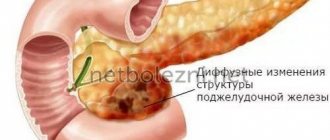
- diffuse - the lesion affects the entire organ.
The most common are diffuse changes, and they are also the most dangerous. With focal inflammation, the entire liver parenchyma is not affected, but this is usually only temporary. For example, these include cysts and abscesses, but often immediately against the background of the appearance of these focal changes, diffuse changes appear that affect the entire organ.
Diffuse liver changes
Pathological changes in the liver parenchyma, called diffuse, are of several types:
- Hypertrophy. It implies an enlargement of the organ, in which the natural liver tissue dies and fibrous tissue grows in its place. Often hypertrophy is one of the signs of hepatitis of various forms. An increase in fibrous tissue is called hepatomegaly.
- Heterogeneous fabric structure. The evenness of the contours of the parenchyma gradually changes, and at first it is difficult to notice this on ultrasound. The diffusely heterogeneous structure is clearly visible with other diagnostic methods - CT, MRI.
- Dystrophic and other structural changes. The liver decreases in size and its functionality suffers. The contours of the organ become uneven. This condition increases the risk of developing chronic liver failure. Dystrophy can be congenital or acquired.
- Fatty parenchymal degeneration. This condition means that there is an excessive amount of fat in the liver tissue. This is often associated with lipid metabolism disorders.
- Reactive changes. They are a secondary disease that occurs against the background of inflammation of other organs of the gastrointestinal tract, poisoning by toxins, malfunctions of the endocrine system, etc.
Normally, the structure of the parenchyma is homogeneous and of low density. Any deviations from this condition mean changes in the organ, which requires urgent diagnosis.
One of the complications of many diffuse changes is cirrhosis, a disease in which liver cells simply do not work.
Causes
The main causes of diffuse changes in the parenchyma are incorrect human habits. In more than 80% of cases of liver disease in young people, their lifestyle is taken into account. Among the possible reasons, the most common are:
- Poor nutrition. The main problems with choosing a diet are eating semi-finished products, fast foods, fried, fatty, and smoked foods. Non-compliance with food intake, overeating, and incorrect diets for weight loss also play an important role. If a child has had such nutritional problems since school age, then by the age of 30-35, if not earlier, the liver will begin to show the first alarming signs.
- Alcohol. Drinking alcohol in large quantities harms the liver. She has to process alcohol, it is broken down by her enzymes. Harmful compounds are produced, as a result of which parenchyma cells die and fat cells appear in their place.
- Ecology. Environmental pollution negatively affects the functioning of many internal organs, among which the liver is especially affected. All toxic substances that enter the body enter the liver through the bloodstream. People living in large cities are most susceptible to negative environmental impacts.
- Side effects of medications. Some medications contain potent components that are difficult to process by the liver. Danger arises when they are used incorrectly or excessively.
- Stress. When a person experiences a strong emotional shock, adrenaline is released. The liver must dissolve it, but for it this hormone is toxic. One-time experiences are not so dangerous, but if a person lives in constant stress, this will affect the functioning of the liver.
Some factors that provoke the development of diffuse changes do not depend on a person’s lifestyle, but they are less common. These include heredity and congenital pathologies.
Symptoms of diffuse changes
Damage to the liver tissue does not have pronounced signs, which creates difficulties with the timely diagnosis of diseases. Typically, patients complain about the functioning of the gastrointestinal tract, without even suspecting the presence of liver problems.
The main symptoms of diffuse changes are:
- pain in the right hypochondrium;
- yellowing of the tongue, bad breath;
- feeling of heaviness after eating food, especially fatty or fried food;
- frequent nausea;
- feeling of weakness;
- fast fatiguability;
- headache.
Lung damage 25-50%
Corresponds to KT-2. Community-acquired pneumonia with a favorable or conditionally favorable prognosis. The level of blood oxygen saturation decreases. If the body cannot cope with a viral infection on its own, pneumonia can progress to the next stage quite quickly. The prognosis depends on the patient’s age, his immune system and some individual characteristics of the body.
If the lungs are affected by 25, 30, 40 percent, intensive therapy is necessary - it is important to prevent further spread of the viral infection through the lung parenchyma. As a rule, with CT-2, patients are treated at home after consulting a doctor. If the symptoms of a respiratory disease intensify and therapy does not bring positive results, the patient may be hospitalized at the discretion of the attending physician or emergency physician.
Ultrasound and increased echogenicity
The main diagnostic method to determine the size of the liver and each of its lobes is ultrasound. But the most important parameter is the echogenicity of the organ. An ultrasound machine generates waves. It is impossible for a person to hear or feel them. The tissue of each organ reacts differently to the action of waves, which is called the echogenicity index. The device records reflected waves, which allows you to draw conclusions about the condition of the organ.
Echogenicity reflects the density of the parenchyma. If it is decreased or increased, these are signs of violations. The normal density for healthy liver parenchyma is 58-61 HU (HU is a unit of measurement for organ density during ultrasound). An indicator of 57 and below is a symptom of low density, above 62 - increased density.
Ultrasound may reveal areas of parenchyma with reduced density - hypoechoic. This zone looks like a dark spot on the monitor screen; using research, its exact dimensions are determined. Hypoechoic zones in most cases turn out to be benign or malignant formations.
Echo signs indicating increased density may indicate the presence of various liver diseases, such as hepatosis, hypertrophy, and others. Diffuse compaction is less common than reduced density.
Degrees of lung parenchymal involvement
The percentage of parenchyma involvement is determined by the number of areas of compaction of lung tissue (for example, by “ground glass”, but not only). On CT images, infiltrates and signs of pneumofibrosis are visualized in a relatively lighter color, since tissue that is denser in texture with excess fluid and cellular components transmits X-rays less well.
- Involvement of the lung parenchyma from 0 to
- Lung parenchymal involvement > 5% –
- Lung parenchymal involvement > 25% –
- Lung parenchymal involvement > 50% –
- Involvement of the lung parenchyma > 75% – severe pneumonia (CT-4).
Precision assessment of the respiratory organ during coronavirus on CT allows you to see all possible complications even with a small percentage of damage, and with a significant percentage, it allows you to timely understand when the patient needs hospitalization in a medical facility.
Pathologies diagnosed using ultrasound
In conclusion, the specialist indicates the homogeneity, density, and size of the organ. Diffuse changes in the parenchyma are not the only pathology that can be diagnosed by ultrasound. Other common phenomena include:
- cysts, other neoplasms - more common in women;
- congenital pathologies - develop in the fetus in the womb due to a failure in the formation of ducts;
- echinococcal lesions - arise due to the destructive effects of parasites, often a tapeworm;
- injuries – often associated with tissue rupture due to a blow, fall, or broken ribs;
- benign or malignant tumors.
The doctor who performed the ultrasound gives an opinion that is only preliminary. Then the attending physician makes an accurate diagnosis, and if necessary, prescribes additional examination.
There is no cause for concern if the size, shape, structure, ducts, echogenicity, location of veins and bile ducts are normal - no additional diagnostics are needed
How is the percentage of lung parenchymal involvement determined on CT?
In 2022, CT diagnostic specialists adopted an empirical scale for visual assessment of the lungs. If the abbreviations KT-0, KT-1, KT-2, KT-3 or KT-4 are indicated in the report, then the doctor examined the lungs for inflammatory foci and infiltrates associated with COVID-19.
The objectivity of CT diagnostics is ensured by modern software. At the stage of computer image processing, the program automatically or semi-automatically identifies areas of ground glass and consolidation, measuring the volume of affected tissue and calculating the density.
During consolidation, the alveoli are filled with liquid, and accordingly the texture of the lung tissue is more dense, while “frosted glass” indicates a less severe violation of pneumatization - liquid exudate is also present in these areas, but in smaller quantities. The difference between consolidation and ground glass can be compared to a wet sponge in the first case and a damp sponge in the second.
To assess the scale of the infectious-inflammatory process and determine the severity of pneumonia, a radiologist examines the lobes of the lungs on cross-sectional tomograms and, depending on the detected areas of compaction (“ground glass”), indicates what percentage of the lung tissue is susceptible to pathological processes. The conclusion indicates the overall average percentage of pneumonia, sometimes a range of values:
- 0 –
- 5 – 25%;
- 25 – 49%;
- 50 – 75%;
- >75%.
Until the ninth version of the temporary guidelines of the Ministry of Health of the Russian Federation (October 2022), the universal scale proposed by the US Center for Diagnostics and Telemedicine was adopted. However, due to the high volume of patients during the second wave of the pandemic, the point scale was abolished. Nowadays, diagnostic objectivity is ensured through computer image processing and examination.
In the description of the conclusion, the radiologist indicates clinically significant information about the condition of the pulmonary segments (10 in the right, 9 in the left; S1-S10) and assesses the likelihood of a “Covid” origin of the pneumonia.
Other diagnostic methods
If after the ultrasound results doubts remain or additional clarification of the condition of the parenchyma is required, additional research methods are prescribed. These include:
Liver examination
- CT. Computed tomography is performed with contrast - the introduction of a substance that will make the changed areas of the organ visible - and without. A procedure with contrast is more often prescribed; it helps to visualize the lobes of the liver, determine the exact size, density of tumors, and anomalies. With the help of CT, even small foci of inflammation can be detected.
- MRI. Magnetic resonance imaging differs from computer technology - it is based not on X-ray radiation, but on magnetic wave research. MRI is considered a safe diagnostic method and can even be prescribed to pregnant women and children.
- Biopsy. Prescribed only after ultrasound, CT or MRI. The main indications for prescribing a biopsy are suspicions of cancerous lesions of an organ, liver metastases in oncology of other organs, unclear anomalies and other abnormalities in preliminary studies.
Lung damage more than 75%
Corresponds to KT-4. The prognosis is the most unfavorable, but the survival rate is higher than the mortality rate. If the lungs do not function at 90%, patients are admitted to intensive care - additional oxygen support and artificial ventilation (ALV) are required.
Without additional oxygen supply, the patient suffers from acute respiratory failure; saturation can drop to 60%, and this leads to cardiac arrest.
The extremely high-risk group includes older people, even without concomitant diseases. Patients who have suffered stage CT-4 pneumonia require long-term rehabilitation after coronavirus.
Treatment
If, when diagnosing an organ, it is found that the structure is unchanged, the density and size are within normal limits, there are no inflammatory processes, and there are no problems with the liver. Any changes in the parenchyma are the basis for treatment. Usually, a specialist, a hepatologist, examines the ultrasound findings, other diagnostics and prescribes treatment.
Considering the extensive list of possible diseases that provoke changes in the parenchyma, there is no general recipe. Each patient receives individual recommendations, taking into account the diagnosis, its neglect, and other factors. The basis of treatment is drug therapy. The table below summarizes treatment options for the most common causes of parenchymal changes.
| Group of drugs | Why are they prescribed? |
| Antiviral | In case of toxic damage to the parenchyma by viruses |
| Hepaprotectors | Hepatocides “protect” |
| Phospholipids | Strengthen tissue cells, reduce necrosis of hepatocytes |
| Amino acids | Have a general strengthening effect on the liver |
| Vitamins B and E | Natural hepaprotectors |
Special therapy is required when changes in the parenchyma turn out to be signs of cancer. In such cases, treatment is individualized and chemotherapy is often required.
Effective drugs for liver diseases are hetaprotectors. They have anti-inflammatory, membrane-stabilizing, antioxidant, and tonic effects. Phosphogliv, Essentiale, Hepa-merz, Heptral are often prescribed.
Diet
An important part of therapy is adjusting the diet. The liver is forced to break down all the foods we eat, so the success of treatment directly depends on nutrition. The diet includes a ban on the following foods:
- fatty, spicy, fried;
- smoked meats;
- canned food;
- mushrooms;
- high fat dairy products;
- flour;
- sweets;
- sugar and sweet fruits (bananas, grapes);
- carbonated drinks.
Alcohol is strictly prohibited for any changes in the parenchyma, since it is one of the factors that provokes many liver pathologies. You should not even drink low-alcohol alcoholic drinks.
In addition to avoiding prohibited foods, adherence to a diet is required: you need to eat at the same time every day, in small portions. All dishes must be warm, hot is prohibited. Even tea and other warm drinks must be cooled to a temperature no higher than 60 degrees.
Prevention of liver diseases
To prevent the development of parenchymal changes in various liver diseases, you must follow simple rules. Such prevention will be useful for the normal functioning of not only the liver, but also other internal organs.
The main recommendations of experts are:
- Eat right. Every day your diet should include lean meats or fish, vegetables, and fruits. Avoid fatty, fried foods. Products with a high fiber content are also useful: cereals, whole grain bread, vegetables.
- Watch your weight. Overeating and, as a result, obesity have a negative effect on the liver. If a person constantly eats a lot, it simply does not have time to break down all the enzymes. However, if you need to lose weight, avoid strict diets, it is better to adhere to the principles of proper nutrition and exercise.
- Don't abuse alcohol. If there are no liver diseases yet, you must adhere to the established permissible alcohol limits. For women – 12 g of ethanol per day, for men – 24 g. If you already have chronic diseases, you must completely give up alcohol.
- Avoid stress. Due to constant stress and adrenaline production, pressure on the liver increases. Try to live a measured life, if necessary, consult a psychotherapist.
Thus, the liver parenchyma or its tissue is the first thing that reacts to the onset of disease development. If diffuse or focal changes begin, it is necessary to diagnose the organ, establish the cause and select appropriate treatment.


