A foreign body of the esophagus is food stuck in the lumen of the organ or some inedible object swallowed accidentally or on purpose. Children, who make up approximately 12% of patients with this problem 1, most often swallow coins, coin-cell batteries, and magnetic balls from construction sets. In adults, the “assortment” is more diverse: bones, dentures, packages with psychotropic substances, nails, needles. The exact statistics of the frequency of foreign bodies entering is unknown, since according to rough estimates, 80% of swallowed objects leave the body naturally. Among patients presenting with complaints characteristic of a foreign body in the esophagus, the foreign object itself is found in approximately 40% of cases. In the rest, only the damage to the mucous membrane caused by it is detected.
Classification of esophageal foreign bodies
Foreign bodies can be organic or inorganic in nature. Among organic foreign bodies, trichobezoars stand out separately - accumulations of hair swallowed by a patient (most often with an unstable psyche). Among inorganic batteries, batteries are especially dangerous - under the influence of a weak electric charge or alkali, necrosis of the wall of the esophagus can occur literally in a matter of hours. By shape, foreign bodies of the esophagus can be round, piercing, cutting, cylindrical, irregular in shape, and so on. By their ability to absorb X-rays: radiopaque and radiopaque non-opaque.
How to avoid choking from objects stuck in the throat?
Approximately 60 percent of choking deaths are caused by food getting stuck in the throat. The most dangerous foods are those whose pieces are close in size to the diameter of the respiratory tract. Encourage children to chew their food well
and make sure your older relatives have properly fitted dentures.
Try not to give your baby drinks and solid food at the same time. Do not allow your baby to eat on the go or while playing. Teach him to sit quietly and not be distracted while eating. Also, only buy toys for your child that are appropriate for their age
. Keep small items that may cause choking hazards locked away.
Symptoms of a foreign body in the esophagus
First of all, the patient complains of pain in the lower part of the throat or behind the sternum. The pain can be stabbing, aching, bursting. It usually intensifies when swallowing, but if changes in the mucous membrane begin under the foreign body, the pain becomes severe and constant. In addition to pain, patients complain of increased salivation. In fact, there is no more saliva, it’s just that the person feels uncomfortable swallowing it. There is discomfort when swallowing and disruption of the passage of food. The severity of these manifestations depends on the volume of the foreign body, its shape and the nature of the food. Usually, liquids “pass” freely, but solid food “gets stuck.” When you try to “push” a foreign body with food, regurgitation and vomiting begin. If the foreign body is located in the transition between the pharynx and the esophagus near the larynx, then breathing problems may occur due to spasm or swelling of the larynx. Breathing may also be impaired due to a large foreign body compressing the trachea.
In both cases, the patient complains of a lack of air, constantly coughs hysterically, his breathing is noisy and wheezing. If inflammation at the location of the foreign body spreads to the entire thickness of the esophageal wall and spreads to the peri-esophageal tissue, the pain increases sharply and begins to be felt a little lower than before . The temperature rises sharply, and swelling (infiltration) appears in the neck area. If a foreign body destroys the anterior wall of the esophagus, a tracheoesophageal fistula can form. In this case, any attempt to eat food or drink water causes a feeling of suffocation, and food entering the lungs provokes inflammation, which is manifested by high fever and cough, often with purulent sputum. If a foreign body damages a large vessel, life-threatening bleeding is possible, which is manifested by profuse vomiting with bright blood, dizziness, and decreased blood pressure. This occurs if the foreign body is sharp and pierces the wall of the esophagus along with the vascular wall, or if a bedsore occurs at the site of its fixation - tissue necrosis that spreads to neighboring organs.
Foreign bodies of the esophagus are a common and dangerous pathology in terms of possible consequences, occurring both in childhood and in adults [1, 2]. Foreign bodies are most often retained in places of physiological and anatomical narrowing of the esophagus, and in 60-80% of cases - in the first narrowing [3-5]. Prolonged presence of a foreign body in the esophagus can cause bedsores, necrosis of the esophageal wall, its perforation, and mediastinitis. We present two observations of long-term presence of foreign bodies in the esophagus, which ended in a favorable outcome.
Sick D.
, 17 years old, resident of Zheleznogorsk, admitted to the ENT department on 02/15/12. I made no complaints. On February 13, while undergoing a commission at the military registration and enlistment office, he was prescribed a chest x-ray, which revealed a foreign body in the upper esophagus (Fig. 1, a).
Rice.
1. X-ray of the chest organs of patient D. (a), foreign body of the esophagus (b). Since the foreign body was a metal plate that secures a champagne cork, the patient remembered that during a meeting in 2011, at the moment when he was drinking Coca-Cola from a glass, he felt a short-term pain when swallowing behind the sternum, which he did not pay attention to; did not seek medical help. In the following months, he did not notice any discomfort or pain when eating.
Taking into account the X-ray data, fibroesophagoscopy was performed on February 14, 2012 under intubation anesthesia, but a foreign body could not be detected during this study. Next, rigid esophagoscopy was performed with tube No. 5. Immediately behind the entrance to the esophagus, with great difficulty, after tightly pressing the endoscope tube to the posterior wall of the esophagus, it was possible to see the edge of a foreign body, the concave surface facing posteriorly, which was captured with forceps. The attempt to remove the foreign body along with the esophagoscope tube had to be abandoned due to the fact that it was tightly fixed to the posterior wall of the esophagus, and there was a danger of its rupture. However, when moving downwards, the foreign body easily separated from the wall of the esophagus and, under the control of an esophagoscope, was displaced into the stomach, from where it was removed after performing fibrogastroscopy (see Fig. 1, b). Contrast radiography of the esophagus was performed; there was no leakage of contrast material into the mediastinal tissue. The next day the patient was discharged home in satisfactory condition. A month later, fibroesophagoscopy was performed - no pathological changes in the esophagus were detected.
Thus, the metal plate remained in the esophagus for more than a year without manifesting any clinical symptoms.
The second case of a long-term presence of a foreign body in the esophagus in a child aged 1 year is due to an incorrect assessment by the attending physicians of the clinical manifestations of the disease, as well as data obtained from an X-ray examination of the chest organs.
Sick Ya.
, 1 year old, resident of Kursk, was admitted to the ENT department on December 24, 2014 with a diagnosis of a foreign body in the esophagus. From the anamnesis it was established that the child fell ill on 12/09/14, when he developed a high temperature, anxiety, and shortness of breath; refused to eat. The patient was taken by ambulance to the infectious diseases hospital, where the diagnosis was made: bilateral community-acquired pneumonia, acute obstructive bronchitis, respiratory failure of I-II degree. Treatment was carried out in the intensive care unit, the child received Invanz, Amikacin, Maxipin, inhalations with Berodual and Ambrobene, Viferon. On admission, a chest x-ray was performed, but the cervical esophagus was not examined. Upon careful examination of the radiograph, it was possible to notice the shadow of the lower edge of the foreign body. On 12/17/14, a chest X-ray was repeated, where a foreign body (hook) of metallic density was visualized on the profile image (Fig. 2, a, b), but this fact went unnoticed by the attending physicians. The patient was observed until 12/24/14. On this day, he was transferred to a regional children's hospital with a diagnosis of bilateral pneumonia and acute bronchitis, where a chest x-ray was immediately taken, which clearly revealed a foreign body in the esophagus, and the child was urgently transferred to the ENT department. On the same day at 18.00, the child underwent a rigid esophagoscopy under intubation anesthesia using tube No. 3 of the Mezrin esophagoscope. Below the entrance to the esophagus, a foreign body was found, which turned out to be a plastic clothespin, with the help of which the curtain is attached to the window cornice (Fig. 3). The clothespin was removed from the esophagus along with the esophagoscope tube. Immediately after removal, a fibroscopy of the esophagus was performed; a fibrinous plaque was found on the right side wall in an area measuring about 1 cm. At 20.30, an X-ray of the esophagus was performed with contrast (urografin), no leakage of the contrast agent into the mediastinum was detected. The next day, the child was transferred in satisfactory condition to the regional children's hospital, from where he was discharged after recovery on December 29, 2014.
Rice. 2. X-ray of the chest organs of patient Ya. from 12/17/14 (a) and from 12/24/14 (b).
Rice.
3. Foreign body of the esophagus (curtain clip). There is no conflict of interest.
Diagnosis of a foreign body of the esophagus
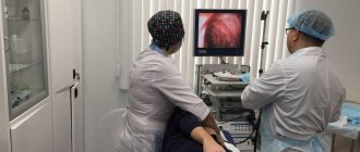
The examination of the patient begins with pharyngoscopy (examination of the pharynx) and mirror laryngoscopy (examination of the larynx using a mirror). The main diagnostic method is esophagoscopy or endoscopic examination of the esophagus. It allows you to see and remove the object, as well as assess the condition of the surrounding tissues. Emergency (immediately after admission to a medical facility) esophagoscopy is necessary if there is a suspicion of a battery or a sharp object in the esophagus. For patients with non-sharp (except coins) esophageal foreign bodies, endoscopy is recommended within 2 hours of presentation to the doctor. If a coin gets into the esophagus, you can postpone endoscopy for up to 12 hours: there is a high probability that it will leave the body naturally.
But since endoscopy may not be available (no endoscopist on duty in a small hospital, etc.), a radiograph of the neck and chest area in two projections can be used to diagnose a foreign body. If the foreign body is radio-opaque (plastic, hydrogel ball, etc.), an x-ray is taken with contrast. The patient is asked to drink a barium suspension that is opaque to x-rays, against which the foreign body becomes visible. If the technical capabilities of the medical institution allow, if inflammation of the peri-esophageal tissue or tracheoesophageal fistula is suspected, the doctor may prescribe an MRI or CT scan, or bronchoscopy.
Causes and course of the disease
Foreign bodies in the esophagus occur in almost all age categories.
This mainly happens when a person eats very hastily, talks while eating, or has the habit of holding any objects in the mouth. Quite often, foreign bodies of the esophagus occur in people who have low sensitivity of the oral mucosa due to the fact that they wear dentures, as well as in elderly people who do not have teeth. In some cases, foreign bodies enter the esophagus from the respiratory tract and stomach. Foreign bodies found in the esophagus are varied, but most often they are fish bones, buttons, badges, coins, etc. Children often come across parts of children's toys, coins, etc.
A foreign body can slip through the esophagus into the stomach or get stuck in the lumen of the esophagus. Whether a foreign body remains in the esophagus depends on its size, nature and shape, consistency, as well as the presence of pathological changes in the lumen of the esophagus.
Most often, foreign bodies get stuck in the cervical esophagus. This happens much less frequently in the thoracic esophagus.
Treatment of esophageal foreign body
As a rule, treatment of a foreign body of the esophagus comes down to its endoscopic removal and the prescription of a short course of antibacterial drugs to prevent inflammation. If endoscopic removal is impossible without severe damage to the wall of the esophagus, as well as if there are signs of bedsores, perforation of the esophagus and the spread of infection to neighboring organs, the foreign body is removed surgically. Antibacterial drugs are also prescribed after surgery. Analgesics are recommended to reduce pain after surgery. To restore the mucous membrane, gastroprotectors based on rebamipide are prescribed. The diet before and after surgery should be as gentle as possible: cool semi-liquid food, maximally balanced in the content of proteins, fats, vitamins and microelements. In the early postoperative period, it is possible to use special nutritional mixtures.
Who is most likely to choke?
The risk of choking is highest in the following groups:
- children under five years of age,
- older people,
- suffering from neurological diseases,
- suffering from chronic acid reflux,
- suffering from acute respiratory diseases,
- people with injuries and anatomical abnormalities that affect the swallowing process (for example, with a cleft lip).
How to choose a toy as a gift?
Find out how to choose a safe toy for your child so that no play ends in an accident.
Some eating habits increase a person's chance of choking:
- eating food too quickly
- eating while standing, sitting in an awkward position or lying down,
- poor chewing of food,
- eating too dry and hard foods.
Treatment
Bronchoscopy is the gold standard for the diagnosis and treatment of lower respiratory tract foreign bodies. The first successful removal of a bronchial foreign body was performed by Gustav Killian in 1897. He removed bone from the bronchi of a 63-year-old man using an esophagoscope, marking the beginning of the era of rigid bronchoscopy. But the flexible bronchoscopy method, introduced into clinical practice in the 1970s, gradually replaced rigid bronchoscopy, and is now the first-line method in most adult patients, allowing in most cases to identify and remove a foreign body of the lower respiratory tract [3].
Although rigid bronchoscopy is still the traditional method in pediatrics. Also, rigid bronchoscopy is preferable in cases of acute respiratory distress (stridor, asphyxia), or when flexible bronchoscopy has failed. Rigid bronchoscopy should always be available as a backup for any case where flexible bronchoscopy is chosen as the initial procedure [5]. Open surgical interventions may be required in some patients, according to the literature in 3.76-10% of cases), usually due to residual effects after the inflammatory process [3].
The advantages of endoscopy over abdominal surgery are obvious:
- There is no abdominal wall incision;
- Minimum risks and no complications;
- General anesthesia is not required;
- Easy tolerability and no rehabilitation period;
- Speed of manipulation (up to 45 minutes);
- Affordable price.
A huge advantage of endoscopy is complete visual control. The doctor sees with his own eyes both the object and the inner surface of the organ, which allows him not only to remove the foreign body, but also to evaluate the mucous membrane for damage and, if any, eliminate them.
How to help someone who is choking?
If a person chokes and coughs, this means that his airway is not blocked. There is no need to help him by hitting him on the back - let the person get rid of the stuck object on his own. Otherwise, your attempt to help may result in the stuck object slipping even deeper. If you can’t clear your throat on your own
, help the person take a more comfortable position - tilt him forward and down, ask him to take a slow, gentle breath and exhale sharply, encouraging him to clear his throat.
A large object stuck in the esophagus causes not only discomfort, but also an increased risk of damage to this part of the gastrointestinal tract. In this condition, urgent medical attention is required. If a person is choking
, unable to cough or speak, this means that a foreign body has entered the respiratory tract.
In this case, you need to urgently call an ambulance and try to help the victim yourself. To do this, the so-called Heimlich method
: 1. Grasp the victim with both hands from behind - place both hands between the navel and the bottom of the ribs. 2. Make one hand into a fist, place it on your stomach, and clasp it on top with your other hand. 3. Sharply bending both arms at the elbows, sharply press your fist on the victim’s stomach. Repeat this procedure until his airway is clear.
You need to know this
A fracture is a serious injury that requires mandatory and urgent medical attention. Find out why bones break and how to provide first aid for fractures.