Microscopic colitis is a rarely diagnosed pathology of the colon, the manifestation of which is diarrhea. When conducting a colonoscopic examination for microscopic colitis, no changes in the mucous membrane of the colon are detected. Only a biopsy of the mucosa reveals specific microscopic changes - this is where the name microscopic colitis comes from.
Since 1984, studies began in European centers, which showed an increase in cases of microscopic colitis. Its prevalence has become comparable to nonspecific ulcerative colitis and Crohn's disease. If patients have chronic diarrhea, along with classical diagnostic signs, it is necessary to exclude microscopic colitis.
It is believed that this is a disease of older people, more often women, burdened with various autoimmune diseases such as diabetes, thyroiditis (thyroid disease), etc.
Typology of colitis
Autoimmune inflammatory bowel diseases
Caused by disturbances in the body's immune response mechanisms. Among them:
- Crohn's disease. Severe chronic disease. It is first diagnosed in adolescents, but may appear later. If it manifests itself in childhood, it can cause growth and development delays;
- Nonspecific ulcerative colitis. A disease with an unclear etiology. It is also chronic. A complete cure, as in the case of Crohn's disease, is impossible.
- Microscopic colitis. They are named so because they are visible only during microscopic examination of tissue. Microscopic colitis cannot be diagnosed with the naked eye (using endoscopy). This type includes:
- Lymphocytic colitis. It affects men and women aged 50-70 years equally. A distinctive feature is the increased content of white blood cells (leukocytes) in the intestinal epithelium;
- Collagenous colitis. It is more common in women aged about 40-50 years. With this pathology, there is a significant proliferation and thickening of the collagen layer under the intestinal mucosa.
- Incomplete colitis. Combines the symptoms of colitis of the two previous types.
Colitis resulting from treatment:
- Diversion colitis. It occurs in people who have undergone surgery to form an ileostomy or colostomy. The duration of the disease is from one month to three years. Symptoms may go away without treatment.
- Chemical colitis. Caused by introducing irritants into the intestines through an enema or other procedure, such as leftover chemicals used to clean a colonoscopy tube.
- Radiation colitis. Occurs during radiotherapy of the abdominal and pelvic organs. There are acute (0-90 days from the start of therapy) and chronic (3 months - a year after the end of therapy) phases of the disease. Most often occurs in elderly patients.
- Ischemic colitis. Develops due to deterioration of blood supply to the intestines. Associated with low blood pressure, vascular atherosclerosis, individual characteristics of blood supply and intestinal vascular thrombosis. It appears mainly in old age.
- Infectious colitis. The main reason is damage to the intestinal mucosa by various microorganisms. These can be bacteria of the type Clostridium difficile or Shigella dysinteriae, as well as parasitic amoebas – Entamoeba histolytica. Without treatment, infectious colitis can last for years.
- Allergic colitis. It appears primarily in infants under one year of age in response to consumption of cow's and goat's milk products, as well as products containing soy. It may also occur in breastfed children whose mothers consumed cow's milk.
Microscopic colitis: “the invisible bowel disease”
Let's start with a brief medical history. A 40-year-old woman consulted a gastroenterologist at the Expert clinic with complaints of abdominal pain, watery diarrhea, and urgency of the urge to defecate. These symptoms have bothered her for the last 2 years, during which the patient changed 3 gastroenterologists. The diagnosis has not been established, although celiac disease, Crohn's disease, and various changes in the intestinal microbiota were suspected. These diseases were not confirmed, and colonoscopy (without biopsy!), performed three times over 2 years, did not reveal tumor and inflammatory changes in the colon and ileum. There was a slight effect from taking bismuth and loperamide.
The doctor suspected that the patient had irritable bowel syndrome (IBS), since this disease is characterized by abdominal pain and diarrhea in the absence of organic intestinal pathology. However, upon careful analysis of the complaints, it turned out that abdominal pain and bowel disorders were predominantly nocturnal, causing awakening. Such symptoms usually make one doubt the diagnosis of IBS and direct the diagnostic search in other directions. In particular, to exclude microscopic colitis.
Microscopic colitis (MC) is a chronic inflammatory bowel disease, which is characterized by typical symptoms and the presence of immune inflammation in colon biopsies. This disease is what is called “unheard of,” but it is not so rare: its prevalence reaches 119 cases per 100 thousand population, and the incidence (the number of new cases of the disease) averages 11.4 per 100 thousand population per year. Despite the fact that MK is mainly characterized by people over 60 years of age, in approximately 25% of cases the disease is diagnosed in people under 45 years of age. MK occurs in childhood, but rather as an exception. Women are more often affected: 52-86% of cases of MC are female; female gender in general increases the risk of developing the disease by 2.5 times or more.
Currently, the term MC is commonly used to designate two separate diseases: collagenous colitis (CC) and lymphocytic colitis (LC) . Among the two types of microscopic colitis, CC occurs much more often - in 2/3 of cases.
Risk factors for developing microscopic colitis
As mentioned above, one of the risk factors for the occurrence of MC is female gender. The risk also increases with age: the greater it is, the higher the likelihood of developing the disease (for people over 65 years of age, the risk increases by 5.25 times).
Taking certain medications is associated with an increased risk of developing urticaria: proton pump inhibitors (especially lansoprazole), non-steroidal anti-inflammatory drugs, statins, antidepressants from the group of serotonin reuptake inhibitors, lisinopril, carbamazepine and a number of others.
Tobacco smoking is another well-known risk factor that increases the likelihood of developing MC by 2.1 times, while smokers are characterized by an earlier age of onset of the disease (by 10 years or more).
Causes and mechanisms of microscopic colitis
The causes of the disease are not completely clear. It is now recognized that MK is a multifactorial disease with a genetic predisposition and the participation of the immune system with the development of an inflammatory reaction in response to intestinal contents.
Currently, the specific gene responsible for the development of MC has not been described; most likely, it is a combination of several genetic markers. Family cases of the disease have been described, but there is no mandatory inheritance of MK.
Clinical manifestations (primarily watery diarrhea) are caused by increased secretion of water and electrolytes into the lumen of the colon. In patients, the secretory type of diarrhea predominates, which can be determined by special studies. The fact that food restriction reduces stool frequency in patients with urticaria suggests a possible osmotic component of diarrhea.
Clinical manifestations (symptoms) of microscopic colitis
The following symptoms are characteristic of MK:
1) Chronic diarrhea with frequent (5-15 times a day) loose, often watery stools. Diarrhea usually develops gradually, but in 40% of cases its sudden onset is described;
2) Urgency (urgency, immediacy) of the urge to defecate, characteristic of 70% of patients;
3) Stool incontinence, which is common (up to 40% of cases);
4) Nocturnal episodes of bowel movements, incl. causing awakening of the patient, are typical for half of people with MK;
5) Abdominal pain in patients with active MK is observed in 50% of cases
6) Loss of body weight (occurs in some patients due to decreased food intake and fluid loss)
Additional (but not obligatory) symptoms may include arthritis and uveitis.
Laboratory and instrumental studies for microscopic colitis
Typically, laboratory tests do not reveal any specific changes that are unique to MK. However, patients may experience mild anemia, a slightly accelerated erythrocyte sedimentation rate, and autoantibodies (antinuclear, antineutrophil, antibodies to Saccharomyces cerevisiae, rheumatoid factor) are often detected, which are not diagnostic methods for this disease.
Stool testing for fecal calprotectin may be elevated, but the use of this “surrogate” marker of inflammation to monitor exacerbation and remission of urticaria is not currently recommended. The same applies to other fecal markers of inflammation.
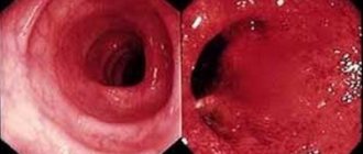
Colonoscopy usually shows a normal picture of the colon mucosa, but nonspecific changes may occur in the form of swelling, redness and blurred vascular patterns in different parts of the intestine. Despite the fact that MC is a disease of the colon only, when performing a colonoscopy, examination of the ileum is mandatory to exclude other pathologies.
Histological examination is the main method for confirming the diagnosis of MC, as well as for identifying its type (lymphocytic or collagenous colitis). During a colonoscopy, biopsies are taken from the right and left parts of the colon and then examined under a microscope.
The main histological criteria for lymphocytic colitis when stained with hematoxylin-eosin are:
- an increase in the number of lymphocytes in the surface epithelium of the colon (so-called interepithelial lymphocytes or IEL). For diagnosis, the number of MELs must be ≥20 per 100 surface epithelial cells;
- increased density of the inflammatory infiltrate in the lamina propria;
- absence of significant thickening of the collagen layer (collagen basement membrane) of the surface epithelium (
Histological signs of collagenous colitis are:
- significant thickening of the collagen layer (collagen basement membrane) of the surface epithelium (≥10 µm, normal 3-5 µm).
- increased density of the inflammatory infiltrate in the lamina propria.
European recommendations on microscopic colitis (2020) also distinguish incomplete LC and incomplete CC.
Incomplete LC is characterized by the histological signs described above, but the number of MEL is >10 and less than 20. The thickness of the subepithelial collagen layer in incomplete LC exceeds 5 µm, but does not reach the required 10 µm.
Differential diagnosis
MK should be differentiated from diseases such as Crohn's disease, celiac disease, irritable bowel syndrome. Crohn's disease is characterized by the presence of erosive and ulcerative lesions of the intestine; markers reflecting the presence of a systemic inflammatory process are often elevated. Celiac disease is characterized by the presence of atrophic changes in the small intestine; the large intestine is not involved. Irritable bowel syndrome is similar in symptoms to urticaria, but the presence of nighttime symptoms (causing the patient to wake up) is not typical for it.
In persons with recently emerging symptoms, it is imperative to exclude an acute intestinal infection, as well as conduct research to exclude giardiasis, helminthiases and other parasitic infestations, and Clostridium (Clostridioides) difficile infection.
The diagnosis of MC can be confirmed by identifying typical histological signs in a colon biopsy in combination with characteristic symptoms.
Treatment of microscopic colitis.
The goal of treatment is to achieve clinical remission, which is defined as having fewer than 3 bowel movements per day with no loose stools for at least one week. Another goal is to improve the quality of life of a patient with MC.
I. Treatment of active disease (stools 3 or more times per day or watery stools ≥1 time per day).
A. General recommendations.
1. Refusal to use non-steroidal anti-inflammatory drugs and (if possible) medications. for which a possible connection with MK has been established (see section “Risk Factors”). Cancellation of therapy is possible only after discussion with a specialized specialist when comparing the benefits/risks.
2. Stop smoking.
B. Symptomatic therapy. Loperamide can be used as sole therapy (for mild symptoms of urticaria) or in combination with basic drugs.
B. Basic therapy.
1. Steroid hormones of local (budesonide) or systemic (prednisolone, methylprednisolone) action.
2. Auxiliary agents, including bile acid sequestrants (cholestyramine) and bismuth preparations. Usually used in addition to hormonal therapy when it is insufficiently effective or separately in people with intolerance to steroid hormones. The advisability of using cholestyramine is due to the frequent combination of MK with hologenic diarrhea. As for bismuth preparations, their effectiveness is still disputed, although there is evidence that they affect the frequency of bowel movements, stool consistency, and also improve the histological picture of the colon mucosa.
3. Immunosuppressants (azathioprine, mercaptopuine) can be used in people with relapse after discontinuation of steroid hormones.
4. Genetic engineering biological therapy with drugs from the group of tumor necrosis factor alpha inhibitors (infliximab, adalimumab, etc.) can be discussed in patients with severe CM and ineffectiveness of steroid hormones.
5. Surgical treatment with removal of the colon or ileostomy (in older people) can be discussed in people with refractory CM. If basic therapy is ineffective, the diagnosis should be reconsidered.
II. Maintaining remission of urticaria.
Achieving clinical remission of the disease is not a reason to discontinue therapy; patients with MK often require maintenance therapy. Topical steroid hormones (budesonide) and immunosuppressants are considered as means of maintaining remission.
Note: 5-aminosalicylic acid preparations (mesalazine) and probiotics have not been shown to be effective in the treatment of urticaria.
Course and prognosis of microscopic colitis
MK is a chronic disease characterized by periods of exacerbation and remission. Remission (i.e., inactive disease) can occur with treatment or on its own, but absence or cessation of therapy often leads to rapid relapses (30-60%).
Despite the fact that urinary tract disease formally refers to a chronic inflammatory bowel disease, it does not increase the risk of developing colon cancer. Frequent relapses or the presence of persistent symptoms reduces the quality of life of patients.
PS The clinical case described at the beginning of the article ended happily. After performing a colonoscopy with biopsy samples, the patient was diagnosed with microscopic (lymphocytic) colitis. The treatment started with topical steroid hormones turned out to be very effective: the stool returned to normal, the urgency of defecation disappeared, the abdominal pain stopped, and the quality of life noticeably increased.
Signs of colon inflammation
The main symptoms of colitis include moderate to severe pain, tenderness at the site of inflammation, increased bowel movements, persistent diarrhea with blood or pus, fecal incontinence, increased gas, loss of appetite, fatigue and unexplained weight loss. Metabolic and electrolyte disturbances occur due to disruption of absorption processes.
More severe illness may include rapid or irregular heartbeat, difficulty breathing, and a high fever.
Less common symptoms include joint pain, mouth ulcers (stomatitis), bloodshot eyes, and skin rashes.
Chronic pain and poor health can cause anxiety and affective mood disorders in patients.
Nonspecific ulcerative colitis and Crohn's disease
Differential diagnosis
The main differences between UC and CD are as follows.
By definition, ulcerative colitis is characterized by ulceration of the intestinal mucosa (remember, an ulcer is a long-term non-healing inflammatory wound area that penetrates into the underlying layers - in contrast to superficial erosion - and causes the irreversible loss of one or another volume of tissue “corroded” by the ulcer. Unlike, again, erosion, an ulcer cannot heal without a trace; it always leaves a fibrous defect). Crohn's disease is pathologically manifested by granulomas - concentrated foci of inflamed connective tissue. An important differential diagnostic point is that in ulcerative colitis granulomatosis never occurs, while in Crohn's disease both ulcers and granulomas are often found. In other words, the presence of transmural (to the entire depth of the intestinal wall) granulomas is an evidence in favor of CD, and the presence of ulcers, etc. crypt abscesses can only partially serve as a differential diagnostic sign in favor of UC.
Next, the most important criterion is localization. If inflammation of the intestine is limited to the thick part of the intestine, it can be either UC or CD, with ulcerative colitis being much more likely. If the inflammation spreads and/or is predominantly localized in other parts, this is definitely Crohn’s disease, which can affect absolutely any part of the gastrointestinal tract, from the oral cavity and the inner surfaces of the cheeks to the anus.
Rectal bleeding is twice as common in UC than in CD. Weight loss is more characteristic of Crohn's disease in its small intestinal localization. Malignization (malignancy), on the contrary, is more common in ulcerative colitis, and in this case the tumor process usually starts over a shorter period than in the case of Crohn's disease.
Fibrous processes, thickening of the intestinal walls due to the proliferation of scar tissue are always detected in Crohn's disease, and rarely in ulcerative colitis.
The final diagnosis is established through a thorough clinical, anamnestic, medical-genetic, laboratory and instrumental examination. The most informative in this case are endoscopic methods (colonoscopy, FGDS, diagnostic laparoscopy), contrast-enhanced radiography, biopsy (histological analysis). Sometimes an additional ultrasound with rectal access, MRI or other studies are additionally prescribed.
Diagnosis of colitis
To determine the area and extent of damage to the large intestine, endoscopy (colonoscopy, sigmoidoscopy) is actively used.
To carry out a differential diagnosis, a biopsy is performed: a piece of mucous membrane measuring several square millimeters is removed and submitted for histological examination under a microscope.
A complete blood count is performed (hemoglobin level, hematocrit, white blood cell count, electrolytes, ESR); stool analysis for blood cell content, bacterial culture to identify pathogenic strains.
How to treat inflammation of the large intestine
When treating colitis, the localization of the pathology, the severity of the disease, the side effects of medications and the presence of concomitant diseases are taken into account. Therapy is symptomatic.
- Aminosalicylates (sulfalazine, mesalazine, olsalazine) are used to relieve inflammation;
- Corticosteroids (prednisolone, budesonide) are also used to relieve inflammation. Not recommended for long-term use due to strong side effects.
- Immunomodulators (6-mercaptopurine, methotrexate) reduce the activity of the immune system. if the body is not able to cope with constant processes of inflammation;
- Antibiotics (ciprofloxacin, metronidazole) help fight concomitant infections.
- To restore electrolyte balance and rehydration, a liquid diet is indicated for a while.
Nutrition for inflammation of the large intestine
There is no formal diet for colitis. The main requirements are the exclusion from the diet of spicy foods that irritate the mucous membranes, fried meat, milk, and foods rich in fiber. Be sure to avoid alcohol and smoking if you have colitis.
We care about the health of our patients and offer to use our programs for the prevention and treatment of diseases of the digestive system. These programs can be completed in 1 visit to the clinic.
The clinic sees five gastroenterologists and five proctologists. We work every day.
You can make an appointment with a gastroenterologist at the Naedine Clinic by phone in Kirov: (8332) 32-7777 or through the form on the website.
Risk factors for microscopic colitis
- Medications. It is assumed that some drugs may be triggers in the development of microscopic colitis, which does not exclude drug-associated colitis. Drugs with a high degree of probability of triggering colitis include the following drugs:
- aspirin,
- lansoprazole,
- non-steroidal anti-inflammatory drugs,
- ranitidine, etc.
- proton pump inhibitors, which reduce gastric acidity,
- non-steroidal anti-inflammatory drugs. Side effects when taking these drugs include diarrhea.
- Smoking. There is little evidence on this issue, but it is known that smokers develop the disease earlier than non-smokers.