Medical statistics are inexorable; one of the most frequently detected helminthiases, both in children and adults, is enterobiasis. About 460 million people in the world are infected with pinworms; in the Russian Federation in 2012, 206,743 cases of the disease were identified among children under the age of 17 years (data from the Consilium Medicum portal).
In the overall structure of helminthiases, enterobiasis occupies about 90-91%, the remaining 10% is accounted for by other types of invasions.
Why is enterobiasis such a common infection? First of all, this is due to the relatively simple life cycle of pinworms, which allows little dependence on environmental conditions and ease of re-infection (autoinfestation). In addition to these factors, the low-symptomatic nature of the disease in the majority of infected people is also of great importance, which allows them not to seek medical help and treatment.
You can become infected with enterobiasis not only by eating unwashed food, as is commonly believed, but also by swimming in bodies of water, by contact with a person or household items on which pinworm eggs have settled, and also while playing in the sandbox. Moreover, eggs can even become infected through airborne droplets, by inhaling dust particles containing pinworm eggs.
Despite the numbers and significant damage to the economies of countries, helminthiasis is included in the list of “forgotten diseases”, and even in endemic countries this pathology is not given due attention.
A special risk group for this helminthiasis are children who, in their infancy, actively explore the world around them (including with their mouths), observe little personal hygiene rules and communicate closely with each other, exchanging toys. Enterobiasis is often detected in babies who sleep with their parents.
The main symptoms of helminthic infestation
Parents need to be very vigilant if the child has complaints of diffuse abdominal pain, nausea and vomiting, increased salivation, allergic manifestations on the skin, itching in the external genitalia and folds around the anus, frequent urinary tract infections, recurrent synechiae in girls , masturbation or enuresis. Some patients experience persistent changes in the general blood test: an increase in the level of eosinophils, a decrease in hemoglobin (normocytic or microcytic anemia).
Indirect symptoms of helminthiasis can be irritability and fatigue, bruises around the eyes, sleep disturbances (screams at night, crying for no reason, frequent waking up), headache, weight loss, frequent ARVI.
Since these symptoms are only indirect signs of enterobiasis infection, special methods that allow confirming the diagnosis play an important role in diagnosis. These include the following analysis options:
- 1Scraping from the perianal folds, performed in several ways.
- 2Fecal microscopy (standard stool analysis for oviworm).
- 3Detection of pinworm DNA in feces using PCR.
Scraping from the perianal folds can be performed in the following modifications:
- 1Classical scraping for enterobiasis (Torgushin method or Kevorkova method);
- 2Print on transparent adhesive tape from the perianal area using the Graham method;
- 3An alternative option is to make an imprint on a sticky transparent blade, with its further microscopic examination (the so-called Rabinovich method).
It is possible to defeat parasites!
Antiparasitic Complex® - Reliable and safe removal of parasites in 21 days!
- The composition includes only natural ingredients;
- Does not cause side effects;
- Absolutely safe;
- Protects the liver, heart, lungs, stomach, skin from parasites;
- Removes waste products of parasites from the body.
- Effectively destroys most types of helminths in 21 days.
There is now a preferential program for free packaging. Read expert opinion.
Read further:
Enterobiasis: how scrapings are taken for analysis in children and adults, interpretation of the results
How to take a stool, blood and scraping test for enterobiasis for an adult and a child
Referral for enterobiasis: in what cases is a referral for scraping for enterobiasis given?
Itching due to pinworms in the anus: how to relieve it and how to eliminate it
Scraping for pinworms in children and adults: how to take a test, what to do if pinworms are found
Worms in the anus in children and adults: who is the cause and how to treat
Which groups should be tested for enterobiasis?
Not only persons with suspected enterobiasis are subject to examination, but also all children attending preschool institutions, schoolchildren in grades 1-4 and staff of children's institutions should be examined once a year.
Food group workers and everyone equivalent to them, persons receiving a certificate to visit the pool, as well as applicants. In addition, all children admitted for treatment in a hospital of any profile and sanatorium-resort treatment are also subject to additional examination for enterobiasis.
If, during the examination, more than 15% of cases of infection were found in children's groups, then no further search is carried out, and all children are subject to mandatory treatment.
The most common mistakes when conducting analysis
For correct and accurate diagnosis, it is very important to avoid mistakes when collecting biological material. The result is influenced by slow delivery of the test sample to the laboratory, determination of helminthiasis during the period of taking medications, such as antibacterial drugs, antidiarrheals or laxatives, antacids. You should stop taking them at the time of testing or, if this is not possible, notify the laboratory.
Unscrupulous and poor-quality work of laboratory technicians (incorrectly prepared material or lack of persistence when examining a slide) often leads to failure in fecal analysis. It is important to remember that microscopy of material during the period when the female stops laying eggs (silent period) will also show a false negative result.
Why are pinworms rarely detected in feces?
The generally accepted analysis of feces for helminth eggs to identify enterobiasis is of little information, due to the morphobiological characteristics of pinworms, since they do not lay eggs in the intestinal lumen. That is why scatological laboratory diagnostic methods rarely detect helminths.
Standard stool microscopy is usually used to detect eggs of other parasitic worms (roundworms, opisthorchids, tapeworms, etc.).
Perianal fold scraping and microscopy are the most effective way to detect eggs of Enterobius vermicularis pinworms. Of course, if adult individuals were found in underwear or the anus during egg laying, then the diagnosis of enterobiasis is beyond doubt.
What is the reason for such a diagnostic search?
Laboratory research is based on the development cycle of the helminth. All processes of maturation and fertilization of the parasite occur in the distal part of the small intestine, after which females with mature eggs descend into the rectum. Actively crawling out of the anus, they lay up to fifteen eggs in the perianal folds, on the skin and die.
The eggs laid are fully mature in an average of five hours. The movement of females during laying causes irritation and severe itching. Children, scratching the perianal area and perineum, contaminate their hands and re-swallow eggs, from which larvae emerge in the duodenum. The lifespan of pinworms is from two to four weeks.
What diseases can be diagnosed?
- Enterobiasis is a parasitic infection in which small roundworms, pinworms, live and reproduce in the human intestine. The length of the female is up to 12 mm, the male is up to 4 mm. Female pinworms emerge from the anus to lay eggs. Most often this happens at night.
- Teniarinhoz is a parasitic disease in which bovine tapeworm develops in the small intestine of a person. The body of a mature worm consists of thousands of segments. They contain eggs that are passed out with feces. They are also found in perianal scrapings. The length of an adult worm can reach 12 m.
- Taeniasis is a parasitic disease in which pork tapeworm develops in the human body. The worm has suction cups on its head, with which it attaches to the wall of the small intestine. The body of the tapeworm consists of many segments containing eggs. An adult worm can reach a length of 7 m.
Rules for preparing biomaterial for research
Now let's look at how to get tested for enterobiasis correctly. When preparing for a classic scraping for enterobiasis, it is important not to wash your face in the morning if it is better not to urinate. Carrying out the sampling procedure after the child’s restless sleep increases the efficiency of the study.
Classic collection of material for research is carried out by medical workers. Before performing the manipulation, nursing staff (less often a laboratory assistant) explains to the parents and child the essence of the study, as well as the sampling technique. It is important that the patient and their representatives understand the safety and nuances of the study. At the same time, the laboratory assistant fills out a referral for the study, if this has not previously been done by the attending physician.
If this is a child, then he is laid on his side, with his back to himself, while slightly raising the upper leg, bent at the knee joint. With the fingers of her left hand, the nurse spreads the buttocks, and with her right hand she makes radial movements around the skin and folds of the anus with a previously prepared loop with a cotton swab moistened with water, an oil solution or 50% glycerin. For adults, the procedure can be performed in a standing position, bending forward.
Kit for performing scrapings for enterobiasis in the classical way
After this, the scraping is placed in a glass or disposable plastic tube, which is tightly closed and labeled. This material can be stored for no more than two hours at a temperature of 4-8 degrees Celsius.
After the tube is delivered to the laboratory, the material is removed and streaked onto a clean, grease-free glass slide with a drop of glycerol for further microscopy (Torgushin method). If the swab is washed in a solution and then centrifuged followed by microscopy, then this modification is called the Kevorkova method.
Thus, a classic scraping for enterobiasis includes the following activities:
- 1Preparing the patient and filling out a referral for the study;
- 2Placement of the patient;
- 3Preparing hands and tools for sampling, putting on gloves;
- 4 Taking material for research and labeling it;
- 5Storage and delivery to the laboratory;
- 6Applying material to a glass slide and its microscopy;
- 7Entering the result in the journal and in the direction;
- 8Delivery of directions to the site.
What types of scraping are equally effective?
Another variation of this study is an imprint on adhesive transparent tape (Graham method) . Transparent tapes for household use are also used in medical practice. This technique, when carried out three times with an interval of 7-10 days, with mandatory fingerprinting in the morning, is effective in up to 90% of cases.
The procedure is identical to the previous one, but instead of a cotton loop, a piece of adhesive tape is used, which must be applied to the perianal area. The adhesive tape then adheres to the grease-free glass slide without creating air bubbles that would interfere with microscopy.
The Graham method is a good alternative to classical scraping, especially if the material is collected directly in a health facility. I would like to note the following nuances of this method:
- 1Patients often take the material at home, independently. If necessary and desired by the patient, this stage of the study can be falsified.
- 2The delivery time to the laboratory is the same as with the classical method, that is, the next 2-4 hours. It is advisable to store the material at a temperature of 4-8 degrees.
- 3It is important to adhere the tape to the slide correctly.
A very convenient and alternative is the Rabinovich method . It is widely used in the examination of decreed groups - food industry workers (tables, cafes, restaurants, etc.), personnel of child care institutions, schools, hospitals.
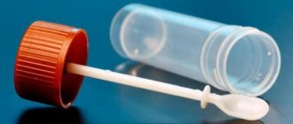
This option involves the use of special pharmacy kits for taking samples for enterobiasis according to Rabinovich. This kit includes a clear plastic spatula with a sticky layer in a labeled tube slightly filled with cleol. When taking the material, a spatula is carried out in the area of the anus and perianal folds, after which it is placed back into the tube and the sample is transported to the laboratory. There should be no feces on the spatula. Microscopy of such a blade is very convenient due to its flat and even surface.
Another type of examination is scraping from the subungual plates, since when scratching, helminth eggs can remain on the hands. The result is not reliable and is used extremely rarely.
Enterobiasis Rabinovich method
Enterobiasis is a helminthiasis caused by worms from the genus pinworms. It is one of the most common parasitic diseases in children. The infection is widespread throughout the world, including in developed countries; according to rough estimates, the infection rate in children is 4-28%.
Pinworm - the causative agent of the disease - is a white worm, 0.5-1 cm in length, in the human body it lives in the cecum and appendix. It feeds mainly on the contents of the intestines. The main clinical symptom, itching in the perianal area, occurs when pinworms lay eggs on the skin during sleep (from 5,000 to 15,000 eggs) and then die. Spread occurs predominantly through the fecal-oral route, for example, eggs can get under a person’s nails when scratching, and then get onto household items and personal hygiene items. In the transmission of the pathogen, the main problem is hand hygiene, so children are more likely to get enterobiasis, especially in crowded places (kindergartens, schools).
The high prevalence of enterobiasis can be explained by the mild clinical picture of the disease, which most often manifests itself in perianal itching. In addition, symptoms such as anxiety, loss of appetite, insomnia, and irritability may be present. In severe and prolonged cases, mental development disorders in children may occur. In rare cases, ectopic enterobiasis may develop, for example in the kidneys or fallopian tubes, which can lead to serious consequences.
Diagnosis is carried out by scraping from the perinanal area (using adhesive tape) followed by microscopy.
In the treatment of enterobiasis, a special role should be given to hygienic measures: washing hands, cutting nails, frequently changing, washing and ironing clothes. To prevent the spread of eggs, zinc ointment is applied topically to the perianal area. Drug treatment consists of taking anthelmintic drugs (contact persons are also treated).
Is it possible to collect material at home?
The Rabinovich method can also be used at home, making sure to follow all the above sampling rules. Instead of a sticky spatula, you can use a cotton swab with glycerin, which will also need to be placed in a test tube that closes tightly. The test tube must include the full name, year of birth and address of the person being studied.
It’s also good to use adhesive tape technology at home. To do this, you need to contact the laboratory to obtain not only the tape itself, but also two clean glass slides. Following the correct technique for taking the material, apply the tape to one glass, and tightly cover the second one on top. Just as when making a print on a transparent spatula with a sticky layer (according to Rabinovich), the material must be delivered to the laboratory within two hours.
It is important for mothers to remember that the correct diagnosis and further treatment depend on their technique for taking material, time and storage temperature.
What does the laboratory technician examine in the resulting smear?
When a scraping is obtained on a cotton loop (in the classical way according to the Kevorkova method), the swab is washed off, rinsed in a solution that is placed in a centrifuge, the resulting sediment is applied to glass and examined under a microscope. When using other methods, including Graham and Rabinovich, microscopy does not require additional effort.
During microscopy of the sample, pinworm eggs are identified in the field of view. They have a double-contour shell, irregularly rounded, with some asymmetry - one side is more convex, the other is flat. The eggs themselves are colorless; inside you can see an immature larva. The laboratory examination takes little time and the very next day you will be able to find out the result.
This is what pinworm eggs look like under a microscope. Dimensions 50-60 microns by 26-30 microns, oval-elongated, asymmetrical shape. The shell is thin, smooth, transparent, colorless.
How to take material at home
Do not forget that the test should be taken in the morning before defecation (and in women, before urination), and you should not wash yourself or use wet sanitary napkins, as this can remove pinworm eggs from the surface of the skin.
To collect the material, the stick with the scraper is removed from the container and approximately 4 drops of glue from the dropper bottle are applied to the scraper so that the scraper is completely covered with glue, and then the stick with the scraper is put back into the container.
Further actions:
- Lay the patient on his side, having previously freed the anus from underwear;
- With one hand, spread the buttocks to gain access to the anus, and apply the scraper with glue applied to it several times to the areas around the anus, trying to cover the entire area around it;
- Place spatula in container;
- On the label pasted to the container, write the patient’s data (full name, age) and the date the scraping was taken using a regular ballpoint pen.
Scraping process
To perform this simple manipulation, no experience is required. Another thing is that it is unlikely that you will be able to take such a scraping from yourself without outside help, so it is better to put aside false modesty and ask someone close to you to help.
It is necessary to deliver the kit with materials to the laboratory in the morning; they are usually received until 10 o’clock.
Such a kit can be stored in the refrigerator for no more than 6 hours, which means that if you do not have time to hand it in that day, then the next day it will no longer be suitable, and the scraping will have to be done again.
How to interpret the data obtained?
The absence of pinworm eggs under microscopy is considered the norm. The negative (negative) result that you get does not always indicate the absence of enterobiasis or a cure. It is possible that the test was carried out during the so-called “silent period”, when the female did not lay eggs, or during the period of early infection, when the female was simply not yet ripe for laying eggs. Therefore, it is recommended that the analysis be repeated up to seven times with an interval of two to three days.
According to research, the information content of a single scraping does not exceed 50%, while three-fold microscopy of the material increases it to 90%.
A positive result, of course, confirms the diagnosis. The disadvantage of our laboratories is that they indicate in the conclusion only information about the presence of pinworm eggs, but do not quantify them, at least with the general phrases “in large quantities” or “insignificant quantities.” Such a conclusion would help in treatment tactics, as well as in prescribing the dose and duration of therapy.
A negative result, like a positive one, does not indicate the absence of other helminths in the body (ascariasis, toxocariasis, diphyllobothriasis, taeniasis, echinococcosis or opisthorchiasis), which are no less common in the Russian Federation. If you receive a negative conclusion, but the symptoms still indicate the presence of helminths, then the best solution would be to conduct a series of repeated stool tests.
The result of the test for enterobiasis is considered valid within a week. This short lifespan for reference is due to the life cycle of pinworms.
Features of use and benefits
As already noted, the set can be called primitive in terms of use, which does not cause difficulty in the process of use by people. The algorithm for taking a sample using Dr. Rabinovich’s method is as follows:
- Wash your hands and wipe dry. Open the container and remove the spatula;
- Apply 2-3 drops of the solution included in the kit to the end of the spatula;
- The glue is distributed in a thin layer, and it should not be located further than 15 mm from the end of the spatula;
- Part of the spatula, lubricated with an adhesive substance, is placed against the anal folds, after which it is placed in a container.
There is a label on the container on which you can indicate your full name and the date of collection of the biological material. The sample can be stored in the refrigerator for several hours, but it is better to deliver it to the laboratory as fresh as possible.
Note: the maximum sample storage time is 6 hours; this period includes delivery time.
The advantages of the pinworm testing kit include accessibility and low cost. The test is ideal for young children, as well as people who do not have the opportunity to spend time in a medical facility or who cannot move due to their illness.
The analysis will show the presence of pinworm eggs during enterobiasis, if the biological material was collected correctly. However, it is recommended to carry out several tests, since any method for diagnosing a parasitic disease cannot give a 100% guarantee.