Is the armor strong?
An ulcer is an open wound on the gastric mucosa, and serious reasons are needed for its appearance. Since the stomach is constantly in contact with an aggressive environment - gastric juice, nature has provided an effective mechanism to protect it. The mucous membrane of the stomach is covered with a dense biofilm consisting of microorganisms living in it. Biofilm reliably protects against low-quality food and potentially harmful agents. Sometimes this protection fails.
Questions and Answers Is it possible to get re-infected with Helicobacter pylori?
It's not just the food that's to blame
For a long time, poor nutrition was blamed for the occurrence of ulcers. Since rough and spicy foods promote increased production of hydrochloric acid, it was believed that such food could “burn through” the gastric mucosa. However, junk food alone is not enough to cause ulcers. One of the causes of the disease was stress. There is even a special term - a stress ulcer, which can appear in a few hours in people due to severe emotional shock.
Another type of ulcer is medicinal. It can occur against the background of long-term use of non-steroidal anti-inflammatory drugs, which, when they enter the gastric mucosa, penetrate directly into the epithelial cells and damage them.
But the main culprit of ulcers is considered to be the bacterium Helicobacter pylori, which has an extremely negative effect on the gastric mucosa.
Helicobacter is the most common bacterium on earth. It lives in the stomachs of 70–80% of Russians. However, not everyone gets a stomach ulcer. For an ulcer to occur, aggravating circumstances are needed - the same stress, poor diet and bad habits: smoking and alcohol abuse.
For gastritis, contagious and inherited. Debunking myths about stomach ulcers Read more
Have you changed your mind?
Have you decided that you don’t need to get the answer to the question of how to get an ulcer through your own experience? Perfect solution.
To prevent stomach-related diseases, you should listen to the recommendations of doctors:
- Proper nutrition is one of the first steps to avoiding diseases.
- Let's leave bad habits in the past.
- Less stress and worries.
These are three “Atlantas” on which the health of the gastrointestinal tract is based.
Feedback
An ulcer cannot appear at once (with the exception of stress ulcers). Usually problems begin with chronic dyspepsia - a disruption of the normal functioning of the stomach, which, if left untreated, develops into gastritis. And he, in turn, is in an ulcer.
The patient may not know about his illness for a long time. The ulcer is often asymptomatic, especially in older people, or makes itself known delicately - moderate periodic nagging pain in the stomach.
Therefore, some patients find out about their diagnosis when they are admitted to the hospital with a perforation - a rupture of the stomach wall. If there is a perforation, surgery is indicated, followed by long-term rehabilitation and a lifelong diet.
That is why doctors urge you to be attentive to yourself and under no circumstances ignore even the most minor warning symptoms.
Let's summarize
So, we looked at the possibility of getting a stomach ulcer. What is needed for this, remember?
- Irregular meals. Eating large amounts of fast food, smoked meats, marinades and pickles. The ulcer also loves spicy foods.
- You should only drink strong black coffee and black tea with lemon. Do not add sugar or milk to these drinks. Don't forget about sweet soda, it will help the ulcer to attack the stomach faster. Especially in combination with fast food.
- Forget about avoiding stress and hassle. To freak out and worry as much as possible.
- In times of stress, it would be good to smoke. And drink a cup of coffee and a cigarette.
- There will be alcoholic drinks! Drink beer in unlimited quantities, snack on chips, crackers and other goodies.
- Continue to engage in sports that can injure your stomach. Or start doing it.
- After gastritis or gastroduodenitis appears, do not pay attention to the doctor’s recommendations. Continue your normal lifestyle. Diet and giving up bad habits? Did not hear.
- The ulcer has finally found its owner. The desired goal has been achieved. Now you need to hold on during times of severe pain and not go to the hospital for examination.
In the rhythm of the ulcer
An attack of peptic ulcer disease is characterized by “dagger” pain in the upper abdomen, in the pit of the stomach. The skin turns pale, cold sweat appears, the pulse slows down, breathing becomes shallow. During an attack, a person assumes a fetal position - lies on his side and tucks his legs towards his stomach.
If you have such symptoms, you should immediately call an ambulance. You can relieve the condition for a short time by drinking a glass of milk with a pinch of soda.
Vomit the color of coffee grounds or black, tarry stools are signs of a perforated ulcer. In this state, you can neither eat nor drink, much less take painkillers. You should drink water with pieces of ice, call an ambulance and mentally prepare for the operation.
Will we get treatment?
Diagnosing an ulcer is not difficult. An experienced doctor will make a diagnosis based on examination and history, but to confirm the diagnosis you will need gastroscopy - an unpleasant procedure, but quick and informative.
To treat peptic ulcers, patients are prescribed several types of antibiotics and antacids - drugs that neutralize hydrochloric acid and form a protective film on the stomach wall. The course of treatment takes two weeks. In this case, relief occurs after 3–5 days; it takes about a month and a half for the ulcer to heal. All this time, the patient must adhere to diet No. 1, which excludes any irritants (chemical and thermal) and stimulants of gastric juice secretion (hot, spicy, fatty foods). The diet also requires eating food at a temperature that is comfortable for the stomach – 40–50 degrees, 5–6 times a day. It is difficult to stay hungry on such a diet. You can eat porridge, mashed potatoes, vermicelli, boiled and stewed meat and vegetables. You will have to give up strong tea and coffee. From smoking and alcohol - even more so.
Article on the topic
How to eat deliciously when you have a stomach ulcer. Menu for dietary table No. 1
What's next?
Can a person feel healthy after an ulcer has healed? This depends on many factors: age - in mature people the disease is prone to relapses due to incompetence of the mucous membrane, gender - ulcers occur more often in men than in women, and even character: explosive temperament is a risk factor for peptic ulcer disease. The risk will decrease if the patient gets rid of Helicobacter, bad habits and eating habits.
Most doctors agree that it is unlikely that it will be possible to completely forget about the disease. In any case, you will have to visit a gastroenterologist and undergo gastroscopy annually.
The statement that a stomach ulcer always turns into cancer is a gross exaggeration. For an ulcer to degenerate into cancer, several factors must come together. According to statistics, this happens only in 2–14% of cases.
We are for an unhealthy lifestyle
All gastroenterologists, when treating the gastrointestinal tract, unanimously advise leaving bad habits in the past. Alcohol and smoking, a sedentary lifestyle and the abuse of junk food are directions for getting an ulcer and more.
However, we are firmly focused on getting stomach ulcers quickly. And we will not listen to specialists in white coats. Nobody forbids their vision on this matter. We continue to eat everything and also follow the following recommendations:
- A candidate for peptic ulcer disease should definitely continue to smoke if he has this habit. And the more cigarettes he smokes a day, the better. And for the stomach, and for the pancreas, and for the lungs.
- Smoked ribs, croutons, chips, etc. are very tasty. And it tastes twice as good with beer. The future patient should not limit himself in the consumption of alcoholic beverages.
- In case of gastric ulcer, abdominal injuries are unacceptable. But anyone thinking about how to quickly get an ulcer should not pay attention to this. Continue to pump up your abs, play sports, focusing on your stomach. And if there is a possibility of injuring him, then that’s a good thing.
How to avoid stomach ulcers
- Wash your hands before eating, do not use other people's utensils, and do not try what others have eaten. This will help avoid many infections, including Helicobacter.
- Don’t be nervous about trifles - the psychological state has a huge impact on the organs and the digestive process.
- Do not torment your stomach with either gluttony or fasting - it is better to eat little by little, 5-6 times a day.
- Do not consume hot food and drinks - this burns the mucous membrane and leads to atrophic gastritis, which is considered a prognostically unfavorable condition. The food should be comfortable, the temperature should be 40–50 degrees.
- Get tested annually for Helicobacter pylori - according to WHO experts, this is stomach enemy No. 1.
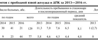
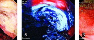
Hard-to-heel gastroduodenal ulcers
Issues of clinical presentation, diagnosis and treatment of intractable ulcers (TRU) of the stomach and duodenum (DU) remain relevant, despite the progress achieved in the treatment of peptic ulcer disease (PU). Their frequency after the introduction of proton pump inhibitors (PPIs) into clinical practice decreased significantly, but the problem of TOC still did not disappear from the agenda. This applies to both therapeutic and diagnostic aspects and to the definition of the very concept of TRN.
According to A. A. Sheptulin, hard-to-scar (torpid, resistant, long-term non-healing) gastroduodenal ulcers include ulcers that do not scar within 12 weeks. According to other data, long-term non-scarring ulcers include ulcerative defects, the healing period of which with standard therapy exceeds 6 weeks when localized in the stomach and 4 weeks when localized in the DC [1]. There is a point of view according to which TRU are considered to be ulcers of the DC that do not scar within 6-8 weeks, and gastric ulcers - within 10-12 weeks [2]. Thus, at present there is no consensus on which ulcers should be considered difficult to scar. This leads to the fact that data on the incidence of TOC vary from 1% to 10% and even 22–23% [3–5]. On the other hand, the definition of TRN, based only on the timing of healing, does not give an idea of the treatment (histamine H2 receptor blockers, PPIs, combination therapy using gastroprotectors), which was ineffective. Currently, the drugs of choice in the treatment of ulcer are PPIs, so resistance to this group of drugs should be included in the definition. The question of the dose of PPI, in the context of refractoriness to therapy, is also debatable: the dose of the drug should be sufficient to suppress gastric secretion and relieve symptoms, and it can be either standard or double or higher. A. Lanas et al. Refractory duodenal ulcers include those that did not heal when using a full dose of H2 blockers in 8 weeks or PPIs in 6 weeks; to refractory gastric ulcers - ulcers that did not heal after 12 weeks of treatment with H2-blockers or 8 weeks of PPI therapy [6]. According to JJ Kim et al. (2007) ulcers that did not heal within 12 weeks of PPI treatment or that quickly recurred after cessation of antisecretory therapy should be considered refractory.
In our opinion, ulcers that have not healed or have not shown significant healing dynamics during endoscopic examination with adequate anti-Helicobacter and antisecretory therapy (PPI) for 4 and 6 weeks (for duodenal and gastric ulcers, respectively) should be classified as TUR, excluding their symptomatic nature .
The last addition seems important, since there are many causes and factors associated with peptic ulcers. Thus, A.H. Soll identifies the following [7] (table).
It seems more common to classify symptomatic ulcers as a separate category (from ulcers). These include:
- stress ulcers;
- combined ulcers with concomitant diseases of the cardiovascular system, lungs, liver, pancreas and kidneys, diffuse connective tissue diseases (scleroderma); systemic vasculitis (polyarteritis nodosa), systemic granulomatosis of unknown etiology (sarcoidosis), Crohn's disease;
- medicinal ulcers;
- endocrine ulcers (with Zollinger-Ellison syndrome, hyperparathyroidism);
- against the background of infectious diseases [1, 8–12].
Symptomatic ulcers should be taken into account in case of poor healing of the ulcer, since in some cases they are characterized by resistance to antiulcer therapy [12].
Numerous data obtained in recent years suggest that Helicobacter pylori infection is a key factor in the pathogenesis of ulcer. The use of modern standardized eradication therapy regimens has contributed to a decrease in the incidence of TOC and relapses in the last decade. It is known that Helicobacter pylori slows down the healing of ulcerative defects by disrupting the migration of epithelial cells, re-epithelialization, increasing the apoptotic activity of lamina propria fibroblasts and reducing blood flow in the edges and bottom of ulcers, activating matrix metalloproteins [13–16]. H. pylori produces a number of enzymes that damage the protective barrier of the mucous membrane, as well as cytokines that activate immunocompetent cells, causing the development of chronic inflammation [17, 18]. A number of authors include among the factors causing refractoriness to therapy: reinfection and greater resistance of coccal forms to environmental influences, superinfection, polymorphism of bacteria, the spread of various mutants with different genetic structures that are resistant to chemotherapeutic drugs [19, 20–23].
There is a point of view according to which the severity of ulcer does not depend on the presence of H. pylori infection (it plays a leading role in the protracted course of the disease), but the mucosal microflora slows down the repair due to the intensification of inflammatory-dystrophic processes in tissues. Mucosal flora (more than 20 species and genera), which has pathogenic properties, is detected in 72.3% of TRON and leads to the formation of pathomicrobiocinosis, corresponding to grade 3–4 dysbacteriosis [13, 32, 38–40].
Also, the influence of a viral infection (herpes virus type 1, cytomegalovirus) on the formation of TRN has not been sufficiently studied, although a certain role of viruses in the formation of ulcers is recognized [7]. Thus, the visceral form of herpetic infection migrates to the upper gastrointestinal tract from the mucous membrane of the oropharynx, into the mucous membrane of the esophagus and further or along the vagus nerve. The nature of the inflammation is chronic and recurrent against the background of developing immunosuppression, which is a provoking factor in the reactivation of the infectious process. Multiple oval erosions with a hyperemic bottom appear on the mucosa, some of which are covered with fibrinous films. Using an X-ray examination with a barium suspension, it is impossible to identify early changes in the esophagus and distinguish herpetic lesions from inflammation caused by other pathogens. During esophagogastroscopy, vesicles and small erosions with steep edges, often covered with fibrinous films, are found on the mucous membrane. Subsequently, the erosions increase in size and merge to form multiple ulcers. To make a diagnosis, a biopsy is needed - regular (from the edge of the ulcer) or brush. The cytological picture shows characteristic signs: balloon degeneration of the epithelium and intranuclear eosinophilic inclusions surrounded by a clearing zone; Giant epithelial cells are found in smears. Isolation of the virus in cell culture is of diagnostic importance, which takes several days. Antiviral drugs alleviate the patient's condition and accelerate the healing of erosions, but an immunocorrective effect is necessary to increase the duration of the remission period. In this regard, the data we obtained when testing the drug Panavir in scarring ulcers in general and scarring long-term non-healing ulcers in particular are of certain practical and theoretical interest.
Panavir is a Russian antiviral and immunomodulatory drug of plant origin, consisting of hexose glycosides, prescribed in a dose of 5 ml of a 0.004% solution for intravenous administration in ampoules every other day for 10 days. For periods of abdominal pain, Almagel was prescribed at a dose of 15 ml 4 times a day 1 hour before meals. The study included 30 patients with ulcers (21 men and 9 women) associated with H. pylori, in the acute stage, gastric ulcer (GUD), 8 patients) with ulcers localized in the body or in the antrum of the stomach (GA) ( 4 patients each), duodenal ulcer (DU, 22 patients). The age of the patients ranged from 18 to 65 years (mean age 42.3 ± 4.1 years). The duration of the disease in patients averaged 6.8 ± 0.5 years. The healing time and HP status of the patients were assessed. It should be noted that before treatment in 4 patients (2 with ulcers in the body of the stomach and 2 with ulcers in the antrum), the ulcers were torpid in nature: therapy with proton pump blockers and gastroprotectors in an adequate dose for 2–4 months did not lead to healing ulcers The administration of Panavir was accompanied by complete scarring of gastric body ulcers after two weeks and gastric ulcers three weeks after the start of therapy. Assessing the effect of Panavir on H. pylori, we found that in 4 patients the disappearance of H. pylori was recorded, and in 6 patients there was a decrease in the degree of H. pylori contamination in the mucous membrane. Moreover, the drug does not have antimicrobial activity, therefore, this effect can be associated with the effect on the immune system. In those patients with persistent H. pylori whose immune system was “ready” to cope with HP infection, the effect of the drug was sufficient to either reduce the degree of H. pylori contamination of the mucous membrane or eliminate H. pylori fully.
Particular attention is drawn to the possibility of using Panavir in the treatment of patients with TRON, in whom ulcer scarring occurred over the next two weeks. This gives us hope that Panavir will be the drug of choice in patients with TUR. A possible point of application of the drug in the treatment of TUR may be the immune system/viral infection, as well as the identified anti-ulcerogenic and reparative properties of Panavir, which will make it possible to accelerate the protective factors of the gastric mucosa at an early stage and significantly reduce the period of scarring of a chronic ulcer.
In the last decade, an increase in HP-negative forms of DU has been noted, while treatment outcomes are significantly worse in HP-negative patients, especially if eradication therapy is prescribed empirically [24]. The prevalence of DU not associated with H. pylori began to increase: from 2–3.0% in Japan and the UK to 45.0% in Australia (cited by V. A. Isakov, I. V. Domoradsky, 2003). In Russia, H. pylori is not detected in 13.0–30.0% of patients with ulcerative disease; 15.0% of patients with PUD and 41.0% of patients with PUD do not have significant titers of all Ig fractions to H. pylori [25, 26].
The reasons for refractoriness may include the ineffectiveness of antisecretory therapy with PPIs due to resistance and rapid metabolization: in 9–18% of patients it is not possible to achieve an adequate reduction in the level of acid formation, which contributes to the persistence of pain and an increase in the duration of scarring of the ulcer [12, 28]. Insufficient suppression of gastric secretion can be observed due to a sharp increase in acid production in gastrinoma, multiple endocrine neoplasia type 1, mastocytosis, hyperplasia/hyperfunction of antral G cells. In this situation, it is recommended to carry out daily pH measurements while taking a PPI with the selection of an adequate dose of the drug, studying the level of gastrin and the number of pyloric G-cells.
Thus, in some patients, the refractoriness of gastroduodenal ulcers may be associated with ineffective eradication due to the formation of resistance to antibacterial agents, slower repair by mucosal flora or viral infection, insufficient inhibition of hydrochloric acid secretion and the formation of HP-negative ulcers.
Factors that slow down the scarring of ulcers include older age and male gender: among patients with TRN, people over 40 years of age predominate - 72.2–80.0%, the ratio of women to men is 1:6–8 [29–31]. This is associated with the high prevalence of bad habits among men (smoking, alcohol abuse) and higher rates of acid-forming function of the stomach and a larger area of the ulcer [31, 33]. Other researchers indicate that in women in 53.8% of cases an atypical course of ulcer is formed with slow scarring of the ulcer in 18.0% of cases during menopause due to the extinction of hormonal activity of the ovaries [34].
Among other factors, an important role in the formation of TRN is given to the concomitant pathology of other digestive organs, respiratory, endocrine and cardiovascular systems. The combination of ulcer with another pathology creates certain difficulties in diagnosis and reduces the effectiveness of basic therapy. When choosing therapy, there is a need to take into account its impact, both positive and negative, on the development of multimorbid cause-and-effect transformation [35–37].
Impaired blood circulation in the serosa of the stomach and DC is considered one of the important pathogenetic factors in the development of long-term trophic disorders in the gastroduodenal zone due to hypoxia [41, 42]. Hypoxic-dystrophic changes are associated with disturbances of central and peripheral hemodynamics, the influence of lipid peroxidation (LPO) products, which can cause an increase in vascular tone, a decrease in blood flow velocity by approximately 24.4%, an increase in existing hypoxia and, as a consequence, a decrease in the level of gastric mucins [ 43]. In patients with TRON of the DC, microcirculation disorders of varying degrees of severity were detected in 86.4% of cases, in patients with TRON of the stomach - in 81.2–90.85% [32].
The traditional approach to the treatment of ulcers does not always solve the problem, as evidenced by the very fact of the existence of long-term non-healing ulcers. At the same time, it is noted that when H. pylori is eradicated, ulcers heal faster and with better quality [44]. In conditions of increasing expansion of mutagenic H. pylori strains that are resistant to the action of eradication agents, a necessary condition for effective treatment is the suppression of Helicobacter pylori infection, which is achieved by using effective PPIs, sometimes in high doses, using sufficient doses of antibacterial drugs and a sufficient duration of treatment [45–47 ].
PPIs occupy a central place in the treatment of ulcers. It seems appropriate to use the following tactics for their application. If the pain syndrome persists on days 3–5 from the start of PPI treatment, it is necessary to conduct 24-hour intragastric pH-metry and a pharmacological test with antisecretory drugs to exclude refractoriness or rapid metabolization of PPIs [12]. If it is impossible to carry out a pH-metric test in patients with persistent pain, it is necessary to change the antisecretory agent and prescribe a drug of the next class [48, 49]. According to another point of view, therapy for TUR should be carried out with higher doses of PPIs until the ulcers are completely healed with maintenance therapy for a year [29].
In order to optimize regenerative and reparative processes, dalargin and various methods of low-intensity laser therapy are used [50–52]. Under the influence of laser radiation, the rheological parameters of gastric mucus increase: the elastic limit of the gel increases by 1.5 times, its effective viscosity by 1.6 times, there is an increase in the phagocytic activity of neutrophils and normalization of humoral immunity with a significant improvement in microcirculation in the area of the ulcer [52, 54, 55]. To overcome resistance and increase the effectiveness of treatment, dysbiosis is corrected with Lactobacterin and Bifiform [53].
With combined pathology, the effectiveness of basic therapy is reduced, the possibility of using monotherapy is excluded, which dictates the need for targeted selection of medications. Calcium antagonists and beta-blockers are considered the most universal drugs, since they have a therapeutic effect not only on the cardiovascular system, but also on the gastrointestinal tract. The inclusion of Corinfar and angiotensin-converting enzyme inhibitors in the therapy, especially in combination with intravenous laser irradiation, is pathogenetically justified and clinically effective, since it accelerates the elimination of pain and dyspeptic syndromes, reduces the time and increases the percentage of scarring of ulcers, promotes the normalization of hemostatic, microcirculatory, secretory and gastric motor function [52, 56]. For PU occurring with arterial hypertension, Anginin, Trental (pentoxifylline), Curantil, Dipyridamole, Tanakan are recommended to improve microcirculation.
In conclusion, the following points should be noted.
There is a need for a generally accepted definition of TRN, which can be developed on the basis of consensus, wide discussion in periodicals, at the next congress of gastroenterologists. This will allow us to develop a specific diagnostic algorithm, compare proposed treatment methods, and develop and evaluate new ones.
Therapeutic and diagnostic approaches at the first stage should be based on the identification and eradication of H. pylori and adequate acid suppression, using 24-hour pH measurements if necessary. If there is no effect from these measures, the second step is to conduct a differential diagnosis with symptomatic ulcers.
Reports about the possibility of using new (drug and non-drug) treatment methods (reparants, probiotics, laser therapy, immunomodulatory, antiviral therapy, etc.) essentially do not go beyond the scope of pilot studies. This does not allow us to recommend them for use outside the framework of such tests.
In the presence of concomitant pathology, the choice of drugs for its treatment should take into account their possible both negative and positive effects on the course of ulcer.
Literature
- Hams AH Diagnosis of diseases of internal organs: T. 1. Diagnosis of diseases of the digestive organs. M., 2002. P. 560.
- Shkatova E. Yu. Mechanisms of formation of the torpid course of gastroduodenal ulcers, development of multifactorial forecasting and pathogenetic treatment. Abstract of thesis. Doctor of Medical Sciences M., 2008. 48 p.
- Remacha Tomey V., Lanas Arbeloa A., Sainz Samitier R. Refractory peptic ulcer: the pathogenic mechanisms Rev Esp Enferm Dig. 1995; 87(6):453–59.
- Rodriguez Hern ndez H., Sanchez Anguiano LF, Quinones E. Eradication of Helicobacter pylori in peptic ulcer and chronic gastritis // A randomized clinical trial Rev Gastroenterol Mech. 1998; 63(1):21–27.
- Baranovsky A. Yu., Nazarenko L. I. Unfavorable variants of the course of peptic ulcer disease. St. Petersburg, 2006. P. 144.
- Lanas A., Remacha B., Sainz R., Hirschowitz BI Study of outcome after targeted intervention for peptic ulcer resistant to acid suppression therapy // Am J Gastroenterol. 2000; 95:513–519.
- Soll AH Gastric, duodenal, and stress ulcer. In: Sleisinger M., Fordtran J. eds. Gastrointestinal Disease. 5 th ed. Philadelphia: WB Saunders; 1993: 580.
- Sheptulin A. A., Khakimova D. R. Algorithm for the treatment of patients with peptic ulcer disease // Russian Medical Journal. 2003. T. 11. No. 2.
- Ponomarev A. A., Kulikov E. P. Unusual ulcers of the stomach and duodenum. Ryazan, 2003. P. 343.
- Guide to gastroenterology. T. 1. Diseases of the esophagus and stomach / Ed. Komarova F.I., Grebneva A.L., Sheptulina A.A.M., 1995. pp. 534–550.
- Metz DC, Fulda GJ, Olsen KM et al. Intravenous esomeprazole pharmacodynam-ics in critically ill patients // Curr Med Res Opin. 2010; 26(5):1141–1148.
- Maev I.V., Kazyulin A.N., Dicheva D.T., Buragina T.A. Risk factors for the development and treatment of hard-to-heal ulcers of the stomach and duodenum // Farmateka. 2010. No. 15, p. 39–43.
- Aruin L.I. Regeneration of gastroduodenal ulcers and Helicobacter pylori. How an ulcer becomes chronic // Experimental and clinical gastroenterology. 2002. No. 1. P. 113.
- Marshall BJ Helicobacter pylori // Amer. J. Gastroenterol. 1994. V. 89 (8). P. 116–128.
- Li H., Mellgard B., Helander HF Inoculation of VacA- and CagA-Helicobacter pylori delays gastric ulcer healing in the rat // Scandinavian Journal of Gastroenterology. 1997. V. 32 (5). P. 439–444.
- Keto Y., Ebata M,. Okabe S. Pharmacological study on the pathological changes of the gastric mucosa in Helicobacter pylori-infected Mongolian gerbils // Nippon Yakurigaku Zasshi. 2001. V. 118 (4). P. 259–268.
- Graham DY Helicobacter pylori in the pathogenesis of duodenal ulcer: interaction between duodenal acid load, bile, and Helicobacter pylori // Am. J. Gastroenterol. 2000. V. 95 (1). P. 87–91.
- Atherton JC et al. Clinical and pathological importance of heterogeneity in vacA, the vacuolating cytotoxin gene of Helicobacter pylori // Gastroenterology. 1997. V. 112 (1). P. 92–99.
- Gebel J. et al. Disinfectant activity against different morphological forms of Helicobacter pylori: first results // J. Hosp. Infect. 2001. V. 48. Suppl AP 58–63.
- Monstein HJ, Jonasson J. Differential virulence-gene mRNA expression in coccoid forms of Helicobacter pylori // Biochem Biophys Res Commun. 2001. V. 285 (2). P. 530.
- Han SR et al. Helicobacter pylori: clonal population structure and restricted transmission within families revealed by molecular typing // J. Clin. Microbiol. 2000. V. 38 (10). P. 3646–3651.
- Lin LF et al Comparative genomics of the restrictions/modification systems in Helicobacter pylori // Proc. Natl. Acad. Sci. USA. 2001. V. 98 (5). P. 2740.
- Megraud F. Epidemiology of antimicrobial resistance implications for treatment failure // H. pylori resistance and management strategis. World Congress of Gastroenterology. Montreal, 2005.
- Sugiyama T. et al. Attributable risk of H. pylori in peptic ulcer disease: does decreasing prevalence of infection in the general population explain the increasing frequency of non-H. pylori ulcers? //Dig. Dis. Sci. 2001. V. 46 (2). P. 307.
- Grinevich V.B. et al. Features of peptic ulcer not associated with Helicobacter pylori // Therapeutic archive. 2002. No. 2. P. 2427.
- Butov M.A. On the etiology and pathogenesis of peptic ulcer // Experimental and clinical gastroenterology. 2003. No. 5. P. 5–9.
- Isakov V. A., Domaradsky I. V. Helicobacteriosis M.: Publishing House Medpraktika-M, 2003. 412 p.
- Labenz J., Leverkus F., Borsch G. Omeprazole plus amoxicillin for cure of Helicobacter pylori infection. Factors influencing the treatment success // Scand. J. Gastroenterol. 1994. V. 29. P. 1070–1075.
- Sokolova G. N. et al. Complicated form of chronic gastric ulcer // Experimental and clinical gastroenterology. 2002. No. 4. pp. 14–20.
- Stupin V. A. et al. Features of the course of peptic ulcer disease in old age // Attending Physician. 2000. No. 3. P. 54–55.
- Molchanova Zh. I. Features of the clinic, diagnosis and treatment of difficult-to-heal gastric ulcers: abstract of thesis. honey. Sci. M., 1989. 25 p.
- Preobrazhensky V.N. et al. The role of Campylobacter pylori and mucosal microflora in the pathogenesis of long-term non-healing gastric ulcers // Therapeutic archive. 1991. No. 2. P. 19–21.
- Sheptulin A. A., Molchanova Zh. I. Difficult-to-heal gastric ulcers // Clinical medicine. 1991. No. 3. P. 34–38.
- Orlovsky V.F., Medvedev V.N. Features of the clinical course and treatment of duodenal ulcer in women // Therapeutic archive. 1991. No. 2. P. 9–12.
- Minushkin O. N. et al. Peptic ulcer disease. M., 1995. 196 p.
- Chorbinskaya S. A. et al. Some features of long-term non-healing gastroduodenal ulcers in patients with concomitant peptic ulcer disease and coronary artery disease // Perspective problems of gastroenterology. M., 1994. pp. 138–139.
- Smirnova L. E. The influence of arterial hypertension on the course of peptic ulcer // Experimental and clinical gastroenterology. 2003. No. 5. pp. 172–173.
- Aruin L.I. Quality of healing of gastroduodenal ulcers: functional morphology. The role of pathogenetic therapy methods // Experimental and clinical gastroenterology. 2006. No. 5. P. 40–49
- Chernin V.V. Peptic ulcer. Tver, 2000. 359 p.
- Bazlov S.N., Sergeev S.A. Features of the microflora of the gastroduodenal zone in peptic ulcer disease in different age groups // Gastroenterology of St. Petersburg. 2005. No. 1–2. P. 9.
- Lazebnik L. B. et al. The role of visceral blood flow in diseases of the digestive organs // Clinical gerontology. 2005. T. 11. No. 9. P. 73.
- Kalia N. et al. Studies of gastric mucosal microcirculation // Gut. 1997. V. 40. P. 31–35.
- Oparin A. G., Oparin A. A. Oxidative stress in the mechanism of implementation of psychosomatic disorders in duodenal ulcers in students // Therapeutic archive. 2005.
- Imaizumi H. et al. Effects of Helicobacter pylori eradication therapy on the healing process of peptic ulcers // Nippon Rinsho. 1999. V. 57 (1). P. 167–72.
- Standards for diagnosis and treatment of acid-related diseases, including those associated with Helicobacter pylori. Third Moscow Agreement, 4 February. 2003. Ed. L. B. Lazebnik and Yu. V. Vasilyeva // Experimental and clinical gastroenterology. 2005. No. 3. P. 1–4.
- Malfertheiner P., Megraud F., O'Morain C. Guidelines for the Management of Helicobacter Pylori Infection // European Gastroenterology Review. 2005.
- Ivashkin V. T. et al. Recommendations for the diagnosis and treatment of peptic ulcer: a manual for doctors. M., 2005. 30 p.
- McKeage K., Blick SK, Croxtall ID et al. Esomeprazole: a review of its use in the management of gastric acid-related diseases in adults // Drugs. 2008, 68(11): 1571–607.
- Katz PO, Castell DO, Chen Y. et al. Intragastric acid suppression and pharmacokinetics of twice-daily esomeprazole: a randomized, three-way crossover study // Aliment Pharmacol Ther. 2004; 20 (4): 399–406.
- Zimmerman Ya. S., Vedernikov V. E. Chronic gastroduodenal erosions: clinical and pathogenetic characteristics, classification, differentiated treatment // Clinical medicine. 2001. No. 6. P. 30–36.
- Kozlov V.I., Builin V.A. Laser therapy using ALT “Mustang”. Ed. OK Skobelkina. M.: “Aspect Press”, 1995. 143 p.
- Afonchikov Yu. V. The use of low-energy laser radiation in the treatment of patients with gastric and duodenal ulcers with concomitant coronary heart disease: abstract. dis.. cand. honey. Sci. M., 1993. 22 p.
- Yakovenko E. P. et al. The influence of the probiotic bifiform on the effectiveness of treatment of Helicobacter pylori infection // Therapeutic archive. 2006. No. 2. pp. 21–26.
- Gorban V.V., Ponomareva E.P. The influence of laser radiation on the functional parameters of the gastric mucosa and the healing of gastroduodenal ulcers // Physiology and pathology of digestion. Krasnodar, 2002. pp. 42–43.
- Matyushichev V.B., Soldatov A.I. Factors of effectiveness of treatment of duodenal ulcers with copper vapor laser light // Russian Gastroenterological Journal. M., 2000. No. 2. P. 37–40.
- Kalinin A.V. et al. Chronic abdominal ischemic syndrome and associated diseases: clinical features, diagnosis and treatment // Clinical perspectives of gastroenterology and hepatology. 2003. No. 6. pp. 19–23.
O. N. Minushkin, Doctor of Medical Sciences, Professor L. V. Maslovsky, Doctor of Medical Sciences, Associate Professor
Federal State Institution Educational and Scientific Medical Center of the Presidential Administration of the Russian Federation, Moscow
Contact information for authors for correspondence