The hyperplastic form of atrophic chronic gastritis differs from others in the pronounced ability of some cells of the gastric mucosa to actively grow (proliferate).
The process is accompanied by the destruction of the epithelium, which is useful for functioning, which produces hydrochloric acid and components of gastric juice. As a result, chronic inflammation in the stomach is maintained and functional connections with neighboring organs involved in digestion are disrupted.
Diagnosis and treatment of atrophic hyperplastic gastritis require assessment of structural changes formed as a result of proliferation and differentiation from malignant growth. According to the International Statistical Classification, the disease is taken into account in the group “Other gastritis” under code K29.6.
Definition
The term "Ménétrier's disease" is often applied to any condition in which the size of the gastric folds is increased [3-6]. This imprecise definition leads to confusion in the literature and does not distinguish between hyperplasia (an increase in the number of cells, as occurs in Ménétrier's disease and Zollinger-Ellison syndrome) and hypertrophy (an increase in the mass of a cell or organ, as also occurs in some conditions). [7]. The best way to classify conditions associated with enlarged stomach folds is to divide them into two broad categories:
- Hyperplastic gastropathy
- Enlarged stomach folds due to other reasons
Hyperplastic gastritis
Various authors offer several classifications of the disease. Thus, L.I. Aruin classifies four types of gastropathy as hyperplastic gastritis, in which the thickness of the mucous membrane is 1.5 mm or more: Ménétrier’s disease, Zollinger-Ellison syndrome, hypertrophic hypersecretory gastropathy. These diseases differ by type of hyperplasia (mucous, glandular, mixed).
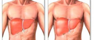
With Menetrier's disease (giant hypertrophic gastritis), a significant increase and lengthening of the pits of the epithelium of the stomach occurs; huge rigid folds are formed in the mucosa that do not straighten when the stomach is inflated with air. In this case, intestinal metaplasia of the epithelium, proliferation and hyperplasia of the mucous glands are noted with almost complete atrophy of the main glands that produce hydrochloric acid. These changes can acquire both focal and diffuse character.
In another form of hyperplastic gastritis - Zollinger-Ellison syndrome - under the influence of increased gastrin production, hyperplasia of parietal cells occurs; they are found in almost all parts of the stomach. The gastric pits are flattened and shortened. Hyperplasia of the mucosa due to parietal cells leads to a significant increase in the production of hydrochloric acid, the formation of many erosions, and then peptic ulcers of the stomach. Zollinger-Ellison syndrome most often develops against the background of a gastrin-producing tumor of the pancreas.
The rarest form, hypertrophic hypersecretory gastropathy, can occur with or without protein loss. With this pathology, glandular (glandular) or foveolar (epithelial) hyperplasia is noted, which is not associated with increased gastrin production. The pits and ridges of the gastric mucosa have a normal appearance. With glandular hyperplasia, cysts formed from enlarged mucous glands may be found in the mucosa. This form is intermediate between Ménétrier's disease and Zollinger-Ellison syndrome.
Based on endoscopic assessment of the degree of epithelial hypertrophy, focal (granular), giant hypertrophic gastritis (a common form of Menetrier's disease), warty gastritis (single growths of the epithelium), polypous hyperplastic gastritis (formation of hypertrophied folds with multiple polyps against the background of complete atrophy of the gastric mucosa) are distinguished.
Hyperplastic gastropathy
Hyperplastic gastropathy refers to conditions limited to the body and fundus of the stomach and associated with an excessive number of epithelial cells of the mucous membrane [8]. There are two main types of this pathology:
- Epithelial hyperplasia involving superficial and foveal cells of the mucosa (foveal hyperplasia); acid-producing cells can be in a normal or atrophied state. These are characteristic changes for Ménétrier's disease.
- An increase in the number of parietal cells without changes in the superficial and foveal cells of the mucosa. These changes are observed in Solinger-Elison syndrome.
A mixed type of hyperplastic gastropathy, in which both mucus-producing and acid-producing cells give a picture of hyperplasia, can also be observed in conditions such as lymphocytic and Hp-associated gastritis.
Most patients who show enlarged gastric folds on endoscopic examination do not have Ménétrier's disease. In one study, for example, endoscopic loop biopsy was performed in 52 patients with large gastric folds (greater than 1 cm in width and persisting with air insufflation) [9]. The main findings were:
- Chronic gastritis/lymphoid hyperplasia - 40%
- Benign tumors - 16%
- Stomach cancer - 12%
- Zollinger-Ellison syndrome - 10%
- Ménétrier's disease - 8%
Types of gastritis
Antral gastritis is a chronic inflammation that affects the antrum mucosa (the outlet of the stomach). The medical name of the disease is gastritis type B. It is caused by the activity of the bacterium Helicobacter pylori. The main symptoms are heaviness and pain in the stomach, belching, nausea.
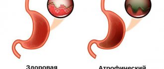
Atrophic gastritis is one of the forms of chronic gastritis. This disease of the stomach is the most insidious, since it is often a sign of a precancerous condition of the organ. The gastric mucosa becomes very thin, the glands that are responsible for the production of gastric juice degenerate. This process disrupts the digestion of food and reduces the overall immunity of the body.
Catarrhal gastritis is also called simple food gastritis. This is an acute inflammation caused by excessive consumption of fatty, spicy foods, seasonings, alcohol, and poor diet. The main symptoms of catarrhal gastritis are the same as any other types of gastritis: vomiting, nausea, heaviness in the stomach, unpleasant taste in the mouth. With proper treatment, these symptoms can be quickly relieved. Otherwise, the disease may become chronic.
Focal gastritis is a local inflammation of the gastric mucosa. Such lesions can vary in shape and size. Focal gastritis is divided into acute catarrhal and chronic. If acute inflammation is easily treatable, then a chronic disease can lead to atrophy of the gastric glands. In this case, it is important to start treatment in a timely manner.
Superficial gastritis is the first, safest stage of the disease. Inflammation affects only the upper layer of the gastric mucosa. In this case, there are no lesions, the deep tissues of the stomach and duodenum are not affected. But this disease should not be ignored. Over time, it can develop into a more serious form.
Reflux gastritis is a disease in which a harmful substance that differs in acidity constantly enters the stomach. This process is called reflux. As a result, the walls of the stomach become inflamed, and gastritis develops against this background. Reflux gastritis can be biliary (bile) and duodenal. The first type comes from the biliary tract. With deodenal reflux gastritis, contents rise from the intestines. Source: Sh.Z. Galiev, N.B. Amirov, O.A. Baranova. Morphological signs of reflux gastritis // Kazan Medical Journal, 2022, v. 98, no. 4, pp. 533-537.
Erosive gastritis is a type of disease in which small wounds – erosions – appear on the gastric mucosa. The main symptoms are bloody vomiting, loose stools, dark stool. One of the causes of the pathology is a burn of the mucous membrane, which is caused by the consumption of spicy, low-quality food, medications, and toxic substances. In addition, the disease occurs due to improper diet, stress, and disruptions in the body.
Autoimmune gastritis is a disease that causes the cells of the gastric mucosa to atrophy. This occurs due to improper functioning of the body's immune system. In other words, the immune system begins to “fight” the digestive system and destroys stomach tissue. The symptoms of the disease cannot be ignored. They mainly appear after eating. This may include belching, heartburn, or rumbling in the stomach. Autoimmune gastritis is a rare disease that is currently poorly understood. Source: Coati I, Fassan M, Farinati F, Graham DY, Genta RM, Rugge M. Autoimmune gastritis: Pathologist's viewpoint // World J Gastroenterol. 2015 Nov 14;21(42):12179-89. doi: 10.3748/wjg.v21.i42.12179.
Hyperplastic gastritis is an inflammation of the gastric mucosa. During the course of the disease, the inner lining of the organ grows, and cysts or polyps may form on it. This type of gastritis is chronic and benign. At first, its symptoms may not be noticeable to the patient. Often the disease is detected only during an endoscopic examination.
Ménétrier's disease
Ménétrier described two types of disorders associated with polypoid growths in the stomach, which he tried to associate with gastric carcinoma [10].
- The first (called “polyadenomes polypeux”) consists of multiple non-confluent gastric polyps, which are currently classified as hyperplastic polyps [11].
- The second (which he called Ménétrier's polyadenomatosis, or multiple sheet-like adenomas) is a new type associated with foveal hyperplasia and should by law bear his name [8].
Pathogenesis
The pathogenesis of Ménétrier's disease is not entirely understood, but may be mediated by transforming growth factor alpha (TGF-alpha). TGF-alpha increases gastric mucus production and inhibits acid secretion [12-14]. The level of TGF-alpha in the cells of the gastric mucosa in patients with Ménétrier's disease is significantly increased [12]. The role of TGF-alpha is further supported by a mouse study in which overproduction of TGF-alpha is associated with significant hyperplasia of gastric mucus-producing cells comparable to that found in humans with Ménétrier's disease [12]. These mice also had reduced basal and histamine-stimulated acid secretion, similar to that observed in humans with Ménétrier's disease [12,13,15]. TGF-alpha may exert its effect by binding to epidermal growth factor receptors. This was suggested based on a report of a patient who experienced clinical improvement after treatment with monoclonal antibodies directed against epidermal growth factor receptors [16]. A similar connection with cytomegolovirus infection is observed in the so-called childhood Ménétrier's disease. Its mechanism can also be realized through TGF-alpha [17]. However, during various types of inflammation and regenerative responses in the stomach, increases in foveal TGF-alpha may also be observed, demonstrating that these findings may be nonspecific [18]. In addition, this pathology may be an example of the incorrect use of the term Ménétrier's disease to other forms of hypertrophic gastropathy.
Clinical manifestation
Patients with Ménétrier's disease may present with a variety of clinical symptoms, including epigastric pain, significant weight loss, nausea, vomiting, gastrointestinal bleeding, diarrhea, and protein-losing gastroenteropathy [4,5,19,20]. In one study of 40 patients without morphological confirmation of Ménétrier's disease, the main complaints were:
- Epigastric pain - 65%
- Asthenia - 60%
- Anorexia - 45%
- Weight loss - 45%
- Edema - 38%
- Vomiting - 38%
In addition, approximately 80% of patients had hypoalbuminemia and increased intestinal protein loss. In other studies, hypoalbuminemia was present in 20-100% of patients, but its presence does not distinguish Ménétrier's disease from other causes of enlarged gastric folds other than Zollinger-Ellison syndrome [4,5,19,21,22]. Basal and stimulated gastric secretion of hydrochloric acid in Ménétrier's disease is usually reduced or normal, but varies depending on the stage of the disease [4,19,22,23]. A slight to moderate increase in serum gastrin concentrations may be present [24]. Additional clinical and laboratory findings are similar to those of other forms of protein-losing gastroenteropathies.
Symptoms of atrophic hyperplastic gastritis
Clinical signs of atrophic hyperplastic gastritis differ somewhat depending on the type of pathology. But the initial symptoms are usually the same and manifest themselves in the form of a feeling of heaviness in the epigastric region after eating fatty meat dishes, spicy seasonings, and pickles.
The disease lasts for a long time without complaints. But during a retrospective survey of the patient, the doctor can reveal:
- frequent heartburn;
- nausea;
- bloating;
- rarely vomiting food eaten;
- the appearance of plaque on the tongue;
- belching with an unpleasant odor.
Since at the initial stage the acidity remains normal or increased, pain in the epigastric region can be cramping in nature (spastic), less often described as aching or pressing
In cases of erosive gastritis, pain intensifies when bending the body or walking. Exacerbations are associated with the spring and autumn periods. Blood is found in stool and vomit. With giant hypertrophic gastritis, symptoms are often absent. Some patients still report nausea, diarrhea, weight loss, lack of appetite, and rare stomach bleeding.
The level of protein (albumin) in the blood of such patients is significantly reduced. This contributes to additional swelling of the stomach tissue. Hyperplastic gastritis is a chronic disease. It occurs with periods of exacerbations and remissions. The following symptoms characterize the exacerbation stage.
Diagnosis
The diagnosis of Ménétrier's disease is based on the identification of extreme foveal hyperplasia with glandular atrophy on biopsy in a patient with significant enlargement of the gastric folds observed on endoscopic examination or barium radiography. A full-thickness or loop biopsy is usually necessary [3,22]. Enlarged folds are limited to the body and fundus of the stomach. The folds are usually symmetrically enlarged, although asymmetrical "polypoid" enlargement may rarely occur.
Treatment
A variety of medications (including antacids, anticholinergics, prednisolone, H2-blockers, proton pump inhibitors, and prostaglandins) are used in the treatment of patients with Ménétrier's disease, but none have been shown to be consistently effective [20,21,25]. One exception may be treatment aimed at eradicating HP, however, such patients may have gastritis associated with HP as a cause of enlarged gastric folds (see below). Based on the results of treatment of a patient with Ménétrier's disease, octreotide (100 mg subcutaneously twice daily) was reported to be effective in reducing enteral protein loss [26]. Another similar report showed improvement after treatment with monoclonal antibodies directed against epidermal growth factor receptors [16]. Surgical treatment is warranted for patients with intractable pain, hypoalbuminemia with edema, bleeding, pyloric obstruction, and those patients in whom malignancy cannot be present [4,22]. Pain is often relieved and hypoalbuminemia corrected after gastrectomy. Although subtotal gastrectomy is the most commonly used procedure, total gastrectomy is also justified because this operation allows you to remove all pathological mucosa and prevent the development of stomach cancer (see below) [22]. In addition, the majority of postoperative deaths occur in patients undergoing subtotal gastrectomy. Many of these deaths are associated with anastomotic failure, possibly reflecting the difficulty of creating a strong anastomosis between normal and hyperplastic mucosa [22].
Forecast
The natural history of Ménétrier's disease is not fully understood. Symptoms may persist for decades [4,5,19]. There are scattered reports of the progression of Ménétrier's disease to atrophy of the gastric mucosa after 4-8 years of illness with a return of serum albumin concentrations to normal values [23,27,28]. However, in light of modern thinking, at least some of these patients may have had HP-associated gastritis rather than Ménétrier's disease. The risk of developing gastric cancer in patients with Ménétrier's disease is not entirely clear and, according to various estimates, varies from 2 to 15% [4,6,19,22,29]. One possible explanation for this discrepancy in numbers is the inaccurate diagnosis of Ménétrier's disease in many cases where the condition was associated with gastric carcinoma. There are only a small number of reports of Ménétrier's disease preceding gastric cancer by one or more years [6,22,29]. The hypothesis that the risk of developing carcinoma in Ménétrier's disease is lower than previously thought arose after observing 43 patients with this disease in whom no cases of cancer were detected for 10 years [5]. Other reports suggest that sepsis and vascular thromboembolic complications may be a greater threat to patients with Ménétrier's disease than malignant transformation [5,22].
Types of disease
The latest classification of gastritis is called Sydney after its adoption. Not all domestic gastroenterologists agree with her conclusions. In the practice of Russian doctors, several types of hyperplastic gastritis are distinguished.
Focal
Another name is “nodular endocrine cell hyperplasia”, benign hyperplasia in the form of a tumor less than 15 mm in diameter. It is based on the proliferation of endocrine cells, which are stimulated by excess gastrin hormone.
It occurs more often in patients with pernicious anemia caused by vitamin B12 deficiency. The mutated tumor suppressor gene MEN1 is recognized as the “culprit” for tumor growth; it is related to multiple endocrine lesions.
Surface
Only the uppermost layer of prismatic epithelium on the gastric mucosa is involved in the process.
Diffuse
The diagnosis is made when hypertrophic changes are multiple in nature, regardless of the etiological factor.
Polyposis
According to the classification, “multifocal atrophic gastritis with focal hyperplasias,” multiple or single polypous growths (focal and diffuse forms), consisting of glandular cells, are found on the mucosa. More often associated with massive Helicobacter pylori infection, autoimmune processes, and low acidity.
Typical for patients over 50 years of age
Erosive-hyperplastic
Otherwise called lymphocytic-erosive gastritis, against the background of leukocyte infiltrates and hypertrophy of the folds, nodules and areas of erosion of mucosal tissue are visible, most often in the area of the pits of the cardiac, pyloric sections and body of the stomach. The acidity of gastric juice may vary.
Hyperplastic granular
Or “granular” - close to a focal lesion, formations appear on the mucosa in the form of growing drops up to 3 mm in size, multiple in nature are possible, the mucosa looks lumpy and swollen. It most often affects the antrum. The muscles become dense and inactive. It is observed in men 40–50 years old.
Hyperplastic reflux gastritis
It necessarily includes reflux and damage to the antral mucosa by the alkaline composition of the duodenal contents. The most significant aggressive agents are bile acids.
Antral
Or rigid antral gastritis is distinguished by sharply disrupted folds in the antrum, they are thickened, change direction, and are covered with polyps on the surface. The pyloric part of the stomach gradually scars and narrows, and peristalsis sharply decreases. The production of hydrochloric acid stops.
Giant hypertrophic
Or polyadenomatous gastritis - Menetrier disease. It is characterized by the growth of folds along the greater curvature of the stomach, the release of the epithelium from the pits with excessive mucus production. Cells that synthesize mucus grow into the muscle layer and form cysts. A decrease in acidity is accompanied by loss of protein and dystrophy.
As you can see, the main differences can be determined only by the appearance of the mucous membrane during fibrogastroscopy and histology of biopsy specimens
Helicobacter pylori
Acute HP-associated gastritis can cause severe inflammation and enlargement of the folds of the stomach, which may even have an appearance similar to that of malignant diseases. In chronic HP associated gastritis, an increase in the folds of the stomach may also be observed, simulating Ménétrier's disease [30,31]. Several reports indicate the presence of enlarged gastric fundus and body folds with glandular hyperplasia, hypersecretion, and the presence or absence of hypoalbuminemia in patients without Zollinger-Ellison syndrome [19,32–34]. In the past, these cases were classified as gastritis with glandular hypertrophy [35], hypertrophic hypersecretory gastropathy [34,36], and Schindler's disease [37]. However, there are two observations indicating the role played by Helicobacter pylori in the development of this pathology:
- An examination of published micrographs of these patients showed that virtually all of them had gastritis consistent with HP infection [34].
- In one study, 18 of 32 patients with thickened gastric folds were infected with Hp [38]. Anti-Hp therapy led to normalization of the size of the gastric folds. Similar findings have been described in other reports [39,40]. It is noteworthy that in two of these reports [38,39], the term “Ménétrier's disease” was inappropriately used to refer to a condition associated with enlarged gastric folds.
Complications
Lack of timely treatment leads to unpleasant consequences of hyperplastic growth:
Functional stomach disorder in children
- the structure of the gastric mucosa is disrupted, more or less severe atrophy appears;
- the participation of the stomach in the digestion process decreases, since the production of gastric juice decreases in parallel with the destruction of parietal cells;
- body weight is lost;
- gastric motility is impaired, which leads to paresis and reflux damage to the esophagus;
- the intensity of protein metabolism decreases, a decrease in albumin affects the restoration processes in all organs and tissues;
- hypovitaminosis is accompanied by anemia;
- Hyperplastic granular and hypertrophic gastritis have the greatest ability to degenerate into an ulcer and a cancerous tumor; with polyposis, every fifth case is transformed.