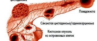
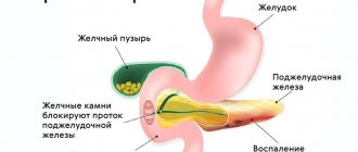
A pancreatic cyst is a cavity that is surrounded by a capsule and filled with fluid. The most common morphological form of cystic lesions of the pancreas are postnecrotic cysts. At the Yusupov Hospital, doctors identify cysts in the pancreas through the use of modern instrumental diagnostic methods: ultrasound, retrograde cholangiopancreatography, magnetic resonance imaging (MRI), computed tomography (CT). Patients are examined using the latest diagnostic equipment from leading global manufacturers.
The increase in the number of patients with cystic lesions of the pancreas is facilitated by the indomitable increase in the incidence of acute and chronic pancreatitis, the increase in the number of destructive and complicated forms of the disease. The frequency of postnecrotic pancreatic cysts is increasing due to the introduction of effective methods of conservative treatment of acute and chronic pancreatitis.
Against the backdrop of intensive therapy, therapists at the Yusupov Hospital are increasingly able to stop the process of destruction and reduce the frequency of purulent-septic complications. Surgeons use innovative techniques for treating pancreatic cysts. Severe cases of the disease are discussed at a meeting of the Expert Council with the participation of professors and doctors of the highest category. Leading surgeons collectively decide on patient management tactics.
Causes of development of adenoma and adenocarcinoma in the pancreas
- Mucinous cystadenoma and cystadenocarcinoma are primary cystic tumors of the pancreas with malignant potential, consisting of mucus-filled cysts that do not communicate with the pancreatic ductal system.
- Adenoma or carcinoma accounts for 1-2% of all exocrine pancreatic tumors
- The average age of patients is 40-80 years
- Adenoma or carcinoma occurs almost exclusively in women.
Causes:
- The stroma is similar in structure to ovarian tissue;
- Lined with mucus-secreting epithelial cells;
- Classified as adenoma, borderline tumor, or carcinoma depending on the degree of dysplasia;
- About a third of tumors are benign at the time of diagnosis;
- Cystadenoma or carcinoma is most often localized in the body and tail of the pancreas;
- The size is 2-25 cm (average 6-10 cm).
Stages
The process of formation of a post-acrotic cyst of the pancreas goes through 4 stages. At the first stage of the appearance of a cyst, a cavity is formed in the omental bursa, filled with exudate due to acute pancreatitis. This stage lasts 1.5-2 months. The second stage is the beginning of capsule formation. A loose capsule appears around the unformed pseudocyst. Necrotic tissue with polynuclear infiltration remains on the inner surface. The duration of the second stage is 2-3 months from the moment of occurrence.
At the third stage, the formation of the fibrous capsule of the pseudocyst, firmly fused with the surrounding tissues, is completed. The inflammatory process is intense. It is productive. Due to phagocytosis, the liberation of the cyst from necrotic tissue and decay products is completed. The duration of this stage varies from 6 to 12 months.
The fourth stage is the isolation of the cyst. Only a year later, the processes of destruction of the adhesions between the wall of the pseudocyst and the surrounding tissues begin. This is facilitated by the constant peristaltic movement of organs that are fused with a fixed cyst, and the long-term effect of proteolytic enzymes on scar adhesions. The cyst becomes mobile and is easily separated from the surrounding tissue.
Which method of diagnosing adenoma and adenocarcinoma to choose: CT, MRI, ultrasound
Selection Methods
- CT, MRI
Pathognomonic signs
- Usually consists of several large cysts (more than 2 cm in size);
- Well-circumscribed tumor with septations;
- Occasionally, thickening of the cyst wall can be detected;
- Does not communicate with the pancreatic ductal system;
- The bile duct and pancreatic duct may be dilated due to compression;
- Peripheral eggshell calcifications indicate malignancy.
What will CT scans of the abdominal cavity show for adenoma and adenocarcinoma?
- The tumor consists of large cysts with septations;
- The cyst wall is enhanced by contrast.
a- d Mucous cystadenoma of the pancreas: a ) CT, arterial phase. Thin-walled partitions are well visualized;
b ) T1-weighted MR image after contrast injection. Partitions are well visualized;
c ) Mucous cystadenoma. T2-weighted MR image. Cysts contain fluid that varies in signal intensity;
d ) MRCP. Displacement of the pancreatic duct in the tail area, high signal intensity can be traced only in individual components of the cyst.
Is an MRI of the abdominal cavity performed for adenoma and adenocarcinoma?
- Hyperintense cysts on T2-weighted images and MRCP
- Partitions are clearly demarcated
- There is no communication with the pancreatic duct.
Why is an abdominal ultrasound performed for adenoma and adenocarcinoma?
- Allows for a biopsy and analysis of the contents of the cyst - mucous fluid with necrosis and hemorrhages. Tumor markers are elevated, amylase levels are normal.
Pathogenesis
They arise as a result of malignancy of mucoid (mucous) cystadenoma.
In 40% of cases, pancreatic cancer is sporadic, that is, the etiology of the disease is not determined.
Risk factors:
• poor nutrition. Constant consumption of fatty foods, dry food, lack of a routine - all this causes digestive problems;
• bad habits (alcohol and smoking). It has been proven that a person who smokes a pack of cigarettes a day is 4 times more likely to develop cancer;
• the presence of a gene that may be involved in the formation of pancreatic tumors;
• hereditary diseases. Diseases that are inherited and contribute to the occurrence of PDA include: adenomatous polyposis, Gardner's syndrome, ataxia-telangiectasia, hereditary pancreatitis. The latter causes cancer in people of retirement age in 40% of cases;
• gastric surgery (gastrectomy or resection). Such interventions affect the digestive system, which disrupts the functioning of the pancreas and increases the risk of developing adenocarcinoma by 3 times;
• exposure to chemicals;
• sedentary lifestyle, excess weight.
What diseases have symptoms similar to pancreatic cystadenoma
Pseudocysts
- consequences of acute or chronic pancreatitis (history)
Serous cystadenoma
- many small cysts configured in the form of a honeycomb
– occasionally there is a central star-shaped “scar” with calcifications
Intraductal papillary mucinous tumor
- occurs mainly in young women
- solid and cystic components
- infiltrative growth and metastases
- hemorrhages
Cystic degenerative tumors
- infiltrative growth and metastases
Clinical manifestations
- Stomach ache.
As the tumor grows, abdominal pain becomes intense, sharp, radiates to the back and intensifies when the body bends forward. Irradiation of back pain indicates tumor involvement in the retroperitoneal region.
- Jaundice.
Tumors localized in the head of the pancreas in 80-90% of cases lead to the appearance of jaundice (as a result of compression of the common bile duct by the tumor). Skin itching, darkening of urine and lightening of stool are also noted.
- Loss of body weight.
This symptom is observed in 92% of patients with tumor localization in the head and in 100% of patients with damage to the body or tail of the pancreas. Weight loss may be associated with steatorrhea (as a result of impaired exocrine pancreatic function).
- Anorexia.
Anorexia is observed in 64% of patients with head cancer and in approximately 30% of patients with tumor localization in other parts of the pancreas.
- Nausea and vomiting.
Nausea and vomiting are observed in 43-45% of cases with cancer of the head and in 37% of cases with cancer of the tail and body of the gland. These symptoms may be the result of compression of the duodenum and stomach by the tumor.
- Development of secondary diabetes mellitus.
Diabetes mellitus as a consequence of cancer is diagnosed in 25-50% of patients, leading to symptoms such as polyuria and polydipsia.
- If the tumor is located in the body or tail of the pancreas, then it contributes to the occurrence of splenomegaly, bleeding from varicose veins of the esophagus and stomach.
- In some cases, a clinical picture of acute cholecystitis or acute pancreatitis develops.
- Metastases to the peritoneum can cause compression of the intestine with symptoms of constipation or obstruction.
Diagnosis of mucinous cystadenoma
For benign ovarian tumors, timely diagnosis of diseases is of great importance, so it is recommended to undergo regular examinations by a gynecologist. Mucinous cystadenoma can be detected using the following research methods:
- Bimanual examination - large tumors are identified that can be palpated through the abdominal wall.
- Ultrasound of the pelvic organs. Using this method, you can not only detect a tumor, but also determine its size, structural features, and location.
- Analysis for tumor markers CA-125+HE4 with calculation of the ROMA index. Allows you to differentiate malignant neoplasms and identify a group of patients who need in-depth examination to exclude oncology.
- MRI and CT scans are performed for large mucinous cystadenomas when there are problems identifying the site of origin of the tumor.
- If malignant transformation is suspected, additional research methods are performed, for example, colonoscopy, FGDS, chest fluorography.
In some cases, the final diagnosis can be made only after surgical excision of the tumor. In this case, the first stage is an inspection of the abdominal cavity, washings and urgent histological examination. Based on its results, the issue of the volume of intervention is decided.
Symptoms
Mucinous cystadenomas of small sizes (up to 3 cm) proceed unnoticed, do not manifest themselves in any way and are often detected during routine examinations, ultrasound of the pelvic organs or examination for other gynecological pathologies.
The main symptoms occur when the cystadenoma reaches a large size, and they are associated with compression of the pelvic organs. In this case, patients may present the following complaints:
- Pain in the lower abdomen, aching or stabbing in nature, which can radiate to the groin or lumbar region.
- Acute pain. They may indicate hemorrhage into the wall of the cystadenoma.
- Increased abdominal volume.
- Feeling of fullness or heaviness in the abdomen.
- Menstrual irregularities.
- If the tumor puts pressure on the bladder, you may experience frequent urination with a feeling of incomplete emptying of the bladder.
- When the rectum is compressed, constipation may occur.
- If compression of large blood vessels occurs, swelling of the lower extremities is possible.
Description example
Descriptive part . The pancreas has transverse dimensions: head x cm, body x cm, tail x cm, has uneven contours, a structure with a moderately pronounced stromal component (and signs of fatty degeneration), without obvious focal changes. In the projection of the head, a volumetric cystic formation is visualized, measuring ..x..x.. cm, with lumpy, sometimes unclear contours. A cystic component of a multi-chamber structure, with septa of uneven thickness (from .. cm to .. cm). The maximum cyst size is ..x..x.. cm, with heterogeneous contents, with uneven internal and external contours. The pancreatic duct is convoluted, unevenly expanded to .. cm, and not differentiated in the projection of the head. Parapancreatic tissue in the head area with signs of infiltration. After IV contrast, there is a heterogeneous accumulation of the contrast agent mainly along the periphery of the cystic formation and thickened cyst walls.
CONCLUSION : MR picture of a cystic formation of the head of the pancreas (more likely cystadenocarcinoma/cystadenoma).
A consultation with an oncologist is recommended, followed by a morphological examination for clarification.
Introduction
Epithelial cystic pancreatic tumors (PCT) represent a group of true cystic neoplasms, the main morphological feature of which is the presence of an internal epithelial lining. Over the past 20 years, thanks to the improvement and emergence of new non-invasive diagnostic methods, the number of patients with cystic tumors of the pancreas has increased significantly. The number of pancreatic resections for cystic tumors in some specialized centers reaches 30% of the total number of organ resections [34]. However, despite the high sensitivity of non-invasive diagnostic methods in detecting cystic tumors of the pancreas, determining the nature of the cystic neoplasm at the preoperative stage remains the main task, which can only be solved if the entire cystic tumor is completely removed. In this regard, the method of choice for treatment of patients with cystic tumor of the pancreas is surgical.
The purpose of the study was to analyze the immediate and long-term results of surgical treatment of patients with cystic tumor of the pancreas.
Material and methods
A retrospective analysis of the examination and surgical treatment of 41 patients with pancreatic epithelial cystic tumor lesions from 1984 to 2009 was carried out. A prerequisite for inclusion of patients in the study was histological confirmation of the diagnosis.
The majority of patients with cystic tumor of the pancreas were women (39, or 94.6%). The average age of patients was 50.7 years (range 29 to 73 years). The average size of cystic tumors of the pancreas was quite large and in all groups, except intraductal papillary mucinous, was about 10 cm (Table 1).
Mucinous cystic tumor was the most common neoplasm in our study and was diagnosed in 30 (73.2%) patients. Depending on the degree of dysplasia of the epithelial lining, the tumors were distributed as follows: mucinous adenoma (absence of any signs of dysplasia) - 18 (60%), borderline type (presence of dysplasia without an invasive component) - 5 (16.7%), cystadenocarcinoma (dysplasia deep degree with an invasive component) - 7 (23.3%).
The anatomical location of cystic tumors in the group of serous and malignant forms was relatively similar in distal or proximal location (see figure).
Figure 1. Distribution of cystic tumors in the pancreas.
Clinical examination of patients in the preoperative period included the study of complaints and medical history. Particular attention was paid to a history of signs of acute or chronic pancreatitis. Laboratory testing included general blood and urine tests, a biochemical blood test, and a study of the level of tumor markers in the blood serum: carbonic anhydrate antigen 19-9 (CA 19-9) and carcinoembryonic antigen (CEA).
Patients with a cystic tumor of the pancreas underwent a comprehensive instrumental study. At the first stage, ultrasound examination was performed in B-mode and duplex scanning with color Doppler mapping of the organs of the hepatopancreatoduodenal zone. At the second stage, patients underwent computed tomography with intravenous bolus contrast of the vessels and abdominal organs.
During the studies, the greatest attention was paid to the following parameters: localization and size of the cystic tumor, the nature of its wall (internal and external contour, thickness, presence of vegetations or papillae), the presence of septa inside the tumor, the presence of areas of calcification in the wall or internal septa of the cyst, contrasting of the septa , the state of the surrounding parenchyma of the gland, the diameter of the Wirsung duct and the presence of its connection with the cystic tumor, the relationship of the tumor with neighboring organs and vessels, the state of regional lymph nodes and the presence of distant metastases in the liver.
If it was difficult to determine the type of cystic tumor or if there was a suspicion of its communication with the ductal system of the pancreas, magnetic resonance cholangiopancreatography or endoscopic ultrasound examination was performed. The morphological diagnosis of a cystic tumor of the pancreas was established after a routine histological examination; it was confirmed in several cases by immunohistochemical examination of a removed macroscopic specimen.
An asymptomatic course occurred in 13 (31.7%) patients with a cystic tumor of the pancreas. Their cystic neoplasm was discovered by chance when performing an ultrasound or CT scan of the abdominal cavity for other diseases. The remaining 28 (68.3%) patients complained of: abdominal pain of varying nature and intensity - 24 (85.7%), weight loss - 9 (32.2%) and diarrhea - 1 (3.6%). The duration of the medical history in patients varied from 1 month to 10 years (on average 24.3±34.47 months).
Surgical interventions were performed in 37 patients with cystic tumor of the pancreas (Table 2).
Three patients with unresectable cystadenocarcinoma received symptomatic treatment; another patient with asymptomatic mucinous cystadenoma was refused surgical intervention due to her advanced age and high risk; she is under dynamic observation.
In the group of patients with serous cystadenoma, its removal was performed in 4, proximal and distal resections of the pancreas were performed in 5, and in another 1 case a median resection of the organ was performed. In the presence of mucinous cystadenoma, due to its more frequent localization in the distal parts of the pancreas, the tail or body and tail were more often resected - 15 patients, preserving the spleen in 2 of them. The volume of removed gland tissue ranged from 50 to 75%. Enucleation of mucinous cystadenoma was performed in 6 cases, pancreatectomy due to the presence of multifocal lesions of the pancreas was performed in 1. All patients with cystadenocarcinoma underwent standard resection of the pancreas in compliance with oncological principles: pancreatectomy (1), distal resection of the pancreas (2), pancreaticoduodenectomy (1). A patient with an IPMN of the head of the pancreas arising from the Wirsung duct underwent standard pancreaticoduodenal resection with urgent histological examination of the remaining organ resection margin.
When analyzing statistical data, average values and standard deviations were determined.
results
Postoperative complications developed in 13 (35.1%) patients. The most common complication of cystic tumor of the pancreas was pancreatitis - 7 (53.8%) patients. A destructive form of pancreatitis occurred in 2 of them. Most patients received conservative treatment of pancreatitis. In 2 cases, relaparotomies were required to sanitation the foci of gland destruction. In one observation, destructive pancreatitis developed in the tail of the pancreas after enucleation of a mucinous cystic tumor, which was the reason for distal resection of the organ; another patient was re-operated after a midline resection of the gland due to the failure of the pancreatojejunostomy for the purpose of sanitation and drainage of the abdominal cavity.
The formation of an external pancreatic fistula was noted in 4 (30.8%) patients. In 2 patients, this complication arose after enucleation of a cystic tumor. In all patients, the fistulas closed spontaneously, without additional surgical interventions. In 1 (7.7%) patient, the development of a colonic fistula was noted, and in another 1 (7.7%) - acute gastrointestinal bleeding from the gastroenteroanastomosis, cured by conservative measures. There were no lethal outcomes in the postoperative period.
Long-term results were observed in 76% of patients (24 patients with benign cystic tumor and 4 with invasive cystadenocarcinoma). The duration of follow-up ranged from 6 months to 10 years (mean 87.3 months).
The results of treatment of patients with benign cystic tumor of the pancreas are good, 5-year survival rate was 100%. In all patients with benign cystadenoma of the pancreas in the long-term period after surgery, there were no signs of local tumor recurrence during control ultrasound or CT scans. This also applies to patients who have undergone tumor enucleation. In patients with cystadenocarcinoma, the 5-year survival rate was 25%; out of 4 operated patients, 2 died 12 and 24 months after the intervention due to progression of the underlying disease. Currently, 2 patients are under observation (one lived for 10 months, the other for 60 months).
Discussion
Cystic tumors of the pancreas are a group of rare neoplasms and, according to the literature, account for 1-1.5% of all primary tumors of the pancreas [8, 10, 13]. This type of tumor has a wide fluctuation in potential up to malignant transformation. Our understanding of cystic neoplasms is based on their classification by the World Health Organization published in 1996 [27].
The majority of patients with cystic tumor of the pancreas we observed were women - 94.6%, whose average age was 50.7 years. This fact is considered as the most typical epidemiological indicator and can serve as one of the diagnostic criteria for this group of neoplasms [7, 30, 34].
The most common cystic tumor of the pancreas is a mucinous neoplasm. It should be noted that at the time of diagnosis, 18% of patients with asymptomatic mucinous tumor already have early or invasive cancer. Invasive cystadenocarcinoma is generally found in 6-36% of cases in patients with a primary diagnosis of mucinous cystadenoma [1, 13, 25, 42]. In our study, mucinous cystic tumor was also observed most often - in 30 (73.2%) patients, while serous cystadenoma was diagnosed only in 10 (24.4%) patients. Among patients with mucinous cystic tumor, 7 (23.3%) were diagnosed with invasive cystadenocarcinoma. One observation revealed an asymptomatic course of a malignant cystic tumor. This fact suggests that the asymptomatic course of the tumor in patients does not exclude the possibility that they have cystadenocarcinoma. The above facts indicate the possibility of transformation of a mucinous cystic tumor from a benign adenoma to a malignant cystadenocarcinoma [48].
Mucinous cystic tumor, according to our data, was most often located in the distal parts of the pancreas - in 83.9% of cases, while serous cystadenoma was most often localized in the proximal parts of the pancreas - in 40%, which correlates with the data of other authors [46, 47].
Clinical manifestations of cystic tumors are scanty and nonspecific. In 68.3% of observations, patients complained of pain or discomfort in the abdomen, weight loss and diarrhea. The presence of symptoms in patients with a cystic tumor of the pancreas increases the risk of malignancy, but the absence of clinical manifestations does not exclude the possibility of malignancy [25]. Physical examination of patients allowed in 46.3% of cases to reveal upon palpation a dense elastic, slightly painful, with clear contours, rounded formation, most often localized in the upper abdominal cavity, while the size of the tumor in all patients exceeded 13 cm.
Indicators of general clinical and biochemical blood tests in patients with cystic neoplasms of the pancreas often remain within normal limits. In the case of malignant transformation of the tumor, an increase in the level of tumor markers (CEA or CA 19-9) in the blood serum may be noted. In our study, high levels of tumor markers were detected in 5 patients. All patients had a mucinous cystic tumor, 3 had mucinous cystadenoma, 2 had cystadenocarcinoma, and 1 had an IPMN. The highest level of tumor markers was observed in patients with cystadenocarcinoma. In most cases, despite the high specificity (>90%) of tumor markers, their level often fluctuates within normal limits and the sensitivity of these tests is extremely low - less than 50% [8, 20].
Unfortunately, clinical, anamnestic, and laboratory research methods have low diagnostic value, so instrumental methods play the main role in the diagnosis of cystic tumors of the pancreas [4, 11]. Visualization of the pancreas during ultrasound examination, due to the deep anatomical location of the organ in the retroperitoneal space and often due to the interposition of gas-filled loops of the small or large intestine, is sometimes a difficult task. The sensitivity of this method in determining the type of cystic tumor is 53% [5], therefore, CT should be considered the method of choice in examining patients with cystic tumors of the pancreas, since this method has greater sensitivity and specificity [3, 23, 44]. Despite its high sensitivity, the diagnostic accuracy of CT in differentiating cystic neoplasms varies from 20 to 90% [8]. In difficult situations, magnetic resonance cholangiopancreatography and endoscopic ultrasound can be used for differential diagnosis.
Cystic tumors of the pancreas are a surgical disease. In the past, many authors advocated an aggressive approach in the treatment of all cystic tumors of the pancreas due to the lack of reliable diagnostic criteria to accurately determine or exclude the malignant nature of the neoplasm [19, 32, 33, 38].
Currently, in many medical centers, tactics have become less aggressive. Modern treatment of cystic tumors of the pancreas should be based on a comparative assessment of the degree of risk and benefit of surgical intervention, which is primarily determined by the risk of malignancy of neoplasms, with the surgical consequences of the operation itself [6, 8, 13].
All patients with mucinous cystic tumor, taking into account its tendency to malignant transformation, require mandatory surgical intervention [14, 18, 25, 41]. Patients with serous cystadenoma less than 3-4 cm in diameter and at moderate or high risk for surgery may be monitored. This approach is associated with the low potential of these cystic tumors for malignant transformation. These patients are subject to active clinical and radiological surveillance.
In case of malignancy of the tumor or its rapid growth, the issue of surgical intervention should be discussed [12, 16, 34, 43]. According to the results of a study by C. Lee et al. [29], even in this group of tumors, hidden malignancy is present in 3.3% of cases, which requires surgery in all patients with suspected cystic tumor. Thus, the issue of monitoring patients with cystic neoplasms of the pancreas continues to be a subject of debate today.
The main principle of surgical treatment of a benign cystic tumor of the pancreas is its removal, which is achieved by organ resection or enucleation. This conclusion is based on good long-term results of both the first and second surgical procedures [24]. The choice of intervention is influenced by the location and size of the tumor, its relationship with the ductal system and vessels of the pancreas.
The location of the tumor dictates the option of pancreatic resection. When the neoplasm is proximally located, a classic pancreaticoduodenal resection is performed, and the localization of the cystadenoma in the body or tail requires distal resection of the organ. Laparoscopic technology can be a good alternative for such gland involvement with a small or medium-sized tumor [25].
The question of the possibility of enucleation of a mucinous cystic tumor remains controversial. There is an opinion that enucleation in such patients can be considered an adequate operation with good long-term results [40]. However, enucleation of a mucinous tumor is controversial from an oncological point of view due to the identification of areas with varying degrees of dysplasia and the lack of diagnostic methods to determine or exclude the presence of foci of malignancy in the tumor itself [30, 35].
In our study, enucleation of mucinous and serous cystic tumors was performed in 10 cases. There were no signs of tumor recurrence in the long-term period. We believe that if the tumor is localized on the surface of the gland, its small size and full confidence in the absence of malignancy, it is possible to plan enucleation of the tumor or local resection of the gland with urgent histological examination of the removed macroscopic specimen. According to our data, similar conditions for cystadenoma enucleation occurred in 27% of patients, inferior to pancreatic resection (73%). It must be emphasized that a relative disadvantage of enucleation of a cystic tumor is the development of external pancreatic fistulas [40].
Surgical treatment of patients with cystadenocarcinoma should include only standard organ resections. The issues of lymph node dissection and adjuvant chemotherapy currently remain unresolved [14]. Monitoring of this group of patients is mandatory using computed tomography or magnetic resonance imaging every 3 months to identify local or distant relapses of the disease and decide on further tactics [2, 41].
Treatment of patients with intraductal mucinous papillary tumor is the most difficult task. Due to possible multifocal damage to the pancreas, not all authors consider organ resection to be a radical operation, since in the long term there is a high risk of developing a relapse of the disease: in benign forms - 7-10%, in malignant forms - 28-30% [15, 31]. In this regard, all patients should be observed in the postoperative period in order to detect tumor recurrence and possible removal of the pancreatic stump [26, 41]. A prerequisite for performing the primary operation is an urgent histological examination of the edges of organ resection and the desire to ensure that there are no tumor cells in the remaining resection edge. Otherwise, the scope of the operation can be expanded up to pancreatectomy [9, 12, 28, 37].
The optimal operation for tumor localization in the proximal parts of the gland should be considered pancreaticoduodenectomy in its various modifications; if the body and tail are affected, distal resection should be considered. If the tumor affects the branches of the Wirsung duct, taking into account the minimal risk of malignant transformation, it is permissible to perform organ-preserving operations, resection of the head of the gland with preservation of the duodenum, or segmental resection of the organ with urgent histological control of the resection margins [12, 28, 31, 39].
Long-term results of surgical treatment of patients with benign serous and mucinous cystic tumors of the pancreas in our study are good and correlate with data from other authors [36, 45, 46]. All patients are alive, with the exception of one patient who died 8 years after surgery from a concomitant disease. Monitoring of patients after radical removal of a benign serous or mucinous cystic tumor is not required, since relapses of such tumors are not observed in the long-term period [14, 21].
The five-year survival rate of patients with invasive cystadenocarcinoma, according to the literature, ranges from 15 to 63% [17, 21, 22]. In our study, out of 4 operated patients with invasive cystadenocarcinoma, 1 died in the first year after surgery and another 1 died in the second year. Currently, 2 patients are under observation (one lived for 10 months, the other for 60 months). In this group of patients, the 5-year survival rate was 25%.
Thus, at present, the possibilities of identifying cystic tumors of the pancreas have expanded significantly, but difficulties remain in determining the type of tumors at the preoperative stage. Mucinous cystic tumors are an indication for surgical treatment. If the size of serous cystadenoma is less than 3-4 cm and there is an average or high risk of surgical intervention, patients can be monitored. The extent of surgical intervention (resection or enucleation) for benign cystic tumors is determined by factors such as size, relationship of the tumor with the parenchyma, ductal system and vessels of the organ. Long-term results of surgical treatment of patients with benign cystic neoplasms are good, while the results of treatment of patients with cystadenocarcinoma remain unsatisfactory. The results of treatment of patients with a benign cystic tumor of the pancreas indicate a real possibility of complete cure, while in the presence of invasive cystadenocarcinoma the results are unsatisfactory, which emphasizes the importance of early diagnosis and selection of an adequate volume of surgical intervention in this group of patients.
Consequences and prognosis
With timely treatment, the prognosis is favorable. But problems with pregnancy may arise. In particular, when one ovary is removed, the likelihood of spontaneous pregnancy remains, although the chances are somewhat reduced. But with bilateral adnexectomy and hysterectomy, menopause occurs, which is accompanied by a decline in sexual function and the inability to get pregnant.
The doctors at our clinic have extensive experience in treating cystadenoma. We use modern treatment protocols and in each specific case we strive to achieve the best possible result.
Book a consultation 24 hours a day
+7+7+78


