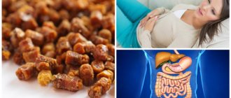
Causes of bile duct cancer and risk factors
Unfortunately, despite many years of study, the exact causes of the development of this type of cancer are unknown. There are a number of factors that can trigger the development of the disease. These include:
- sclerosing primary cholangitis is a chronic disease of unknown etiology;
- Caroli's disease is a congenital pathology in which a large number of intrahepatic cysts form;
- liver hepatosis;
- adenomas;
- diabetes;
- parasitic liver diseases;
- work in hazardous production;
- HIV infection;
- Lynch syndrome is a hereditary pathology that increases the risk of developing cancer of the digestive system;
- hepatitis B, C;
- Crohn's disease is a severe chronic disease in which inflammation of the mucous membranes of the digestive tract occurs;
- cirrhosis of the liver;
- bad habits.
Symptoms of bile duct cancer
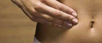
One of the main symptoms of cholangiocarcinoma is obstructive jaundice, which is accompanied by severe skin itching. The patient's stool is discolored. There is usually no pain before jaundice, and attacks of colic are rare. When the lumen of the duct is quickly blocked by a tumor, attacks of pain may occur before jaundice.
Symptoms and signs of bile duct cancer also include dyspepsia, loss of appetite, and weight loss. The body temperature of some patients remains within normal limits, while others increase. As a result of stagnation of bile, liver function is disrupted. It is painless on palpation, enlarged, and has a smooth edge.
One of the methods for differential diagnosis of jaundice is palpation of the gallbladder. In most cases, the tumor is not palpable, since it is located deep in the abdominal cavity and is small in size. When the portal vein is compressed by a neoplasm, hydrocele of the abdominal cavity develops.
Liver bile
Hepatocytes produce bile constantly, 0.5–1 liters per day. 95–98% of liver bile consists of water and has an alkaline reaction. It contains bile salts, bilirubin, cholesterol, fatty acids, lecithin, ions Na+, K+, Ca2+, Cl-, HCO3-, etc. The color of bile is due to bile pigments (bilirubin, etc.), which are formed from the breakdown products of red blood cells. It is bile pigments that color the intestinal contents brown. The role of bile in digestion is reduced to emulsification and breakdown of fats, which facilitates their digestion and absorption. Bile enhances intestinal motility.
Stages of cholangiocarcinoma
There are four stages of the disease:
First stage . The neoplasm is located below the confluence of the hepatic ducts, without involving the place of their connection (confluence) in the pathological process.
Second stage . The tumor is still below the confluence of the ducts, but has spread to the junction of the ducts.
Third stage . The tumor continues to grow and gradually affects the left and right hepatic ducts.
Fourth stage . Segmental ducts are affected.
According to the TNM classification, the following types and degrees of the disease are distinguished:
| T0 | There are no visible signs of a neoplasm |
| Tis | The lesion is localized within the bile ducts |
| T1 | The neoplasm is still within the affected organ and begins to grow into muscle and fibrous tissue |
| T2 | The lesion spreads beyond the ducts, the tumor begins to grow into nearby liver tissue |
| T3 | The tumor spreads to the hepatic arteries |
| T4 | The tumor grows into the liver ducts or nearby vessels are involved in the pathological process |
Survival prognosis
The average five-year survival rate for all patients diagnosed with bile duct cancer is 8%. This low rate is due to the fact that tumors are often diagnosed at late stages, many of them have an “inconvenient” location, and because of this they are difficult to remove.
Forecast of five-year survival at different stages:
- If the tumor is within the bile ducts and does not spread deeper: 24%.
- If the tumor has grown into surrounding tissue or spread to regional lymph nodes: 6%.
- If there are distant metastases: 1%.
The prognosis for bile duct tumors is not the most favorable. These types of cancer are very difficult to fight. But this does not mean that you need to give up. Euroonko uses the most modern treatment methods. We always know how we can help, and for us there are no hopeless patients.
Diagnosis of cholangiocarcinoma
Clinical signs of the disease are not specific, so it is not possible to diagnose biliary tract cancer based only on history and clinical examination.
The patient is additionally prescribed the following laboratory and instrumental diagnostic tests:
- Blood test (biochemical). Provides information about the presence of a pathological process in the liver, but does not allow making an accurate diagnosis. The level of alkaline phosphatase and bilirubin in the blood is increased. Indicators of AST, ALT, as well as the concentration of albumin in this type of oncology are usually within normal limits.
- Analysis for tumor markers of cholangiocarcinoma. Has great diagnostic value. The CA 19-9 antigen is detected in patients (this antigen can also be synthesized in cholangitis and pancreatic cancer). If the marker level in patients with chronic cholangitis increases significantly (up to 100 U/ml or more), then most likely this indicates the development of cholangiocarcinoma.
- Ultrasound of the gallbladder and liver . Prescribed at the initial stages of diagnosis. Using this method, it is possible to detect pathological expansion of the ducts in some places, as well as identify a large tumor.
- Dopplerography of liver vessels . Blood flow disturbances in the lesion are detected, but small tumors cannot be detected using this technique.
- CT scan of the biliary tract . A more informative diagnostic method. Using CT, you can see small tumors and diagnose enlarged regional lymph nodes.
- MSCT . Determine the degree of blockage of the bile ducts.
- PET CT . Improved technique. With its help, you can see nodular-type neoplasms measuring less than one centimeter. But infiltration forms of the disease are difficult to identify using this technique.
- Endoscopic retrograde cholecystography . Used to clarify the diagnosis. With its help, you can accurately determine the location of blockage of the bile ducts and take a tissue sample for histological examination.
- MRI . Today it is the most informative method for diagnosing oncological pathologies of the bile ducts and liver. This study does not use contrast agents; the method is non-invasive and therefore safe for the patient’s health. On MRI, you can see the vessels and bile ducts in a three-dimensional image, correctly assess the stage of the disease, identify small neoplasms, determine further treatment tactics and give a prognosis.
Diagnosis and treatment of functional disorders of the biliary tract
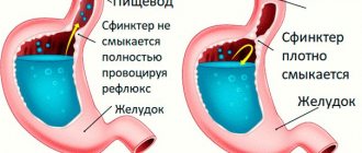
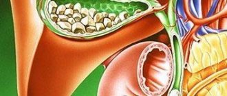
The bile capillary, the initial part of the biliary system, is formed by the outer surface of the apical part of the cytoplasmic membrane of adjacent hepatocytes and tight contact complexes located at the points of contact of hepatocytes. Each liver cell participates in the formation of several bile canaliculi. At the periphery of the lobule, the bile canaliculi merge into intralobular canaliculi, which, emerging into the interlobular connective tissue, pass into the interlobular canaliculi. Next, the interlobular tubules, merging, form interlobular ducts of the 1st and 2nd order, which are lined with prismatic epithelium, and, finally, large hepatic ducts are formed that leave the liver. A direct continuation of the intrahepatic ducts are the bile ducts: the right and left hepatic ducts, the common hepatic duct. The cystic duct, which drains bile from the gallbladder, and the common hepatic duct join to form the common bile duct. Regulation of bile flow is carried out by several sphincters. The sphincter of Miricia is located in the common bile duct. At the junction of the bladder neck and the cystic duct, the muscle fibers take a circular direction, forming the sphincter of the gallbladder duct (Lutkens). The main role in the regulation of bile secretion belongs to the sphincter of Oddi, which is represented by the sphincters of the common bile duct, papilla of Vater and the main pancreatic duct. The sphincter of the common bile duct consists of circular and longitudinal muscle fibers. Throughout its entire length, the circular muscular layer of the sphincter of Oddi is independent of the muscles of the duodenal wall. The circular muscle fibers of the sphincter of the common bile duct begin in its pancreatic part, and then surround the intramural part of the common bile duct and pass to the ampulla. The longitudinal muscle fibers of the sphincter of the common bile duct are located in both ascending and descending directions. In this case, the ascending fibers surround the supraduodenal part and represent a continuation of the longitudinal layer of the muscles of the duodenum. Descending fibers are independent of the duodenal musculature. The pancreatic duct at the confluence with the common bile duct is surrounded by circular, semicircular and longitudinal smooth muscle fibers. The outer longitudinal layer is represented by fibers originating in the muscles of the duodenum. Circular and semicircular fibers are located in the inner layer and belong to the duct itself. If there are sphincter fibers of the ampulla of Vater's nipple, they are located unevenly: there is an accumulation of them at the base of the nipple (sphincter of the base of the nipple) and at its mouth (sphincter of the orifice of the nipple of Vater). Thus, the concept of “sphincter of Oddi” includes a number of sphincters located in the area of the choledocho-pancreatoduodenal junction, namely the sphincter of the common bile duct, the sphincter of the papilla of Vater (sphincter of the mouth and base of the nipple), the sphincter of the main pancreatic duct. The sphincter of the common bile duct is the most constant (100%). The presence of other sphincters depends on the type of entry of the common bile and pancreatic ducts into the lumen of the duodenum [10]. Bile secretion occurs continuously; a person secretes from 0.5 to 2 liters per day. The direction of bile movement is determined by the pressure gradient between the gallbladder, bile ducts and duodenum and the coordinated activity of the sphincters of the biliary apparatus. With the Mirizzi sphincter closed and the Lütkens and Oddi sphincters open, contraction of the gallbladder leads to the release of bile into the duodenum. From the hepatic ducts and the common bile duct, bile enters the gallbladder at the moment the sphincter of Oddi closes (it plays a decisive role in creating the pressure gradient). The sphincter of Oddi outside of digestion is not always closed, and small portions of bile constantly enter the duodenum. After the end of the digestive phase, bile enters the gallbladder for 3 or more hours. Outside of digestion, the bile pressure in the common bile duct is 60–185 mmH2O. During eating, when the gallbladder contracts, the pressure rises to 150–260 mm of water column, which ensures the release of bile into the duodenum, where the pressure is lower. With a sharp increase in pressure in the biliary system (more than 300 mm of water column), the secretion of bile by the liver may stop. At a pressure of about 280–300 mm water column. pain usually appears and the level of alkaline phosphatase and leucine aminopeptidase in the blood increases due to the penetration of bile into the space of Disse, and from there into the sinusoids. The main volume of bile is secreted after eating. Fasting bile secretion is about 20–30% of postprandial bile secretion, it occurs synchronously with the contraction phases of the stomach and duodenum and prevents excessive thickening of bile. Under physiological conditions, the bile in the gallbladder is heterogeneous, and the difference in different layers can reach 2.5 times (this creates the phenomenon of “layered” bile). Most researchers believe that the extrahepatic bile ducts are never at rest and their active peristalsis is considered from the point of view of regulation of bile flow. The tone of the duodenum affects the release of bile, and it has been established that the entry of acidic chyme into the intestine and irritation of the large duodenal nipple can cause its spasm for 4–10 minutes or more. The motor reaction of the gallbladder and sphincter of Oddi significantly depends on the quantity and quality of food, as well as on emotional influences [5,8]. The work of the entire biliary system is strictly coordinated. This is ensured by both nervous and humoral regulation. Motor innervation is carried out by the parasympathetic and sympathetic nervous systems. Nerve plexuses are present in all layers of the biliary system. Sensitive fibers of the gallbladder can only perceive stretching. It has been shown that moderate irritation of the vagus nerve causes coordinated activity of the gallbladder and sphincters, and severe irritation causes spastic contraction with delayed bile evacuation. Stimulation of the sympathetic nerve helps relax the gallbladder. Of the gastrointestinal hormones, the leading role belongs to cholecystokinin - pancreazimin (CCK-PZ), gastrin secretin, motilin, and glucagon. The maximum effect is exerted by CCK-PZ, which, along with contraction of the gallbladder, promotes relaxation of the sphincter of Oddi. The incentive for the production (release of CCK-PZ) is fatty foods, and the volume of the gallbladder decreases by 30–80%. Hormonal influences are stronger than nervous ones, especially since CCK-PZ also acts as a neurotransmitter. Until now, many regulatory mechanisms are not entirely clear, especially since new regulatory components are being discovered (in particular, endogenous peptides of the endorphin group). It is only clear that the influence of various gastrointestinal hormones is largely determined by the self-regulation of organs, which is complex in physiological conditions and is not entirely clear in the case of various pathologies of this system [6,9]. Functional diseases of the biliary tract are a complex of clinical symptoms that develop as a result of motor-tonic dysfunction of the gallbladder, bile ducts and sphincters. According to the latest International classification (Rome Consensus, 2006), dysfunctional disorders of the biliary tract are usually divided into two types: E1 - gallbladder dysfunction and E2 - sphincter of Oddi dysfunction. In the latest International Classification of Diseases (ICD-10), under the heading K 82.8, only “dyskinesia of the gallbladder and cystic duct” and under the heading K 83.4 “spasm of the sphincter of Oddi” are highlighted. There are primary and secondary dysfunctional disorders. Primary ones are rare and average 10–15%. In this case, a decrease in the contractile function of the gallbladder may be associated both with a decrease in muscle mass and with a decrease in the sensitivity of the receptor apparatus to neurohumoral stimulation. Secondary dysfunctional disorders of the biliary tract can be observed with hormonal disorders: treatment with somatostatin, premenstrual disorder syndrome, pregnancy, after gastric resection, anastomosis, vagotomy, systemic diseases, celiac disease, diabetes, hepatitis, cirrhosis of the liver, jejunostomy, and also in the presence inflammation and gallstones; Psycho-emotional overload, stressful situations, and general neuroses are important. The majority of patients who have undergone cholecystectomy are characterized by insufficiency of the sphincter of Oddi with continuous flow of bile; spasm is less common; after distal gastrectomy, a decrease in hormone production occurs with resulting motor disorders. The classification of dysfunctional disorders is presented in Table 1. Clinical manifestations are quite well known: with hyperkinetic disorders, colicky pain of varying intensity occurs without irradiation or with irradiation to the right, to the back, sometimes to the left half of the abdomen (with the involvement of the pancreatic ductal system). With hypokinesia - dull pain in the right hypochondrium, a feeling of pressure, fullness, which intensifies with a change in body position with an increase in intra-abdominal pressure. Common to different forms of dysfunction are bitterness in the mouth, bloating, and unstable stools. The central symptom of dysfunctional disorders is biliary pain. Diagnostic criteria for gallbladder dysfunction 1. Repeated episodes of moderate or severe pain localized in the epigastrium or right hypochondrium and lasting 20 minutes. or more for at least 3 months. (pain is defined as moderate when it interferes with the patient's daily activities, and severe when it requires immediate medical advice or medication management). In addition, pain may be combined with one or more of the following symptoms: • nausea, vomiting; • irradiation of pain to the back or right shoulder blade; • pain after eating; • the occurrence of pain at night. 2. Impaired gallbladder function. 3. Absence of structural abnormalities that explain these symptoms. A very important objective symptom of impaired motility of the gallbladder is the ultrasonic phenomenon of sludge, which, according to our data, can be presented in two variants: a) diffuse; b) near the wall. The parietal variant, depending on the clinical situation, can be characterized as “inflammatory” or without inflammation, but then the sediment elements that form it are quite large. Yet the presence of sediment does not explain the symptom of pain. In addition to this analysis, the entire clinical symptom complex must be subjected to analysis. Diagnostic criteria for DSO a) Biliary DSO type 1. An attack of pain of the “biliary” type in combination with the following three signs: A. an increase in AST and (or) alkaline phosphatase two or more times with a 2-fold study; B. delayed removal of contrast agent during ERCP (more than 45 minutes); B. expansion of the common bile duct more than 12 mm. b) Biliary DSO type 2. An attack of pain of the “biliary” type in combination with one or two of the following signs: A. an increase in AST and (or) alkaline phosphatase by 2 or more times during a 2-fold study; B. delayed removal of contrast agent during ERCP (more than 45 minutes); B. expansion of the common bile duct more than 12 mm. c) Biliary DSO type 3. Only an attack of pain of the “biliary” type is observed. d) Pancreatic type DSO In its most obvious form (similar to biliary type 1), pancreatic type DSO can be represented by classic pancreatitis with epigastric pain, which often radiates to the back and is accompanied by an increase in serum amylase or lipase. The absence of traditional causes of pancreatitis (alcohol abuse and stones) often leads to a diagnosis of idiopathic recurrent pancreatitis. In less obvious forms (like type 3 biliary DSO), the pain is of a similar nature, but there is no rise in pancreatic enzymes; In many such patients, the symptomatology may be a manifestation of functional pain syndrome. In cases where ERCP excludes the absence of a stricture, manometry of the biliary and pancreatic sphincters is indicated. Additional instrumental and laboratory diagnostic approaches. As can be seen from the presented diagnostic criteria of the III Rome Consensus, the diagnosis is based mainly on clinical manifestations. The World Congress of Gastroenterology (Bangkok, 2002) determined that evidence-based medicine does not require consensus, but evidence. It was also stated there that DSO should not be classified as a clearly defined disease, but as a condition with a variable “dysfunction-symptom” relationship; It is unclear whether delayed gallbladder emptying can be considered a specific clinical problem (nosological form). For this reason, none of the tests used are standard, and their diagnostic value remains controversial. Nevertheless, the presence of these complaints presupposes a certain examination algorithm with screening and clarifying tests. Liver function tests - elevated liver enzymes and/or bilirubin coinciding with at least two episodes of biliary pain suggest DSO. Pancreatic enzymes in the blood and urine, a significant increase in amylase or lipase, coinciding with pancreatic pain, suggest pancreatitis due to DSO. Pain provoking tests - the use of morphine (prostigmine), which provokes DSO, is significantly limited by its sensitivity and specificity. Ultrasonography (ultrasound) – to assess the function of the gallbladder, an ultrasound examination is performed with a test breakfast. The contractility of the gallbladder is considered normal if its volume by 30 minutes. decreases by 30–70% from the original. In patients with a removed gallbladder, ultrasound scanning can be used to determine the diameter of the common bile and/or pancreatic ducts before and after the administration of provocative agents (cholecystokinin, morphine, fatty foods). Dilation of the common bile duct over 8 mm may indicate an obstruction to the outflow of bile through the sphincter of Oddi (SO), but these data are diagnostically insignificant, since a similar phenomenon is observed in 34% of asymptomatic patients after cholecystectomy. The diameter of the bile ducts is measured at intervals of 15 minutes. within 1 hour. Normally, the diameter of the bile ducts does not change. If the diameter increases by 2 mm or more compared to the original, the presence of DSO can be assumed. The sensitivity and specificity of provoking tests (fatty food intake, CCK test, secretin test to confirm pancreatic DSO) for unmasking a partially narrowed common bile duct are not known. Endoscopy with examination of the Vater's nipple is also included in the patient examination program to exclude duodenal pathology (diverticulum, papillitis). Multifractional minute-by-minute duodenal intubation can be used to determine not only the volume of various segments of the biliary system (gall bladder, common bile duct), but also the functional state of the gallbladder and individual sphincters of the biliary tract (the technique is described in the monographs of V.A. Maksimov et al. , 1998, 2008) [5,6]. Among other instrumental laboratory tests, dynamic cholescintigraphy should be mentioned, which is based on selective absorption from the blood by hepatocytes and excretion of Tc-99 radiopharmaceuticals labeled in bile. The value of the method lies in the possibility of continuous long-term monitoring of the processes of redistribution of radiopharmaceuticals in the hepatobiliary system under physiological conditions, which makes it possible to indirectly judge the functional state of hepatocytes, quantify the evacuation ability of the gallbladder, and also identify disorders of bile outflow associated with both a mechanical obstacle in the biliary system, and spasm of the sphincter of Oddi. Cholescintigraphy results have been shown to correlate with sphincter of Oddi manometry data [12]. Choledochoscintigraphy is a useful screening method for selecting patients after cholecystectomy for sphincter of Oddi manometry. Magnetic resonance cholangiopancreatography (MRCP; preferably with the introduction of secretin) is a safe way to examine the biliary and pancreatic ducts, which makes it possible to exclude other diseases of the pancreas and biliary tract that cause a similar pain syndrome (chronic pancreatitis, blockage of the duct with a calculus, duct strictures, tumors of the papilla of Vater etc.). The use of this technique is advisable in patients with DSO types 2 and 3, in whose management it is recommended to avoid invasive examinations (ERCP and endoscopic manometry SO) whenever possible [13]. Endoscopic retrograde cholangiopancreatography (ERCP): ERCP data, such as the diameter of the Holedoch of more than 12 mm and slowly release of contrast (more than 45 min.), Are diagnostic criteria for DSO. Additional signs are the expansion of the pancreatic duct by more than 5 mm and slowly eliminating contrast from the pancreatic ducts (more than 10 minutes). However, premedication, insufficient standardization of the method and the risk of pancreatitis develop significantly limit the value of the method. Transendoscopic manometry CO is the most reliable method for studying the function of the Oddi sphincter. It includes the determination of the basal pressure of the sphincter, followed by the study of phase and wave -wave changes - amplitude, frequency and direction of the spread of phase waves. A normal indicator of pressure in the common bile duct is a pressure that exceeds that in the duodenum by 10 mm Hg. The pressure in the CO, which is 18±4 mmHg under normal conditions, increases during its spastic contractions to 110±25 mmHg. [3.16]. Signs of DSO during manometric examination are; a) increase in basal pressure in the lumen of the sphincters; b) increase in the amplitude and frequency of phase contractions (tachyodia); c) increase in the frequency of retrograde contractions; d) paradoxical response to the administration of cholecystokinin analogues. Manometry is not shown to patients with the 1st DSO, since the endoscopic sphincterotomy is effective in more than 90% of cases (even with normal manometry results). In the 3rd type of DSO, pathological changes in the function CO are rarely detected, and the risk of complications as a result of the study is quite high. In patients with a 2nd type of DSO, a manometric study is most justified, since in 50% of cases they have an increased level of basal pressure of the sphincter. In patients with a pancreatic type of disease, the probability of the development of pancreatitis associated with the study is a high probability [14,15]. Increased basal pressure is a sign of either stenosis, or spasm of CO. In case of spasm, the pressure of C is reduced after the appointment of drugs related to smooth muscles. For this reason, the prescription of manometry CO should be based on the severity of clinical manifestations and effectiveness of conservative therapy. Manometry allows an accurate diagnosis to be made before more radical treatment methods are applied. A comprehensive psychological study allows us to identify the presence of psychoemotional disorders in patients with functional diseases of the gallbladder, to evaluate the structure and severity of these disorders. In the study of the mental state of patients, psychometric methods occupy a significant place for a better understanding of the patient’s personality and his own understanding and understanding of his disease [2.7]. Thus, the existing complex of examination of patients with biliary dysfunction today allows you to separate the group with organic pathology and take the necessary complex of treatment (often surgical plan) with functional pathologies that need effective pharmacotherapy. The main goal of the treatment of patients with the dysfunction of the biliary tract is to restore the normal current of bile and the secretion of the pancreas through the biliary and pancreatic ducts. Based on the main goal, the tasks of treatment should be: a) restoration, and if it is impossible to replenish the products of bile (with the development of chronic biliary failure); b) an increase in the contractile function of the gallbladder (with its insolvency); c) a decrease in the contractile function of the gallbladder (with its hyperfunction); d) restoration of the tonic of the sphincter system; e) the restoration of pressure in the duodenum (on which the adequate pressure gradient in the biliary tract depends) [8,9,11]. Until now, diet therapy has a noticeable role in the system of therapeutic measures. Adequate diet with frequent techniques of small amounts of food (5-6 - prayer power) contributes to the normalization of pressure in the duodenum and regulates the emptying of the gallbladder and ductal system. Alcoholic drinks, carbonated water, smoked, fatty and fried dishes and seasonings are excluded from the diet due to the fact that they can cause sphincter of Oddi sphincter. The dietary diet takes into account the influence of individual food substances on the normalization of the motor function of the gall bladder and biliary tract. So, with a hyperkinetic type of dysfunction, products should be sharply limited to stimulating the contraction of the gallbladder - animal fats, vegetable oils, rich meat, fish, and mushroom broths. With gallbladder hypotension, patients are usually well tolerated by certain meat and fish broths, cream, sour cream, vegetable oils, soft eggs. Vegetable oil is prescribed by a teaspoon 2-3 times/day. in 30 min. Before meals for 2-3 weeks. To prevent constipation, it is recommended that products contribute to the bowel movements (carrots, pumpkin, zucchini, greens, watermelons, melons, prunes, dried apricots, oranges, pears, honey). This is especially important because the normally working intestine is normal intra -abdominal pressure and normal passage of bile in the duodenum. The use of food bran (with a sufficient amount of water) is important not only and not so much for the intestines, but also for motor skills of the biliary tract, and especially the gall bladder that has a precipitate. Of the drugs, drugs affecting the motor function of the gastrointestinal tract are used, which is determined by the activity of smooth muscle cells. At the same time, a violation of the motor function plays a significant role not only in the formation of pain, but also for most dyspeptic disorders (a sense of overflow in the stomach, belching, heartburn, vomiting, flatulence, diarrhea, constipation). Most of the above symptoms are observed both with hypotonic and hyperkinetic types of dysfunction. Thus, with spastic dysfunction of any digestive tract, an increase in intra -world pressure and a violation of the promotion of the contents along the floor is observed, which creates the prerequisites for the occurrence of pain, the intensity of which is proportional to the increase in pressure. DSO is characteristic of patients with a remote gallbladder. This serves as an indirect confirmation that the gall bladder serves as a reservoir “extinguishing” excessive surges in the pressure in the entire gallstone. After removing the gallbladder, even a moderate reduction in the sphincter of Oddi can lead to a significant increase in pressure in the entire gallstone, and, as a result, pain may appear, which is confirmed by experiments with the introduction of morphine, which increases pressure in the gallway. In the 1st DSO, the conduct of papillosfinchesterotomy, the 2-3rd type is shown - the conduct of drug therapy. A group of funds affecting the tone of smooth muscles includes: anticholinargic substances, nitrates, calcium channel blockers, myotropic antispasmodics, intestinal hormones (CCC, Glucagon) [4,8]. Anticholinargic agents. They can be used as non -election (handsome preparations, atropine, metacin, platifillin, etc.), and selective M1 - cholin blocks (Pyrenzepine, etc.). Side effects - dry mouth, difficulty in urination, visual impairment - limit the use of drugs of this group. An attempt to increase the safety of therapy with m - cholinoblocators was the creation of hyoscin butylbroomide related to selective m - cholin blockors. It acts on M2– and M3 - subtypes of receptors [17], localized mainly in the walls of the upper sections of the gastrointestinal tract, gall bladder and biliary ducts. Unlike atropine, the drug does not penetrate through a hematoencephalic barrier and has a low (8–10%) systemic bioavailability. A small part of the dose of bilelbromide hyoscin, absorbed in the bloodstream, also has a ganglocum -reinforcing effect. Hyoscine Butilbroomide (Buscopan) accumulates in the smooth muscles of the gastrointestinal tract, excreted from the body unchanged by the kidneys. It acts mainly on the stomach, duodenum, gall bladder. Despite the fact that hyoscin buttilbromide does not penetrate the central nervous system, it can cause side effects typical for M - cholinolytics, which pass independently (compared to placebo 4% more often). Therefore, it is contraindicated in glaucoma, benign prostate hyperplasia, organic stenosis of the gastrointestinal tract, tachyarrhythms [1]. The blockade of muscarin receptors on postsynaptic membranes of target organs leads to a decrease in the intracellular concentration of calcium ions and muscle relaxation. At the same time, the degree of muscle relaxation depends on the tone of the parasympathetic nervous system, which determines the significant differences in the individual effectiveness of the drugs of this group. We studied the effectiveness and safety of the course treatment with a booscopan at a dose of 10 mg 3 times/day. within 14 days. The study included 60 patients (10 men, 50 women, average age - 47.4 ± 2.7 years). Of these, with dysfunction of the gall bladder and/or sphincter Oddi there were 35 patients, with postcholecystectomy syndrome - 25 patients. Before and after treatment, the severity of the symptoms in the points on the Likert scale, the contractile ability of the gallbladder was carried out according to the results of ultrasound and intestinal motility according to electromyography. Used the following effectiveness criteria: excellent efficiency - normalization of clinical and instrumental indicators; good - their noticeable improvement; satisfactory - slight improvement; Bad - absence or negative dynamics of clinical and/or instrumental indicators. The results of the study showed that the initial intensity of the main symptoms (pain in the right hypochondrium, nausea, bitterness, a sense of severity, a feeling of rapid saturation) was from 2.5 to 3.7 points. After treatment, a reliable decrease in the intensity of symptoms from 1.1 to 1.9 points was noted. The terms of stopping the symptoms were 3-7 days. The normalization of the contractile ability of the gallbladder was observed in 80% of cases. According to electromyography, a tendency to increase power at the frequencies of the duodenum when restoring the rhythm coefficient, which indicated the improvement of the passing ability of the duodenum. This effect of Buscopan, apparently, can be explained by the gangloning effect of the drug. Thus, the excellent and good efficiency of Buscopan in the treatment of patients with biliary tract dysfunction was 91.6% of cases. The tolerance of the drug was good in 95% of cases. The high efficiency of the drug can be explained by a double mechanism of action - relaxing to the sphincter of Oddi and stimulating the duodenum to motor skills. Nitrates. It is possible to use nitroglycerin, nitroxorbid. The mechanism of action of nitrates is the formation of free nitrogen oxide in the smooth muscles of free radicals, which activate guanilatziclase and increase the content of the CGMF, which leads to their relaxation. Severe cardiovascular effects, side effects and the development of tolerance make them unsuitable for quite long therapy. Calcium channel blockers. Non -? Calcium channels (nifedipine, verapamil, diltiazem, etc.) close the calcium channels of cell membranes, prevent the entrance of calcium ions into the cytoplasm and cause smooth muscles. However, this group of drugs acts primarily on the cardiovascular system; To achieve gastroenterological effects, high doses are required, which practically eliminates their use. Selective calcium channel blockers (Pinaverum chloride, Pinaverum bromide) mainly act at the level of the colon. A small amount of the drug (5-10%) acts, in all likelihood, at the level of the biliary tract, but this side of their action and the establishment of a place in the treatment of this pathology require further studies, although the indirect effects associated with a decrease in intra -vital pressure facilitate the passage of bile . The sodium channel blocker (furniturer) reduces the permeability of the membrane for extracellular sodium, which violates the intake of sodium into the cage, slows down the depolarization processes and blocks the entrance of calcium into the cell through slow channels. As a result, phosphorization of myosin ceases and there is no reduction in muscle fiber. In addition, the furnitureurin prevents the replenishment of intracellular calcium depot, which ultimately leads to the short -term exit of potassium ions from the cell and its hypopolyarization, which prevents the development of constant relaxation of the muscle cell. Thus, the furnitureurin leads only to the removal of spasm without the development of hypotension of the smooth muscles [4]. The effect of traditional antispasmodics, such as drotaverin, is based on the suppression of the activity of phosphodesterosis (FDE). The FDE is represented in the smooth muscle cells of the intestines, bile and urinary tract, its blockade with a drotaverine has a spasmolytic effect, regardless of the degree of contract and the cause that caused it. Drotaverin has decongestant and anti -inflammatory effects, since the type IV FDE is actively involved in the development of inflammation. Currently, among myotropic antispasmodics, the drug Himecron, which has a selective antispasmodic effect on the sphincter of Oddi and the sphincter of the gallbarm, and also enhances the formation and separation of bile [9]. Botulinnic toxin is a strong acetylcholine release inhibitor: when used in the form of injections in C, it reduces its pressure, improves the current of bile and brings symptomatic relief, but the response to the treatment is transient. Speaking about the hypofunction of the gallbladder, the main campaigns to the treatment should be considered: 1. Preparations that enhance the motility of the gallbladder (choleginetics) - cholecystokinin, magnesium sulfate, olive oil, etc.; 2. Prokinetics (domperidon, metoclopramide) to normalize pressure in the duodenum and improve the pressure gradient in the biliary tract; 3. drugs that reduce visceral hypersensitivity or inflammation (NSAID); 4. Cholecystectomy. With a combination of hypomotor dysfunction of the gall bladder and hypertonus, the Oddi sphincter is advisable to use a bukopan or the addition of hylexinics to the Himecron. Thus, the existing diagnostic tests (ultrasound, ERPHG, the manometry of the spifter of Oddi, MRPHG) make it possible to distinguish patients with organic and functional pathology of the biliary system and determine the corresponding therapeutic approach. The existing set of drugs for correcting violations of motor skills of the biliary tract is sufficient to optimize its work in more patients. Buscopan normalizes the tone of the ducts and sphincters and regulates the pressure gradient, restoring the normal passage of bile. According to our data, Buscopan is the drug of choice and provides the maximum positive effect.
Literature 1. Sphincters of the digestive tract / Ed. prof. V.F. Baitinger. Tomsk, 1994. 207 p. 2. Maksimov V.A. et al. Functional disorders and acute non-infectious diseases of the digestive system. M., 2009, 383 p. 3. Corazziari E, Shatter EA, Hogan WJ et al. Functional Disorders of the Biliary Tract and the Pancreas // Rome II. The Functional Gastrointestinal Disorders. Diqgnosis, Pathophysiology and Treatment, Second Edition, 1999 – P. 433–481. 4. Cohen S, Bacon BR, Berlin JA, et al. National Institutes of Health State–of–Science Conference Statement: ERCP for diagnosis and therapy, January 14–16, 2002. Gastrointest Endosc. 2002;56:803–809. 5. Smith MT Dysfunction of the sphincter of Oddi // Secrets of gastroenterology: Trans. from English – M.; St. Petersburg: BINOM, Nevsky dialect, 1998. – pp. 357–372. 6. Leishner U. Practical guide to diseases of the biliary tract. – M.: GEOTAR Medicine, 2001. – 264 p. 7. Vishnevskaya V.V., Loranskaya I.D., Malakhova E.V. Biliary dysfunctions – principles of diagnosis and treatment // Breast Cancer. 2009. – T. 17. – No. 4. – P. 246–250. 8. Makhov V.M., Romasenko L.V., Turko T.V. Comorbidity of dysfunctional disorders of the digestive organs // RMJ. – 2007. – T. 9. – No. 2. – P. 37–42. 9. Geenen JE The efficacy of endoscopic sphincterotomy after cholecystectomy in patients with sphincter of Oddi dysfunction // New Engl. J. Med. – 1989. – Vol. 320. – P. 82–87. 10. Lehman GY, Sherman S. Sphincter of Oddi dysfunction // Int. J. Poncreatol. – 1996. –Vot.20. – P. 11–25. 11. Yakovenko E.P., Grigoriev P.Ya. Chronic diseases of the extrahepatic biliary tract. Diagnosis and treatment. Methodological manual for doctors. – 2000. – 31 p. 12. Maev I.V., Samsonov A.A., Salova L.M. et al. Diagnosis and treatment of biliary tract diseases: textbook. – M.: GOU VUNMC Ministry of Health of the Russian Federation, 2003. – 96 p. 13. Kurygin A.A., Bagaev V.A., Kurygin A.A., Sysoeva L.I. Motor function of the small intestine is normal and in some pathological conditions. St. Petersburg: NAUKA, 1994. – 202 p. 14. Belousov Yu.B. Antispasmodics // Pharmacist. Vestn. – 2001. – No. 36 – p. 235. 15. Minushkin O.N., Maksimov V.A. Biliary-hepatic dysfunction. Concept, classification, diagnosis, treatment approaches and the place of Odeston in treatment. M., 2008, 26 p. 16. Minushkin O.N. Dysfunctional disorders of the biliary tract. Pathophysiology, diagnosis and treatment approaches. M., 2003, 23 p. 17. Tytgat G. Hyoscine butylbromide: a review of its use in the treatment of abdominal cramping and pain. Drugs 2007; 67(9):1343–1357.
Treatment methods for cholangiocarcinoma
The choice of treatment tactics for gallbladder duct cancer depends on the degree of advanced disease, the location of the tumor, the patient’s age, his general health, and the presence of concomitant diseases.
Surgical methods
Surgery for liver cholangiocarcinoma is the only option to achieve a complete recovery of the patient. But radical surgery is only possible if the pathology was detected at an early stage, the tumor is small in size and has not spread beyond the ducts. Unfortunately, at this stage the disease is diagnosed extremely rarely and usually by chance, during an examination for another pathology.
The type of surgical intervention depends on the location of the lesion. For intrahepatic bile duct cancer, partial liver resection is performed. The remaining part of the organ continues to function.
For cancer of the extrahepatic bile ducts, removal of lesions, lymph nodes, as well as the gallbladder, part of the liver, duodenum, and pancreas is indicated. This is a complex operation that can lead to severe complications. Therefore, it should be performed by a surgeon with extensive practical experience in performing such surgical interventions. If one small node is diagnosed, but the functioning of the liver is severely impaired (for example, due to cirrhosis), then a transplant is performed.
Palliative operations
They are carried out if radical intervention is not possible. This may include installation of stents in the bile ducts, external-internal and external drainage. The specialists of our clinic have extensive practical experience in performing such operations.
Radiation therapy
It can be prescribed before or after surgery to reduce the size of liver bile duct cancer and prevent recurrence of the disease. In advanced oncology, radiotherapy can be used as an independent method in palliative treatment aimed at making the patient feel better and improving his quality of life. Most often, external radiation sources are used. In some cases, intrabiliary brachytherapy is used (a special probe is inserted into the patient's bile ducts for a short time, which is a source of radiation).
Chemotherapy
It can also be prescribed before or after surgery, or independently, in the last stages of cholangiocellular cancer to alleviate the patient’s suffering. Chemotherapy can be systemic or intra-arterial, where drug solutions are injected directly into the hepatic artery. As a result, a small amount of the drug enters the bloodstream, so the dosage can be increased without the risk of side effects.
Chemoradiation therapy
A combination of the two above methods. This allows you to more effectively destroy tumor cells. But with this treatment there is a high risk of developing serious side effects.
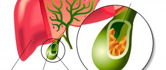
Structure of the liver
Liver and gallbladder
The performance of numerous functions is associated with the peculiarities of the internal structure of the liver. The dense membrane covering the liver under the peritoneum goes deep into the organ and divides it into prismatic lobules with a diameter of about 1.5 mm. The number of such hepatic lobules in humans reaches 500 thousand; they are the structural and functional unit of the liver (Fig. 2). In the lobule, liver cells (hepatocytes) are grouped in the form of radial plates, between which there are wide blood capillaries (sinusoidal), converging to the liver). Inside the radial plates, between two adjacent rows of hepatocytes, slits are formed, called bile ducts: bile produced by hepatocytes enters them.
Each liver cell is in contact with the wall of the blood capillary on one side, and with the lumen of the bile duct on the other. This structure allows hepatocytes to work in two directions: secrete bile into the bile ducts and send glucose, proteins, fats, vitamins, urea, etc. into the blood. Raw materials for the production of bile and numerous substances also enter through the capillaries with arterial and venous blood. As already mentioned, arterial blood arrives in the liver through the branches of the hepatic artery, and venous blood through the branches of the portal vein. In the wide capillaries of the hepatic lobules, arterial blood mixes with venous blood and flows very slowly, which promotes the exchange of substances between blood and hepatocytes. The capillary wall also contains special cells - stellate macrophages, which perform a protective function. They can capture from the blood and destroy various foreign particles, microorganisms, and damaged cells. Blood saturated with waste products of hepatocytes flows from the capillaries into the central vein of the lobule, and from there into larger veins, through which it is carried out of the liver and enters the inferior vena cava, i.e. returns to the general bloodstream.
Prevention
There is no specific prevention of the disease. You can reduce the risk of developing cancer of the bile ducts by following these recommendations:
- proper nutrition;
- weight control;
- compliance with the work and rest regime;
- avoidance of stressful situations;
- giving up a sedentary lifestyle, regular walks in the fresh air;
- rejection of bad habits;
- compliance with safety regulations when working in hazardous industries;
- vaccination against viral hepatitis;
- Regular preventive examinations, at least once a year, will help identify possible pathological changes in the bile ducts at an early stage, which will significantly facilitate treatment and improve the prognosis.
Information verified by an expert
Mikhailov Alexey Gennadievich operating oncologist, doctor of the highest qualification category, Ph.D. experience: 21 years
The information in this article is provided for reference purposes and does not replace advice from a qualified professional. Don't self-medicate! At the first signs of illness, you should consult a doctor.