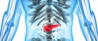
Achalasia cardia (achalasia of the esophagus) is a disease characterized by the lack of opening of the lower esophageal sphincter in response to the passage of food or liquid through the esophagus, due to a violation of the conduction of nerve impulses to the muscles of the esophagus and cardia. It is necessary to immediately clarify the correct name of the disease, because people call both achalasia of the cardia and achalasia of the esophagus. Previously, some doctors believed that it was correct to say achalasia of the esophagus, but the international classification of diseases of the 10th revision put an end to these disputes; it indicated achalasia of the cardia, which means this is the correct name for this disease. Achalasia affects equally often both men and women, more often only at the age of 30 - 45 years. This disease occurs in 3 - 20% of all diseases of the esophagus. Previously, it was believed that achalasia occurs in 1 person per 100,000 population, but with improved diagnosis and accumulation of data, reports began to appear that it occurs with a frequency of 5 and some authors believe that even 10 people per 100,000 population
The concept of achalasia cardia, the prevalence of the disease
The frequency of this disease in European countries is 1 per 100 thousand people, in the structure of all diseases of the esophagus it accounts for from 2 to 25%.
Achalasia cardia is the inability of the esophageal outlet to relax as a result of the congenital absence of specific nerve fibers in the intermuscular layer. With achalasia cardia, the esophagus becomes as if deprived of innervation in a small area. Against this background, its increased sensitivity to gastrin and cholinomimetics (local regulation hormones) is formed. Due to this, over time, a place of persistent narrowing of the esophagus forms before the entrance to the stomach. With this disease, food enters the stomach not through a reflex (during swallowing), but through mechanical opening of the cardia against the background of hydrostatic pressure exerted by a column of liquid food that has accumulated in the esophagus by this time. At first, the muscle strength of the upper esophagus copes with pushing food through this narrowed area, and then the muscle weakens, and the esophagus stretches, taking on an S-shape.
Classification of achalasia cardia
There are four stages in the development of the disease:
- The first (functional) stage
- there is a temporary disruption of the passage of food through the cardiac sphincter due to the cessation of its relaxation during the process of swallowing - The second stage
is accompanied by moderate dilatation of the esophageal wall - The third stage
is characterized by persistent suprastenotic dilatation of the esophagus and the presence of persistent narrowing in the lower part of the esophagus due to the formation of scar changes - The fourth stage
is characterized by a pronounced clinical picture, which develops against the background of an S-shaped dilatation of the esophagus and complications in the form of esophagitis, periesophagitis, fibrous mediastinitis, chronic bronchitis, chronic pneumonia and atelectasis in the lungs (against the background of constant night reflux of esophageal contents and aspiration of food into the respiratory tract mass)
Symptoms and clinical picture of achalasia cardia
The first complaints of patients are difficulty swallowing (dysphagia), a lump in the throat, followed by pain in the chest, regurgitation of food, and cough.
The development of achalasia cardia occurs rather slowly, but the severity of the main symptoms of the disease is constantly increasing.
Dysphagia occurs when consuming a wide variety of foods, both liquid and solid, usually within a few seconds of the start of the act of swallowing. In this case, food is retained not in the throat, but in the chest, so the sensations are localized retrosternally.
Regurgitation
- reverse reflux of food from the lower to the overlying sections (into the oral cavity). Increased casting is observed when the torso is tilted forward, after a heavy meal, and also at night, when the patient assumes a horizontal position. In this case, food debris often gets into the respiratory tract with the development of corresponding complications and clinical manifestations.
Odynophagy
- this is pain in the chest area associated mainly with spasm of the smooth muscles of the esophagus and the overflow of its lumen with food masses.
With the development of the fourth stage, stagnant symptoms appear due to the prolonged presence of swallowed food in the esophagus (bad breath, nausea, rotten belching). Against this background, due to the formation of a large amount of lactic acid, heartburn and inflammation of the walls of the esophagus occur - esophagitis, which can be erosive and ulcerative.
To identify achalasia cardia and choose the correct surgical treatment tactics, you must send me a complete description of gastroscopy, X-ray of the esophagus and stomach with barium, preferably an ultrasound of the abdominal organs, to my personal email address, you must indicate age and main complaints. In rare cases, if there is a discrepancy between complaints, X-ray data and FGS, it is necessary to perform esophageal manometry. Then I will be able to give a more accurate answer to your situation.
Diagnosis of achalasia cardia
Puchkov K.V., Ivanov V.V. and others. Technology of dosed ligating electrothermal effects at the stages of laparoscopic operations: monograph. - M.: ID MEDPRACTIKA, 2005. - 176 p.
Puchkov K.V., Bakov V.S., Ivanov V.V. Simultaneous laparoscopic surgical interventions in surgery and gynecology: Monograph. - M.: ID MEDPRACTIKA, 2005. - 168 p.
Diagnosis of this disease is based on data obtained during a survey (the patient presents the above complaints), physical examination of the patient, as well as the results of instrumental research.
The main radiological sign of achalasia cardia is a narrowed distal section of the esophagus, which has smooth, clear contours, and an expanded section of the digestive canal located above it. In this case, the narrowed part is often located eccentrically and there is an overhang of the walls of the esophagus above it - a symptom of a “writer’s pen”.
The fibroendoscopic method allows you to visually assess the condition of the terminal esophagus and gastroesophageal junction. In most cases, it is possible to pass the fiberscope through the narrowed part of the esophagus, which is more common with the functional nature of the changes, and speaks in favor of achalasia cardia.
Manometry of the esophagus makes it possible to objectively assess the motor activity of the organ and judge the quality of work of the upper and lower esophageal sphincters. Violations of the contractility of the esophagus and cardiac sphincter occur much earlier than the development of clinical symptoms of the disease, in connection with this, manometry becomes the main method of early diagnosis of the disease, when FGS and fluoroscopy do not yet have clear data for cardial achalasia.
What are the complications?
With achalasia, irreversible changes occur throughout the body. Among the most common complications associated with esophageal disease are:
- squamous cell carcinoma;
- bezoars;
- volumetric formations on the neck;
- exfoliating submucosal layer;
- distal diverticulum;
- phlebeurysm;
- lung disease;
- purulent pericarditis;
- pneumopericardium, etc.
With prolonged progression of the disease, the esophagus expands and its walls become thinner, which becomes the cause of complications. In most cases (85%) those suffering from achalasia experience noticeable weight loss.
Treatment of achalasia cardia. Drug therapy and cardiodilation.
It should be noted that pharmacotherapy (calcium channel blockers, nitrates, myotropic antispasmodics, anesthetics) is effective only in the first stage, in which mild and rare transient dysphagia is noted.
The generally accepted method of treatment is endoscopic bougienage of the esophagus, aimed at expanding the narrowed area, but, unfortunately, this occurs through tissue rupture, with the formation of rough scars in this area, which in the future only worsens the situation. The effect of bougienage of the esophagus is short-lived, repeated manipulations are required and after 4-5 procedures relief does not occur. In this situation, an operation is proposed - esophagomyotomy (dissection of part of the wall of the esophagus to the submucosal layer without opening the lumen of the organ), which allows you to restore normal swallowing and return the patient to a normal lifestyle.
At the same time, the endoscopic dilatation technique can be useful for the treatment of patients in the older age group and people with relapse of achalasia cardia who have previously undergone surgery. That is, for those patients for whom, due to concomitant diseases, surgical intervention is contraindicated or its implementation is technically extremely difficult.
Also, one of the minimally invasive methods of treating achalasia cardia is endoscopic intrasphincteric injection of botulinum toxin A. It is considered as a fairly effective alternative option for people who have absolute contraindications to endoscopic hydro- or pneumatic cardiodilation and surgical intervention, especially when it comes to patients in the older age group , about the presence of pronounced, severe concomitant pathology of the cardiovascular and bronchopulmonary systems. Treatment with botulinum toxin is safe, but it should be borne in mind that the effects on the lower esophageal sphincter often last only 2-3 months, and therefore additional repeated administration of this drug may be required.
Early surgery gives very good results, since in the narrowed area there are no scars from previous cardiodilation, and the muscle strength of the esophageal wall has not yet been depleted. But, even with a distended esophagus, good results are possible when using an adequate laparoscopic method.
Very important!
A feature of my approach to choosing treatment tactics for patients with achalasia cardia is the earlier determination of indications for surgical treatment using the laparoscopic method (through several punctures of the abdominal wall), without waiting for complications to develop and without wasting time on useless conservative treatment methods.
Sample menu
From permitted products, you can create a complete diet.
Each patient, together with the doctor, can create a suitable menu for themselves for several days and alternate dietary products in such a scheme as they wish. In general, the recommended diet is as follows:
- Breakfast - pureed semi-liquid millet porridge, boiled in water, a hard-boiled egg, a glass of green tea with a few crackers.
- Second breakfast - fresh salad of cucumbers and tomatoes, seasoned with sunflower or linseed oil.
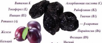
- Lunch – vegetable soup with croutons, buckwheat porridge with boiled chicken, a glass of rosehip broth and several dried fruits.
- Afternoon snack – carrot soufflé.
- Dinner – cottage cheese, seaweed salad, a glass of jelly.
In some cases, the doctor allows deviations from a strict diet if the prescribed diet causes the patient even greater problems with swallowing food. This may be a consequence of the influence of a psychological factor. In any case, only a doctor can give recommendations, and independent prescriptions are unacceptable. By adhering to an established diet plan after curing achalasia cardia, it will be possible to achieve competent prevention of relapses, since healthy food will not allow the problem to arise again.
—
Surgical methods for treating achalasia cardia
Puchkov K.V., Rodichenko D.S. Manual suture in endoscopic surgery: monograph. - M.: MEDPRACTIKA, 2004. - 140 p.
Of the many surgical methods proposed for the treatment of esophageal achalasia, currently only those interventions based on extramucosal cardiomyotomy are mainly performed. One of the main ones is the method of extramucosal cardioplasty, performed according to Heller (E. Heller). The essence of the method is a longitudinal section of the muscular lining of the distal esophagus along its anterior wall for eight to ten centimeters. In this case, the myotomy should cover not only the area of narrowing and the cardiac part of the stomach, but also a partially expanded area of the esophagus. In this case, the edges of the dissected muscular membrane diverge in opposite directions, as a result, the underlying intact mucous membrane begins to bulge into the resulting defect.
According to the literature, good results after this type of surgery are observed in up to 90% of cases. The disadvantages of this technique are associated with leaving the esophageal mucosa unprotected over a large area (8-10 cm x 1 cm). Therefore, in places where the muscular lining of the esophagus is cut, diverticula and scars that deform the cardia can form, causing a relapse of the disease.
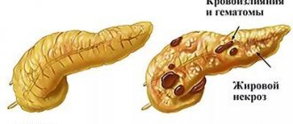
Mortality after Heller surgery averages about 1.5% (a number of authors give the figure 4%). The main cause of death is usually overlooked damage to the esophageal mucosa, which leads to mediastinitis (inflammation of the mediastinal tissue), pleurisy (inflammation of the pleura) and peritonitis (inflammation of the peritoneum). Trauma to the mucosa is observed in 6-10% of all operations, therefore, to prevent severe complications, it is extremely important to conduct a very thorough inspection of this area. If the surgeon discovers damage to the mucous membrane, it must be sutured. Also, unsatisfactory results are associated with the destruction of the cardinal sphincter and the development of peptic reflux esophagitis. And due to the possibility of closure and fusion of the dissected walls of the muscle layer, there is a recurrence of the disease, dysphagia and impaired emptying of the esophagus.
Very important!
To reduce mortality and the number of postoperative complications, it is necessary to cover the surgical defect of the esophageal wall with one’s own tissues, which is rarely performed by surgeons due to the technical complexity of this stage of the operation.
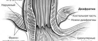
Today, there are a variety of ways to cover the mucous membrane. What can the omentum, the anterior wall of the stomach, and a section of the diaphragm be used for? It should be noted that options for closing the defect in the muscular lining of the esophagus with any synthetic materials are not advisable due to the development of bedsores in this place.
Very important!
To prevent the development of cardiac sphincter failure and reflux esophagitis, preserve the natural anatomical relationships of the esophagus, stomach and diaphragm as much as possible.
What contributes to the disease achalasia
Experts do not name the specific cause that causes the development of the disease. It is associated with infections, compression of the esophagus, inflammation, tumors of malignant origin, infiltrative lesions, etc. In childhood, esophageal achalasia is usually detected after five years of age.
The first manifestations of the disease go unnoticed, so it is diagnosed late. If a child has dysphagia and starts vomiting after eating, you need to sound the alarm. These are common symptoms characteristic of achalasia.
Laparoscopy in the surgical treatment of achalasia cardia and its complications
Laparoscopic operations for diseases of the esophagus and stomach are distinguished by their delicacy and functionality. The enlarged image on the monitor allows you to perfectly visualize all the finest anatomical formations in this area - the vagus nerve, gastric vessels and fascial spaces for gentle surgery. Laparoscopic access allows to reduce the duration of the intervention, shortens the postoperative rehabilitation period and facilitates its course, and also helps to significantly reduce the number of complications and provides a good cosmetic effect.
We have improved the technique of laparoscopic surgery in the treatment of achalasia cardia, which can reduce the number of relapses to 0.2%, and the number of complications to 1%.
My tactics in approaching surgical treatment of patients with achalasia cardia are as follows:
- Earlier determination of indications for surgical treatment, without waiting for complications to develop and without wasting time on useless conservative treatment methods;
- Using only laparoscopic access for surgery - better visualization, less trauma, better cosmetic effect and faster recovery;
- Performing extramucosal cardioplasty with mandatory dissection of the muscle and scar layers at a distance of 8-10 cm, which ensures guaranteed elimination of stenosis;
- In order to prevent complications from the exposed mucous membrane, perform a thorough revision of this area, if necessary, suturing it and obligatory covering it with the anterior wall of the stomach;
- In order to prevent relapse of the disease (fusion of the dissected muscular membrane), cover this area with the anterior wall of the stomach, with mandatory suturing it along the entire perimeter of the mucosal defect using our own method, only with synthetic absorbable threads on an atraumatic needle;
- In order to prevent insufficiency of the cardinal sphincter (as a result of dissection of the muscle layer) and the development of peptic esophagitis, strengthening this area with the anterior wall of the stomach (fundoplication at 120 degrees);
- When isolating the esophagus and stomach, I use a modern device for dosed electrothermal tissue ligation “LigaSure” (USA), which makes it possible, without damaging the surrounding structures, to “weld” the vessels and with thin electrodes to dissect the muscular lining of the esophagus without damaging the mucous membrane;
- The technique I developed allows the transgastric tube not to be left after surgery (which greatly facilitates the postoperative period), and the patient to eat liquid food the next day.
You can watch videos of operations performed by me on the website “Video of operations of the best surgeons in the world.”
As a result of using our method, the vast majority of patients (97%) report good results. In this case, the positive effect is confirmed by data from control fibroesophagogastroduodenoscopy, X-ray examination and esophagomanometry. According to the results of manometric measurements in all operated patients, it was possible to achieve a decrease in the basal tone of the esophagus. In addition, there is a noticeable improvement in the contractile activity of the esophagus and an increase, although insignificant, in the length of the cardia due to the formation of a fundoplication cuff. According to the indicators obtained during 24-hour intraesophageal pH-metry, pathological gastroesophageal refluxes (reflux of stomach contents into the esophagus) after the Heller operation, supplemented by anterior esophageal fundoplication according to Dor in our modification, are recorded at the physiological level.
My experience includes more than 600 operations for hiatal hernia and reflux esophagitis and more than 100 operations for achalasia cardia. It is summarized in three monographs: “Hiatal hernia”, “Hand suture in endoscopic surgery” and “Technology of dosed ligating electrothermal effects at the stages of laparoscopic operations”, as well as in more than 60 scientific publications in various professional peer-reviewed scientific publications in Russia and abroad.
After the operation, 3-4 incisions 5-10 mm long remain on the skin of the abdomen. From the first day, patients begin to get out of bed, drink, and the next day take liquid warm food. Discharge from the hospital is carried out on days 1-3, depending on the severity of the disease. The patient can begin work in 2 - 3 weeks. A strict diet should be followed for one and a half to two months, a softer one - for six months. Further, as a rule, the patient leads a normal lifestyle - without medications or diet.
At the request of patients, the clinic can undergo a full examination before surgery to determine the optimal treatment tactics and choose the method of surgical intervention.
Achalasia cardia is a neuromuscular disease of the esophagus (NMSD), characterized by the absence of reflex opening of the cardia during swallowing, accompanied by impaired peristalsis and decreased tone of the thoracic esophagus [1-4]. Together with diffuse esophagospasm and other motor disorders, it is classified as a group of neuromuscular diseases of the esophagus [5].
This disease was first described by the English physician T. Willis in 1672 in a patient with constant progressive vomiting; It was possible to restore the patency of his esophagus using a sponge attached to a whalebone [5-7].
The prevalence of achalasia cardia, according to world statistics [7-9], is 0.6-2 cases per 100,000 population (regardless of gender), in the USA - 1 case per 100,000 population. The share of esophageal achalasia in the structure of all diseases of this organ is 3-20%.
Over the entire period, more than 25 classifications of achalasia cardia have been adopted [10–13], for example, the classification of T.A. Suvorova (1966), in which two types of esophageal achalasia were identified. A three-stage classification of cardiospasm was proposed by G.D. Vilyavin in 1978. In 1987, the classification of T.A. Suvorova was changed and supplemented by A.L. Grebenev [7]. In our country, the most common classification is B.V. Petrovsky (1962), according to which four stages are distinguished: I (initial) - the esophagus is not dilated, the cardia opening reflex is preserved, but the motility of the esophagus is strengthened and discoordinated; II - the cardia opening reflex is absent, the esophagus is dilated to 4-5 cm; III - significant dilation of the esophagus up to 6-8 cm, retention of fluid and food in it, lack of propulsive motility; IV - sharp expansion (more than 8 cm), elongation and s-shaped curvature of the esophagus with atony of the walls, prolonged fluid and food retention [2, 5, 7, 12]. This classification is based on the results of radiological and endoscopic examination methods.
In X-ray contrast examination of the esophagus, typical signs of achalasia cardia are expansion of the lumen of the esophagus, absence of a gas bubble in the stomach, delayed release of the esophagus from the contrast agent, absence of normal peristaltic contractions of the esophagus, narrowing of the terminal esophagus (“candle flame”) [9, 10].
During an endoscopic examination, first of all, attention is paid to the degree of dilatation of the lumen of the esophagus, the presence of food debris, liquid and mucus in it. At the same time, the condition of the mucous membrane of the esophagus is assessed, its thickness, color, shine, and the presence of peristalsis is determined. Typical signs during esophagogastroduodenoscopy: weakened peristalsis of the esophagus, lack of adequate relaxation of the lower esophageal sphincter (LES), narrowing of the esophagus in the area of the LES and its suprastenotic expansion; slight resistance when passing the endoscope through the LES [14, 15]. In the case of esophagitis, thickening of the folds, hyperemia of the mucous membrane, erosion and ulceration are observed.
Currently, the Chicago classification of esophageal motility disorders, first proposed in 2008 and revised in 2011 and 2014, is generally accepted throughout the world. (SS v3.0) [4, 16-18]. This classification is based on the interpretation of objective data recorded by High-Resolution Manometry (HRM). HRM is now the generally accepted standard for diagnosing esophageal motor function.
According to this classification, three types of achalasia are distinguished depending on the predominance of certain motor disorders of the esophagus, such as LES hypertension, diffuse esophageal spasm, nonspecific or ineffective esophageal motility (see figure).
Manometric picture of achalasia cardia. a - type I; b - type II; c - type III. In type I achalasia cardia (classical achalasia), in 100% of wet swallows there is no peristalsis of the thoracic esophagus - this is severe hypokinesia. In type II, there is no normal peristaltic wave of contraction, but there is a uniform spasmodic contraction of moderate intensity (>30 mmHg) along the entire length of the esophagus from the upper to the LES in more than 20% of wet swallows. In this type, peristalsis of the thoracic esophagus is preserved. Type III is characterized by the absence of a normal peristaltic wave, the presence of isolated episodes of peristalsis in the distal esophagus, or premature spasmodic contractions (distal esophagospasm) recorded in more than 20% of wet swallows - this is a hyperkinetic type of achalasia cardia. With it, non-relaxation of the LES is accompanied by an extremely powerful contraction of the thoracic esophagus [4, 19-21].
Clinical manifestations of achalasia cardia are most often observed between the ages of 20 and 40 years [1, 5, 7]. The main symptoms of the disease are progressive dysphagia, regurgitation and chest pain associated with incomplete emptying of the esophagus and chronic esophagitis [22-24]. Other symptoms of the disease include weight loss, hiccups, nausea, increased salivation, bad breath, and occasionally heartburn [9].
Patients with achalasia cardia may experience the above clinical symptoms for a long time, and the symptoms may be wave-like. Periods of increased clinical manifestations can be abruptly replaced by periods of satisfactory well-being.
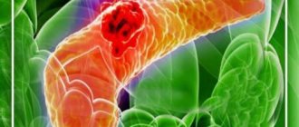
Despite the fact that achalasia cardia is a relatively rare disease, it carries a risk of severe consequences, such as, for example, adenocarcinoma of the esophagus [11, 25, 26]. Achalasia of the cardia, according to various authors [25, 27], can be classified as a precancerous disease, since it is known that cancer develops in 3-8% of patients with cardiospasm and achalasia of the cardia and the likelihood of its occurrence increases with increasing duration of the disease, and not only in the cardia, but also in the altered esophagus. In this regard, its timely detection and treatment is necessary.
Conservative treatment for achalasia cardia includes drug therapy with diet correction [7, 12]. Pharmacological therapy as a stand-alone method is the least effective, rarely leads to long-term and lasting positive results, does not reduce symptoms and is often accompanied by side effects, so it is now receiving less attention than other treatment methods [3, 28].
Over the past 40-50 years, at the initial stage of treatment of patients with achalasia cardia, it was considered advisable to use non-operative methods, and the main and most accessible of them has always been step-by-step balloon pneumatic dilatation (PD) under fluoroscopic control [7, 12].
After dilatation and to monitor the condition after treatment, which must be carried out once a year, patient complaints can be assessed using the Eckardt scale (see table)
Eckardt scale Note. * 0-3 points - remission, more than 4 points - ineffectiveness of PD. [29, 30].
In addition to regression of the clinical manifestations of the disease, a prognostic indicator of long-term remission after PD is the achievement of an LES pressure of less than 10 mm Hg, measured at the end of the procedure. After P.D. It is necessary to conduct a control X-ray examination of the esophagus with contrast.
In recent years, with the development of endoscopic technologies, many authors [7] propose to refrain from using PD to prevent the formation of scars in the area of the cardioesophageal junction, which subsequently complicates the performance of endoscopic operations.
The first reports in foreign literature [31] on the use of botulinum toxin A for the endoscopic treatment of cardiospasm and achalasia cardia appeared back in 1994. The procedure is based on intramural endoscopic injection of botulinum toxin A into the LES at a dose of 80-100 units, with 1 ml The drug (20-25 units) is injected into each of the four quadrants of the LES under visual control. The effectiveness of therapy is about 80% during the 1st month of observation, 70% after 3 months, 50% after 6 months and about 40% after 1 year, and therefore sometimes requires a second injection [7].
Over the entire history of treatment of patients with NMZP, more than 60 surgical techniques have been developed, for example, operations by J. Mikulicz-Radeck, G. Marwedel and W. Wendel, anastomosis of the esophagus with the stomach as modified by N. Geyrovsky, S.S. Yudina, E.L. Berezova and others, which, due to the occurrence of frequent relapses and the development of severe postoperative complications (reflux esophagitis, esophageal stricture), are practically not performed in our time and are of only historical interest [13]. The unsatisfactory results of such operations contributed to the search for new approaches to the surgical treatment of achalasia.
The idea of myotomy for the treatment of NMCD was first proposed by G. Gottstein in 1901 [32, 33]. In 1913, E. Heller [34] performed the first such operation and described it in his article “Extramucosal cardioplasty for chronic cardiospasm with esophageal dilatation”; the operation received worldwide recognition and became the basis for most surgical interventions performed [32]. Esophagocardiomyotomy according to E. Heller, which consists of dissecting the muscular membrane of the lower end of the dilated segment of the esophagus and the cardial part of the stomach along the anterior and posterior walls, was complicated by the development of gastroesophageal reflux disease in 15% of cases [7].
Many modifications have been proposed, for example, an organ-preserving cardioplastic operation developed at the Russian Scientific Center for Surgery of the Russian Academy of Medical Sciences, the basis of which is esophagocardiomyotomy according to E. Heller with incomplete fundoplication [5]. Due to the fact that organ-preserving operations could not always be performed, the development of complications in the form of recurrent reflux esophagitis and peptic strictures of the esophagus forced subtotal resection of the esophagus with simultaneous esophagoplasty with an isoperistaltic gastric tube with anastomosis in the neck [35].
Proposed in 1972 by B.V. Petrovsky’s method of plasty of the esophagogastric junction with a diaphragm flap on a pedicle from a left-sided transthoracic approach often led to relapse of dysphagia caused by the development of a “scar block” around the esophagus and severe peptic reflux esophagitis [36]. To avoid unwanted complications, students of B.V. Petrovsky - A.F. Chernousov and E.N. Wangqian proposed pneumatic stepwise cardiodilatation (as the main method of treatment of achalasia cardia stages I-II), and in patients with stages II and III of achalasia cardia - organ-sparing cardioplastic surgery, which is based on a modified cardiomyotomy according to E. Heller with incomplete fundoplication, which helps prevent reflux -esophagitis and formation of esophageal diverticulum [37]. However, after this operation, some patients may develop residual dysphagia associated with “hyperfunction” of the fundoplication valve.
Progress in the treatment of achalasia cardia is associated with the introduction of laparoscopy and thoracoscopy into clinical practice. Laparoscopic methods in modern surgery have taken one of the leading places in helping patients with NMCD [2, 38]. In terms of effectiveness, minimally invasive interventions are not inferior to open surgical operations - 94 and 84%, respectively, and the incidence of postoperative complications is lower when they are performed [7].
A meta-analysis of the results of treatment of patients with achalasia cardia, performed by W. Hoogerwerf and P. Pasricha [39] in 2002, showed the greatest effectiveness of laparoscopic myotomy among all methods of treating this disease.
In the terminal stage of the disease, extirpation or subtotal resection of the esophagus is performed with simultaneous gastric tube plasty, including the use of laparoscopic and thoracoscopic techniques [40]. Indications for choosing a method of surgical intervention are determined individually [41]. At the same time, in patients with contraindications for radical surgery, organ-preserving techniques are possible [42].
The active development of minimally invasive intraluminal endoscopic surgery has led to the creation, based on two techniques - myotomy and endoscopic dissection in the submucosal layer - of a new minimally invasive method for the treatment of achalasia cardia - oral endoscopic myotomy (POEM). Since the 2000s, the submucosal layer of the gastrointestinal tract has become a working space for operative endoscopy [43]. The next step, which made it possible to get closer to POEM, was the development of the method of transluminal endoscopic surgery (Natural Orifice Translumenal Endoscopic Surgery (NOTES) - intraluminal endoscopic surgery through natural openings) - exit from the wall of the gastrointestinal tract using a tunnel displacement of the entry point for safe exit into the cavity body, in this case the overlying mucosal flap served as a flap as a protective sealant [44]. This technique is based on the formation of a submucosal tunnel with access from the lumen of the esophagus through a small incision in the mucous membrane of the thoracic region to the area of increased muscle tone and subsequent dissection of the circular muscle fibers of the distal esophagus, LES and cardia of the stomach [45, 46].
In 2007, P. Pasricha et al. [47] proposed and successfully tested the technique of endoscopic myotomy in an experiment on animals - control manometry as a result of the operation revealed a significant decrease in pressure in the area of the LES, and when observed for 7 days, no signs of peritonitis and mediastinitis were noted. Further studies [48] showed that the effect on LES pressure was comparable to any previously used treatment for achalasia cardia.
In humans, the endoscopic myotomy technique was first developed and performed in 2008 by H. Inoue and colleagues (Yokohama, Japan). He also proposed a name for the new technique: POEM [45]. At the initial stage, POEM was performed in 17 patients with achalasia cardia. The technique consisted of creating a long submucosal tunnel (average length about 10-12 cm), followed by endoscopic myotomy of circular muscle fibers 8 cm long (6 cm in the distal esophagus and 2 cm in the cardia of the stomach). As a result, the symptoms of dysphagia were relieved in all patients and the resting pressure of the lower esophageal sphincter decreased on average from 52.4 to 19.9 mm Hg. [49].
Currently, POEM is used in all patients with symptomatic achalasia of all types [50]. Initially, the use of POEM was limited to patients under 18 years of age, but it has now been successfully used in patients over the age of 3 years [51].
The results of the first operations using the POEM method indicate that the new technique can be effective and be an alternative for the treatment of achalasia cardia with classical methods, however, its role in patients with previous operations, age restrictions and concomitant diseases has not yet been fully studied [52].
The main advantages of this technique are the absence of the risk of uncontrolled perforation of the esophagus, minimal invasiveness and low trauma of the surgical operation, preservation of the ligamentous apparatus of the esophagus, short recovery and rehabilitation periods [53].
In 2015, H. Inoue [54] published a report on the largest current experience of using POEM - 500 patients with achalasia cardia, with the following results: endoscopic myotomy was successfully performed in all patients, side effects of the operation were noted in 3.2% of cases , 2 months after POEM, when questioning patients, a significant decrease in scores on the Eckardt scale was recorded - from 6 to 1 point, LES pressure from an average of 25 mm Hg. Art. before surgery it decreased to 13 mmHg. Art. Gastroesophageal reflux disease was detected in 16.8% of patients. Enrollment has been completed for a randomized controlled trial comparing endoscopic and laparoscopic myotomy for achalasia. It includes 240 patients from the Czech Republic, Germany, Italy, the Netherlands and Sweden. The expected completion date of the study is December 2022. There are currently several systematic reviews and meta-analyses based on retrospective data published in recent years. R. Talukdar et al. [46] concluded that there is no difference in Eckardt score between POEM and laparoscopic myotomy. However, it was noted that the duration of the operation was significantly reduced with POEM.
In 2022, the Mayo Clinic (Rochester, USA) shared its experience of successfully treating 28 patients from April 2012 to May 2015 using a combined method that included POEM and POEM supplemented with laparoscopic E. Heller myotomy with incomplete fundoplication . The authors noted a decrease in Eckardt scores from 6 to 0, and only 3 patients continued to complain of dysphagia in the postoperative period [55].
L. Marano et al. [56] systematically analyzed data from 486 patients (196 in the POEM group and 290 in the laparoscopic myotomy group). The study showed that the difference in postoperative clinical manifestations, duration of surgery and side effects was not significant. Patients' length of hospital stay was significantly shorter with POEM.
In Russia, POEM is just beginning to be actively introduced into clinical practice; data on its use are extremely scarce. In 2015 E.D. Fedorov [2] presented the results of using POEM in 4 patients with achalasia cardia and concluded that this method was immediately effective and relatively safe in the early stages. In 2016 K.V. Shishin published the results of the successful use of the method in 15 patients with achalasia cardia, indicating its effectiveness and safety.
Despite the positive reviews about the new method of treating patients with achalasia cardia, there are a number of problems and questions regarding the features of the operation [57]. In the process of performing POEM, such technical aspects must be resolved as the choice of the level of access in the esophagus for the formation of a submucosal tunnel, identification of the LES in the formed tunnel, the length of the myotomy, the issue of the full-thickness of the myotomy - limit ourselves to only dissecting the internal circular layer or perform myotomy and external longitudinal muscle fibers [ 58].
Thus, the improvement of technical aspects, the emergence of a minimally invasive surgical technique of POEM, the development of indications and contraindications for this, and the assessment of immediate and long-term results of the intervention are promising areas for further scientific research.
The authors declare no conflict of interest.
The authors declare n conflicts of interest.
Information about authors
Gasanov A.M.
. — https://orcid.org/0000-0002-1994-2052
Aliev N.A
. — https://orcid.org/0000-0002-9772-1351
Danielyan Sh.N
. — https://orcid.org/0000-0001-6217-387Х; e-mail
Gasanov AM
— https://orcid.org/0000-0002-1994-2052
Aliev N.A.
— https://orcid.org/0000-0002-9772-1351
Danielyan Sh.N.
— https://orcid.org/0000-0001-6217-387Х
Questions most often asked by patients with achalasia cardia
What anesthesia do you use for laparoscopic treatment of achalasia cardia?
If you would like to learn in more detail about the methods of pain relief used in the surgical treatment of achalasia cardia, please carefully read the information posted on the website.
Is it necessary to somehow prepare for surgery for laparoscopic treatment of achalasia cardia?
If you are planning surgical treatment for achalasia cardia, I ask you to carefully study the preoperative preparation section.
Is it possible to simultaneously perform laparoscopic surgery for achalasia cardia and, for example, fibroids?
Minimally invasive surgery techniques allow two and sometimes three operations to be performed simultaneously during one anesthesia by a team of several surgeons. The issue of simultaneous operations is discussed in more detail in a special section of the site. Simultaneous performance of several surgical interventions will reduce the load on the body, reduce hospitalization time, and speed up the body’s recovery compared to performing several operations with an interval of 5-6 weeks.
Where can I undergo surgery for achalasia cardia?
I conduct the initial consultation with all patients in Moscow at the Swiss University Clinic. Get acquainted in more detail with my main clinical sites in Moscow and Switzerland.