The cardia, or cardiac part of the stomach, is a cylindrical segment covering the esophagogastric junction and the space 2 cm above and below it. Cancer of the gastric cardia (cardioesophageal cancer, CER) differs in anatomical and clinical features, and requires a special approach to treatment. Many experts even identify it as a separate nosology, and there are several reasons for this:
- Tumors of this location tend to spread to the esophagus much more often than cancer of the underlying parts of the stomach.
- Such tumors metastasize to the abdominal and mediastinal lymph nodes.
- In addition, they have a poorer prognosis than gastric or esophageal cancer.
Cardioesophageal cancer is characterized by aggressive growth and a tendency to metastasize to the mediastinal lymph nodes.
- Causes
- Symptoms of gastric cardia cancer
- Classification
- Diagnostics
- Treatment of gastric cardia cancer
- Complications and relapses
- Prognosis and prevention
Causes
The causes of cardiac cancer are not reliably known. Currently, it is customary to talk about risk factors - circumstances under which the likelihood of developing a particular pathology increases. For stomach cancer they are as follows:
- Helicobacter pylori infection.
- The presence of atrophic gastritis of the proximal stomach.
- Hereditary predisposition.
- Smoking and alcohol abuse.
As for gastroesophageal reflux and Barrett's esophagus, these conditions are the main predisposing factors for the development of esophageal cancer, but they do not affect the incidence of cardia cancer.
Treatment
The main method of treatment for cancer of the gastric cardia is removal of the tumor and surrounding tissues.
The method and extent of the operation depends on the prevalence and type of neoplasm. So, when a patient is diagnosed with type Cardioesophageal cancer (CER I), a proximal gastrectomy and subtotal cutting of the esophagus are performed. If tumor types KER II or KER III are identified, gastrectomy (complete removal of the stomach) with resection of the lower thoracic esophagus is indicated.
At the same time, lymph nodes are removed. Doctors currently disagree about the required extent of the operation. Some believe that it is enough to cut off the nearest lymph nodes of groups 1 and 2. Others advocate removing the entire lymphatic system of the stomach. The latter option is more traumatic, but there is no reliable evidence of an improvement in the patient’s survival prognosis after such an intervention.
The final choice of the type of intervention remains with the attending physician. To improve treatment results, surgery is combined with other therapeutic methods.
Radiation and chemotherapy
Chemotherapy for cancer of the gastric cardia is of two types:
- non-adjuvant - prescribed before surgery to stop tumor growth and metastases;
- adjuvant - reduces the risk of relapses, destroys remaining malignant cells. It is used mainly after non-radical interventions.
To increase effectiveness, chemotherapy is combined with radiation exposure. Radiotherapy alone is rarely used, only if the use of chemotherapy is not possible. Radiation is also given before surgery to reduce the likelihood of potential relapses. After the intervention, radiation therapy is carried out once.
The combination of two methods improves the patient's survival prognosis by 15-20%. For inoperable forms of gastric cancer, chemoradiotherapy is the main treatment. In this case, operations are performed only to relieve life-threatening conditions and improve the patient’s quality of life.
Esophageal stenting
According to medical statistics, about 60-70% of patients with cardia cancer consult a doctor when surgical intervention is no longer possible. The patient is prescribed palliative treatment. To eliminate the main symptom of a cardiac tumor—dysphagia—esophageal stenting is performed.
This is a minimally invasive procedure to install a thin metal tube, which acts as a frame, restoring the lumen of the esophagus. This helps ensure normal passage of food and liquid through the gastrointestinal tract. The installed stent returns the ability to eat properly in 80-90% of patients.
Symptoms of gastric cardia cancer
Cardia cancer is most characterized by weight loss, food aversion, pain and dysphagia. The pain is usually localized in the epigastric region, is constant, pressing in nature and does not depend on food intake. Sometimes it radiates to the lumbar region or behind the sternum. In the latter case, the disease can simulate heart pathology, such as angina.
The severity of dysphagia depends on the size of the tumor and obstruction of the digestive tract. It is mainly manifested by malnutrition, which leads to hypovolemia and changes in electrolyte balance.
Characteristic symptoms
A characteristic sign of a cardia tumor is the obstruction of food and liquid through the esophagus (dysphagia). The symptom appears when a malignant neoplasm covers more than 50% of the lumen of the esophagus. The condition is extremely poorly tolerated by patients, leading to complete refusal to eat, exhaustion (cachexia), and death.
Other signs of cardia neoplasms include:
- belching food;
- rapid weight loss;
- increased salivation;
- nausea;
- satiation with a small amount of food;
- feeling of damage to the esophagus, the presence of a foreign body;
- bloating, heaviness in the abdomen;
- anemia;
- expectoration of mucus;
- weakness, fatigue;
- vomiting of undigested foods;
- increased body temperature (usually observed in the later stages of the disease due to general intoxication of the body);
- change in taste preferences (more often an aversion to meat or fish products develops);
- depressed mood, apathy.
As for such a symptom as pain, doctors distinguish three forms of cardia cancer:
- painless has gastrointestinal symptoms, but there is no pain;
- latent - occurs without noticeable manifestations. The tumor becomes an accidental discovery during routine examinations when it grows to a large size;
- painful - accompanied by constant pain of varying intensity, intensifying with physical activity or after eating.
Pain from neoplasms of the cardia often radiates to the sternum or lumbar region, due to which the disease is disguised as angina pectoris and other heart pathologies for a long time.
The symptoms of cardia neoplasms vary from person to person; patients are often diagnosed with a latent or painless form of the pathology, which significantly complicates diagnosis in the early stages.
Classification
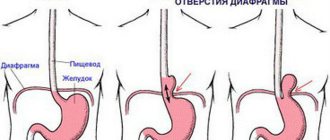
The classification of cardia cancer is based on the location of the center of the tumor. The anatomical reference point is the Z-line - the line of the esophagogastric junction.
- Type I cancer - the center of the tumor is located at a distance of 1-5 cm above the Z-line towards the esophagus.
- Type II cancer - the center of the tumor is located within 1 cm orally and 2 cm below the Z-line.
- Type III cancer - the center of the tumor is located at a distance of 2-5 cm below the Z-line.
Diagnostics
- Endoscopic examination of the stomach and esophagus. Allows you to examine the tumor and take a biopsy for morphological examination.
- Endoscopic ultrasound. It is carried out to determine the degree of tumor invasion, its growth into neighboring organs and structures, as well as to search for regional metastases.
- Abdominal ultrasound. It is carried out to search for metastases.
- X-ray contrast study. Allows you to determine the extent of the tumor and assess the degree of stenosis of the cardia and esophagus. The method is especially relevant for diffuse-infiltrative forms of cancer, when false-negative biopsy results can be obtained due to submucosal localization.
- CT scan is used to look for distant metastases.
- Diagnostic laparoscopy. Allows you to detect the spread of the tumor to the serous membranes, as well as diagnose intraperitoneal dissemination.
- Determination of tumor markers - CEA, CA 19-9, CA 72-4.
Treatment of gastric cardia cancer
Treatment of cardia cancer requires a combined approach and includes chemotherapy and surgery. For unresectable tumors, the main treatment method is chemotherapy, and operations are performed for life-threatening indications.
Surgical treatment
The choice of the extent of the operation and surgical approach is determined based on the type of cancer and its local prevalence. For type I CER, proximal gastrectomy is performed
| KER type I Proximal gastrectomy, subtotal esophageal resection. | KER type II Gastrectomy (the entire stomach is removed), resection of the lower thoracic esophagus | KER type III Gastrectomy, resection of the lower thoracic esophagus. |
The minimum volume of lymph node dissection involves the removal of 1st and 2nd order lymph nodes (extended lymph node dissection is performed in the volume of D2). In CER I, bilateral mediastinal lymph node dissection is performed.
Chemotherapy
Chemotherapy is carried out using one of the following options:
- Perioperative chemotherapy. Performed in CF or ECF mode for 8-9 weeks before and after surgery.
- Adjuvant chemotherapy. It begins 4-6 weeks after surgery, if there are no severe complications, and is carried out for 6 months. XELOX or CAPOX schemes are used.
- Adjuvant chemoradiotherapy. Currently, it is rarely used and only in case of non-radical surgery.
Treatment of unresectable and disseminated tumors is carried out using chemotherapy according to the CF, CX, XELOX regimens in 6-8 three-week courses, or 9-12 two-week courses. After this, treatment is stopped and observation is carried out until the disease progresses. For HER2-positive gastric cancer, treatment with trastuzumab is possible.
After disease progression, chemotherapy is resumed, and the treatment regimen is selected taking into account the existing medical history.
Metastasis
With CER types I and II, metastasis mainly occurs in the lymphatic collectors of the paracardial zone, celiac trunk and lesser curvature of the stomach. In type I CER, the mediastinal nodes may also be involved in the process. Such features of metastasis are taken into account when planning surgery and lymph node dissection.
Structure and function of the stomach
The stomach is located mainly in the upper floor of the abdominal cavity between the esophagus and duodenum. The inlet of the stomach, which is a continuation of the abdominal segment of the esophagus, is called cardiac. The esophagus enters the stomach at an angle - the angle of His. The outlet of the stomach, which passes into the duodenal bulb, is called the pylorus.
According to the anatomical classification, the stomach consists of the cardiac part, the fundus, the body and the pyloric part. The latter is divided into the pyloric cave (antrum) and the pyloric canal. It is generally accepted to distinguish between the anterior and posterior walls of the stomach, which pass into each other. The upper, shorter and concave edge of the stomach is called the lesser curvature, and the lower, convex and longer edge is called the greater curvature. On the lesser curvature there is an angular notch, which is the boundary between the body and the pyloric part of the stomach.
The shape and boundaries of the stomach are not constant and depend on the amount of contents, the functional state of the stomach, body position, physique, diet, condition of surrounding organs, tone of the abdominal muscles, breathing phase and other factors. Conventionally, two extreme forms of the stomach are distinguished - the horn shape (in hypersthenics) and the hook shape (in asthenics), which are extreme options, i.e. The longitudinal axis of the body of the stomach can be located almost horizontally or vertically. With a normosthenic physique, the longitudinal axis of the stomach has an oblique arrangement. The capacity of an adult's stomach is about 1.5-3 liters.
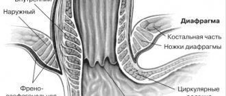
The angle of His, which is one of the factors of the antireflux mechanism, has individual differences and depends on the anatomical features of the stomach. Approximately 80% of people have an angle of His (cardiac notch) of less than 90°. Corresponding to the apex of the cardiac notch, the gastric mucosa forms a cardiac fold, which functions in healthy people as a shutter device—the Gubarev valve. When the stomach contracts, the cardiac opening is closed by this fold.
The stomach is located predominantly in the left hypochondrium (fundus, cardiac part and part of the body) and its smaller part (part of the body and pyloric region) - in the epigastric region, passing to the right beyond the midline. The stomach is covered with peritoneum on all sides. From above and to the right, the anterior wall and lesser curvature of the stomach is adjacent to the visceral surface of the left lobe of the liver. The cardiac part of the stomach is covered by the left lobe of the liver. The lesser curvature of the stomach is connected to the lower surface of the right lobe of the liver using the lesser omentum, which is formed by the hepatogastric and hepatoduodenal ligament. The hepatogastric ligament is located between the lesser curvature of the stomach and the porta hepatis. Above and to the left, the fundus of the stomach, the cardiac part and the body are adjacent to the diaphragm. The fundus of the stomach is located in the dome of the left half of the diaphragm, somewhat lateral and dorsal from the place of projection of the heart. When the patient is in an upright position, there is a gas bubble in the fundus. The quadrate lobe of the liver is in contact with the pylorus when the stomach is moderately full. On the right, adjacent to this part of the stomach, as well as to the initial part of the duodenum, is the gallbladder. When the stomach is empty, the pylorus moves closer to the midline; with significant filling, it moves away from the medial plane by 6–7 cm. The lower parts of the anterior wall of the stomach are adjacent to the left costal arch and the anterior abdominal wall. On the left, behind the fundus and body of the stomach, is the spleen. The gastrosplenic ligament connects the greater curvature of the stomach and the spleen. Behind the stomach, separated by the cavity of the omental bursa, are located: the upper pole of the left kidney with the adrenal gland, the celiac trunk and its branches, the aorta, the pancreas, the mesentery of the transverse colon. With average filling of the stomach and the patient standing, the greater curvature is located approximately at the level of the navel or 1 - 2 cm below. Below the stomach lies the transverse colon and its mesentery, to the left and below is the left flexure of the colon. The gastrocolic ligament, located between the greater curvature of the stomach and the transverse colon, is the superior part of the greater omentum.
The wall of the stomach consists of the mucous membrane, submucosal layer, muscular and serous membranes. The gastric mucosa is the innermost, covered with epithelium, which is a single-layer prismatic glandular (superficial pitted). The superficial pitted epithelium is represented by a single-layer cylindrical (prismatic) epithelium (mucocytes), on which there is a layer of mucoid secretion. The cytoplasm of mucocytes is filled with mucin-containing granules, which push the nucleus toward the basement membrane. Mucin contains neutral mucopolysaccharides (fucoglycoproteins). The superficial foveal epithelium is capable of synthesizing PgE2 and PgF2, and this function, in accordance with biological expediency, is stimulated primarily by damaging factors, including hydrochloric acid.
The mucous membrane contains its own layer of loose connective tissue. In the lamina propria of the mucous membrane there are gastric glands: main (fundic), cardiac and pyloric. The main gastric glands themselves consist of main (peptic, zymogenic), parietal (parietal) granulocytes and accessory, as well as endocrine cells. The third layer of the mucous membrane is the muscular plate. The thickness of the normal mucous membrane is 0.25 - 1.5 mm. The mucous membrane forms folds, which along the lesser curvature have a longitudinal direction, and in the area of the bottom and body - transverse, oblique and longitudinal.
Microscopically, three zones are distinguished in the gastric mucosa: cardiac, fundal and pyloric (antral), which approximately correspond to the anatomical sections, but do not coincide with them. Each zone corresponds to the presence of glands characteristic of it. The boundaries of the zones are unclear, the width of the intermediate zones is about 1 cm.
The surface of the gastric mucosa is 500-800 cm2, the volume is 16-45 cm3, the number of glands is about 4-25 million. There are up to 60 gastric pits per 1 mm2 of the mucous membrane, and for each pit there are 4-5 glands.
The stroma of the gastric mucosa consists of cells of the lamina propria, intercellular substance, reticular, precollagen and collagen fibers. The stroma contains blood and lymphatic vessels that form the microvasculature. The stroma performs a supporting function. In the proper layer of the gastric mucosa there are the following cells: fibroblasts, reticular, mast, plasma cells, lymphocytes of varying degrees of maturity and granulocytes.
The gastric mucosa is well supplied with blood. 67-72% of all blood flowing through the stomach passes through it, while only 13% passes through the submucosa, and 15% through the muscle layer. There are a large number of arteriovenous shunts that provide regulation of blood flow in accordance with needs. The capillaries of the gastric mucosa come close or are adjacent to the basement membrane, on which the superficial pitted and glandular epithelium is located. The number of arterial capillaries significantly exceeds the number of venules draining blood from this capillary network. Blood flow increases along with increased secretion of gastric juice. Histamine, prostaglandins and NO increase blood flow into the gastric mucosa.
The muscularis propria is formed by three layers of smooth muscle. The serosa forms the outer covering of the stomach.
The blood supply to the stomach is carried out through arteries starting from the celiac trunk and its branches. Two arteries pass along the lesser curvature: to the left of the celiac trunk - the left gastric artery and to the right of the proper hepatic artery - the right gastric artery. The arteries of the lesser curvature anastomose with each other. On the greater curvature there are: the right gastroepiploic artery, which arises from the gastroduodenal artery, which originates from the common hepatic artery; the left gastroepiploic artery is from the splenic artery and the short gastric arteries are from the splenic artery. The arteries of the greater curvature, anastomosing among themselves, form an arterial ring on the greater curvature. The veins of the stomach pass next to the arteries and flow into branches that are tributaries of the portal vein.
The external innervation of the intestinal tube is carried out by the parasympathetic and sympathetic parts of the nervous system. Internal innervation is represented by its own nerve plexuses, functioning autonomously, regardless of external innervation. Both types of innervation regulate the motor and secretory functions of the intestinal tube. The parasympathetic and sympathetic nerves contain peptidergic nerve fibers that are part of the metasympathetic innervation system.
The main functions of the stomach are the chemical and physical processing of food received from the oral cavity, the deposition of food masses and their gradual evacuation into the intestines. In addition, the stomach takes part in interstitial metabolism, excreting metabolic products, including protein metabolism products, which are then utilized by the body. The stomach plays a major role in hepopoiesis and water-salt metabolism. The actual digestive activity of the stomach is ensured by gastric juice, under the influence of which hydrolysis of proteins occurs, denaturation of a number of substances and cellular structures of food.
Share link:
Complications and relapses
The main complications of cardioesophageal cancer:
- Tumor disintegration and bleeding. In this case, urgent endoscopic examination and bleeding control are required. If the problem cannot be solved endoscopically, surgical intervention is performed.
- Tumor stenosis. This complication leads to dysphagia and, as a consequence, to the development of nutritional disorders. To eliminate it, recanalization, balloon dilatation, and stenting are used. In severe cases, bypass anastomosis or gastrostomy removal is indicated.
- Pain. To reduce pain, radiation therapy, drug treatment and, in some cases, locoregional anesthesia are used.
- Ascites is an accumulation of fluid in the abdominal cavity. To eliminate it, diuretics and intracavitary chemotherapy are prescribed. When large volumes of fluid accumulate, laparocentesis is indicated - evacuation of the contents through puncture.
Prognosis and prevention
The 5-year survival rate for any type of CER averages 23.6%. Type III tumors have the most unfavorable course, since they are predominantly represented by poorly or undifferentiated neoplasms. The 5-year survival rate for this location is about 14%. The prognosis is significantly worsened by the presence of metastases in the mediastinal lymph nodes.
Euroonko employs doctors with extensive experience in treating stomach cancer. We are guided by current European standards. For this, the clinic has everything necessary - from the latest medications to modern equipment, thanks to which we are ready to provide assistance in the most severe cases.
Book a consultation 24 hours a day
+7+7+78
Forecast
The diagnosis of cardiac cancer has an unfavorable prognosis. This is primarily due to the fact that approximately 70-80% of patients consult a doctor at stages 3 or 4 of the disease. The overall five-year survival rate is 20-25%. Patients with inoperable forms of tumors live from three months to one year.
The exception is a small number of patients whose treatment was started at stages 1 and 2 of cardia cancer, before the appearance of metastases. In this case, 95% of patients survive for more than five years.
Cardiac cancer of the stomach can be cured if you carefully monitor your health and consult a doctor at the first sign of discomfort. Even in advanced cases, do not despair. Modern oncology has a large selection of tools and methods to achieve long-term remission. The psychological attitude of the patient is very important in the fight against cancer.
has been organizing high-quality individual medical care for many years.
Over the years of work, we have accumulated statistics on leading foreign clinics and are ready to recommend to patients for pregnancy management after breast cancer only those medical centers where they will truly provide the most effective care.
![Rice. 2. Clinical stigmas of chronic atrophic gastritis [4] 2. Clinical stigmas of chronic atrophic gastritis [4]](https://zakam.ru/wp-content/uploads/ris-2-klinicheskie-stigmy-hronicheskogo-atroficheskogo-gastrita-4-fig-2-330x140.jpg)