Our body is a well-thought-out holistic system. Each organ in one way or another depends on its neighbors in the body. And violation of the integrity of one or more areas of the skin or mucous membranes can lead to dire consequences.
Perforations or perforations in the digestive system are especially dangerous. The fact is that the organs of the gastrointestinal tract system are most often hollow and filled with aggressive media - gastric or intestinal juice, bile, feces. If intestinal or stomach contents enter the peritoneum without adequate treatment, it can be fatal. You need to know the symptoms of intestinal perforation.
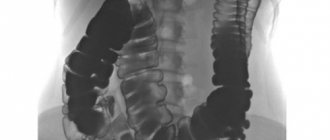
Intestinal perforation, classification, patient management methods, prognosis
Perforation can occur in both the small and large intestines
The intestine is a collective concept. Symptoms of the pathology develop acutely, but may differ from the location of the ulceration. In this case, there is always severe pain and other signs of shock.
The diagnosis is established using instrumental methods. A distinctive diagnostic sign is the presence of free air in the peritoneal space. There are:
- Perforation of the small intestine
- Perforation of the large intestine
Therapeutic tactics for perforation of any area of the intestine are as follows:
- Surgical intervention without options
- Antibiotic therapy
- Infusion of drugs, blood, plasma depending on the severity of the patient
The prognosis for recovery depends on the severity of the injury, the patient’s condition before, during and after surgery, and the degree of penetration of intestinal or stomach contents into the peritoneum. The likelihood of death is extremely high.
Symptoms of the disease
- the presence of a small, painless wound - an external fistula opening on the skin of the perineum or buttocks.
- purulent, mucous or bloody discharge from the fistula opening
- itching, burning, feeling of fullness and discomfort in the fistula area.
- the presence of an internal opening of the fistula, detected upon examination of the mucous membrane of the anal canal (in approximately 30% it is not possible to find the internal opening during examination)
With an incomplete fistula, skin manifestations may be blurred or absent, and symptoms can vary from mild discomfort, a feeling of tightness or fullness, the presence of a lump in the anus, to discharge from the rectum.
When the disease worsens, a painful thickening is observed in the area of the external opening on the skin, the pain is often pulsating, bursting in nature. This may be accompanied by an increase in temperature and general weakness - these are the symptoms of acute abscess or paraproctitis.
Important! Acute paraproctitis should be opened as early as possible, in a surgical hospital!
Sometimes patients mistake the painful process for an exacerbation of hemorrhoidal disease and begin self-medication, which in no case should be done until the correct diagnosis is established!
Perforation of the small intestine, causes of pathology
According to medical statistics, perforation of the small intestine is most often caused by:
- Blunt injuries to the peritoneal area - falling from a height, blows with a stick.
- Penetrating wounds - knife, fall on reinforcement, wooden stakes, gunshot.
- Ingress of sharp objects - pins, toothpicks, fish bones, fragments of bird bones - perforation occurs from the inside.
- Obstruction - a section of the intestinal wall stretches to critical values. There is a violation of integrity.
- Infectious diseases - viral diseases, tuberculosis, impaired immunity, typhoid fever.
How common is rectal fistula?
The incidence in European countries is about 10-25 people per 100 thousand population. Mostly people of working age suffer from rectal fistulas. Men get sick 2-3 times more often than women.
Important! This disease, being benign, can dramatically reduce a person’s quality of life. A serious and most common complication of inadequate treatment of perianal fistulas is impaired holding function due to damage to the muscular apparatus of the anal canal.
Intestinal perforation, treatment features
A diagnosis of intestinal perforation automatically entails surgical intervention. Preoperative preparation lasts from 2 to 4 hours. The duration and intensity depend on the diagnosis, the patient’s condition, and the location of the perforation. For patients who are in shock or in agony, preoperative preparation is not carried out. They should fall into the hands of the surgeon immediately after hospitalization. The choice of intervention technique depends on:
- Patient conditions
- Diagnosis
- Degree of development of peritonitis and concomitant diseases
- The degree of blood loss and blood clotting levels in the patient
- Decisions of the operating doctor and anesthesiologist-resuscitator accompanying the intervention.
The operation is performed under full endotracheal anesthesia.
In rare, exceptional cases, when the patient refuses surgery, conservative treatment is used. The procedure is carried out according to the Taylor technique. In this case, under the control of an X-ray machine, a probe is inserted into the perforation area. Every 15 minutes, the contents of the stomach and intestines are sucked out using a syringe at the site of the perforation.
At the same time, an intensive course of antibiotic therapy is administered to relieve peritonitis. After the patient’s condition improves, a contrast agent is injected into the abdominal cavity, and an X-ray examination is performed to monitor the degree of tissue scarring. It is important to understand that the rate of failure when treating a patient with the Taylor method is very high. Therefore, there is practically no alternative to surgical intervention.
Discussion
Perforation is one of the complications of DC and LC. The frequency of its occurrence in our study (DC - 0.009% and LC - 0.03%) was lower than according to the literature (from 0.016 to 0.8% for DC and from 0.02 to 8% for LC [1]) , however, it should be noted that these results are presented only by two institutions that are centers of expertise in performing colonoscopy.
If we consider all the presented cases of perforation in DC (14 observations), it can be noted that the vast majority of them were localized in the rectosigmoid region (50%) and sigmoid colon (43%), only in one case in the right half of the colon (cecum) , which corresponds to the literature data on a higher risk of perforation with DC in the left half of the colon [4–6].
Perforation in DC in this study occurred in elderly people (mean age 72.4±6.1 years), and female patients predominated (90.1%). Diverticular disease and severe adhesions were identified as the main causes of this event. When conducting multifactor analysis, B. Bielawska et al. also noted that older age and female gender are risk factors for perforation during colonoscopy [7]. According to other researchers, risk factors for perforation included age over 75, female gender, diverticular disease and a history of abdominal surgery [8].
LC perforations in this study occurred during EMR, ESD, stenting, and balloon dilatation. The European Society of Gastrointestinal Endoscopy (ESGE) and the International Association of Emergency Surgery classify colonoscopy as a procedure with an increased risk of colon perforation [1, 9].
According to the data obtained, the size of the perforation hole was statistically significantly larger in the DC group than in the LC group (21.6 ± 13.3 mm compared to 8.6 ± 10.1 mm), which practically coincides with the results published by D. Yang and al. (19.3±12.8 and 5.8±2.9 mm, respectively) [10].
The vast majority of perforations in this work were diagnosed within 3 hours, while according to the literature, only 68% of perforations are detected on the day of the procedure [11], and the average delay time for diagnosing this event is 0.36 days after DC and 1.5 days after LK [4].
An attempt at endoscopic closure of perforation was made in 70% of cases of its occurrence with direct LC and 9% with DC, which is lower than according to a number of foreign authors (82-84% with LC and 31-60% with DC) [2, 12 , 13].
However, according to the results of a systematic review published in 2022, including 19 studies (744 cases of colon perforation), endoscopic clipping was used in only 15% of cases [14]. ESGE recommends using endoscope clips to close small perforations (up to 20 mm) and over-the-scope clips (OTSC) to close larger perforations [9]. In this study, only the first type of clip was used, which reflects both the small average size of the perforation hole and the high cost and low availability of OTSC type clips.
The need for surgical treatment after an attempt at endoscopic clipping occurs, according to the literature, in 14.7% of cases [15], which is 2 times higher than according to our data. This fact, combined with a lower proportion of attempts at endoscopic closure of the perforation, can be explained by a more conservative choice between the indications for endoscopic and surgical treatment by the physicians participating in the study.
Delayed perforation and perforation during diagnostic endoscopy are associated with the need for surgical treatment, which is consistent with literature data [2, 9].
A stoma is created in approximately 30% of patients who require surgical treatment for perforation. In the literature, the incidence of ostomy varies widely, reaching 59.7% [16].
Caring for the patient after intestinal perforation
It is extremely rare to do without surgery
For postoperative patients after intestinal perforation, care is carried out according to the general rules for patients with peritonitis:
- Bed rest
- Complete lack of power
- Regular emptying of stomach contents using a tube
- Bed in Fowler position
- Caring for catheters, drains, surgical wounds or fistulas
- Anesthesia
In the future, in each individual case, patient management will differ depending on the diagnosis and severity. Symptoms of perforation of the hollow organs of the peritoneum are accompanied by severe pain. If such a condition occurs, you should immediately contact a medical facility. Delay in this case means only one thing - death.
What is a fistula
A fistula is a pathological, normally non-existent, passage in the form of a tube connecting the lumen of a hollow organ with the external environment or the lumen of another organ. A rectal fistula, a kind of “tunnel,” connects the lumen of the rectum with the skin of the perineum, buttocks or (rarely) other hollow organs, sharply reducing the patient’s quality of life. This pathological process can involve a large portion of sphincter muscles, which initially characterizes such a fistula as complex and does not allow for “minimally invasive” treatment. In such a situation, a careful assessment of the degree of involvement of muscle structures and planning of special techniques for economical excision of the fistula with subsequent plastic closure of the resulting defect are required.
Important! Such operations must be performed by expert surgeons with extensive experience in such interventions, otherwise the likelihood of relapse (recurrence of the disease) increases several times.
Fistulas have the following structure:
- internal opening: it is usually the affected anal crypt (that is, the most terminal part of the gland duct). The crypt is connected by a duct to the anal gland, located in the space between the sphincters. It is the inflammation of this gland that in the vast majority of cases leads to the development of the disease (Fig. 2).
Figure 2. Stages of fistula formation: a) - anal gland with an excretory duct, b) - inflammatory process in the gland, with a forming abscess (abscess) c) - formed fistula tract.
- fistula tract, which can be tortuous, have cavities and branches
- external opening: but most often located on the skin of the perineum near the anus (Fig. 3), sometimes in the vagina or urethra (urethra).
Important! If you have the slightest suspicion of a rectal fistula, you should immediately consult a specialist. If treatment is untimely or inadequate, the disease can be complicated by acute inflammation and additional purulent leaks, which significantly worsens the prognosis!
However, it also happens that the fistula tract ends blindly in the pararectal (peri-rectal) tissue, tissues of the perineum, buttocks, that is, it has only an internal opening of the fistula, which opens from the mucous membrane of the rectum and has no “exit” to the outside.
Figure 3. External opening of the fistula
What studies should be performed in preparation for surgical treatment?
Before the operation, you need to perform a number of standard tests, however, if the disease worsens with the formation of an abscess, the operation is performed for emergency reasons, so waiting for test results should not delay surgical treatment.
In addition to general clinical standard laboratory tests, if a complex fistula or deep-seated abscess is suspected, a number of additional diagnostic procedures may be needed: MRI of the pelvis, ultrasound examination of the pelvis and perineal tissue.
As a rule, the external fistula opening is detected during examination, the internal one - during digital examination of the rectum or anoscopy. However, in some cases - with recurrent, complex fistulas - there is a need to trace the fistula tract along its entire length, evaluate its relationship with the structures of the perineum, and determine the presence of additional tracts or leaks. Today, only MRI of the pelvic organs has this capability, due to its high specificity and sensitivity. That is why this study is the main method for diagnosing complex rectal fistulas.
To objectively assess the functional state of the sphincter (that is, how well it contracts and performs its function), in some cases it is necessary to perform anorectal manometry. The indications and need for it in your case will be determined by your attending physician.
In addition, the patient is usually asked to fill out a special questionnaire, the results of which can be used to determine the degree of continentness (retention of solid and liquid stools and gases) before surgery.
In women, it is often necessary to conduct a vaginal examination to exclude communication between the fistula tract and the vagina when the fistula is located anteriorly.
To exclude other diseases of the colon (in patients over 45 years of age, in cases of colon cancer in relatives, in the presence of two or more fistula tracts), a colonoscopy may be necessary.