pharmachologic effect
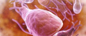
The structure of the medicine is a polypeptide and is prepared from the lung tissue of cattle. Inhibits the kallikrein-kinin system both in general and its individual components. Blocks proteases that are actively involved in fibrinolysis, plasmin , trypsin and other enzymes.
Contrical inhibits fibrinolysis and is used in coagulopathies as a hemostatic agent.
1 antitrypsin unit corresponds to 1.33 kallikrein inactivating units (KIU).
Indications for use of Contrikal
- pancreatitis in the acute phase;
- pancreatic necrosis;
- surgery on the pancreas to prevent autolysis ;
- bleeding caused by activation of fibrinolysis (postoperative, postpartum, post-traumatic, polymenorrhea );
- all types of shock (traumatic, hemorrhagic, toxic, burn);
- prevention of postoperative mumps;
- Quincke's edema;
- massive soft tissue damage due to trauma;
- prevention of postoperative bleeding and pulmonary embolism .
There are also indications for the use of Contrikal in the complex therapy of coagulopathies and bleeding.
Use of the drug Contrikal 10,000
For Contrikal 10,000, the following dosage regimen is used. In acute pancreatitis: immediate intravenous administration (slowly) to the patient of 200,000–300,000 ATred of Contrical 10,000, then over 24 hours another 200,000–300,000 ATred is injected intravenously. Such treatment is carried out until the clinical picture of the disease and laboratory parameters normalize. To prevent inflammation of the pancreas after surgery: Contrikal 10,000 is administered intravenously slowly at a dose of 200,000 ATRED per day as a preventive treatment. For the treatment of shock conditions : first, Contrical 10,000 is administered intravenously slowly at a dose of 200,000–300,000 ATRED, and then at a dose of 140,000 ATRED every 4 hours. To prevent fat embolism : initially, Contrical 10,000 is administered intravenously at a slow dose 200,000 ATRED, then daily slow IV at a dose of 200,000 ATRED as an intermediate treatment. For bleeding : first, Contrical 10,000 is administered intravenously slowly at a dose of 300,000 ATRED, then every 4 hours slowly IV at a dose of 140,000 ATRED. If hemostasis is impaired during childbirth: the initial dose for intravenous administration is 700,000 ATRED with an interval of 1 hour until bleeding stops. For children, Contrikal 10,000 is administered in daily doses of 14,000 ATRED per 1 kg of body weight. The contents of the injection bottle are dissolved in an isotonic sodium chloride solution. This solution is administered to a patient in a supine position intravenously (slowly, maximum 5 ml/min) or by short-term or long-term drip infusion.
Side effects
- Heart and blood vessels: tachycardia and arterial hypotension .
- CNS: hallucinations , psychotic disorders, disturbances of consciousness.
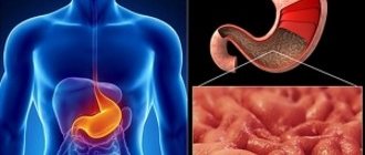
- Digestive system: Nausea and vomiting may occur if administered rapidly.
- Manifestations of allergies: conjunctivitis , urticaria , bronchospasm , anaphylactic shock .
- In the case of prolonged infusions - phlebitis and thrombophlebitis .
Side effects of the drug Contrikal 10,000
When using aprotinin, hypersensitivity reactions of varying severity may develop. Rarely, a severe reaction can lead to anaphylactic shock. Repeated administration of aprotinin is especially dangerous. Although even with the first administration of aprotinin, patients may experience phenomena that are reminiscent of a hypersensitivity reaction to the drug. They are manifested by skin rashes, tachycardia, pallor, blue lips, shortness of breath, profuse sweating, heart rhythm disturbances with a feeling of anxiety, and nausea. With repeated administration, thrombophlebitis may develop at the injection site. If necessary, generally accepted measures are taken to eliminate these complications, for example, single or multiple intravenous administration of adrenaline at a dose of 0.05–0.1 mg, intravenous administration of prednisolone at a dose of 250–1000 mg, and a complete blood transfusion.
special instructions
The instructions for Contrikal indicate the need for a skin test before administering the drug to exclude allergies. If the patient has previously had allergic reactions, glucocorticosteroids and antihistamines are administered before treatment.
For DIC syndrome and hyperfibrinolysis, it is administered after eliminating all disorders and simultaneously with Heparin.
muscle relaxants 2-3 days before the start of therapy , Contrical is used with caution.
Treatment protocol for patients with acute pancreatitis
Classification of acute pancreatitis, formulation of diagnosis.
Etiology of the disease:
1. Acute alcoholic-alimentary pancreatitis.
2. Acute biliary pancreatitis (cholelithiasis, parafaterial diverticulum, papillitis, opisthorchiasis).
3. Acute traumatic pancreatitis (due to trauma to the pancreas, including in the operating room or after ERCP).
4. Other etiological forms (autoimmune processes, vascular insufficiency, vasculitis, medicinal, infectious diseases), allergic, dishormonal processes during pregnancy and menopause, diseases of nearby organs.
Severity of the disease:
1. Mild acute pancreatitis. Pancreatic necrosis does not form in this form of acute pancreatitis (edematous pancreatitis) and organ failure does not develop.
2. Severe acute pancreatitis. It is characterized by the presence of organ and multiple organ failure, peripancreatic infiltrate, the formation of pseudocysts, and the development of infected pancreatic necrosis (purulent-necrotic parapancreatitis).
Pancreatic necrosis:
- Prevalence of the process: small focal pancreatic necrosis (the volume of damage to the pancreas according to ultrasound and CT < 30%; large-focal pancreatic necrosis (the volume of damage to the pancreas according to ultrasound and CT from 30 to 50%)); subtotal pancreatic necrosis (the volume of damage to the pancreas according to ultrasound and CT > 50 - 75%; total pancreatic necrosis > 75 (damages of the entire pancreas according to ultrasound and CT).
- Localization of the process: head (right type), isthmus and body (central type), tail (left type).
- Flow phases:
- phase (enzymatic toxemia) – endotoxicosis, organ failure, enzymatic peritonitis, omentobursitis, parapancreatitis; period - 7-10 days from the onset of the disease;
- phase (aseptic sequestration) – formation of sequesters of the pancreas and parapancreatic tissue, delimited parapancreatic fluid accumulations (pseudocyst); period 10-21 days from the onset of the disease;
- phase (purulent-septic complications) - abscess of the omental bursa, purulent parapancreatitis, retroperitoneal phlegmon, purulent peritonitis, arrosive and gastrointestinal bleeding, digestive fistulas, sepsis; period - later than 21 days from the onset of the disease.
Diagnosis of acute pancreatitis.
The diagnostic program includes: clinical, laboratory and instrumental verification of the diagnosis of acute pancreatitis; stratification of patients into groups depending on the severity of the disease; constructing a detailed clinical diagnosis.
Verification of the diagnosis of acute pancreatitis includes: physical examination - assessment of the clinical and anamnestic picture of acute pancreatitis; laboratory tests - general clinical blood test, biochemical blood test (including a-amylase, lipase, bilirubin, ALT, AST, alkaline phosphatase, urea, creatinine, electrolytes, glucose), general clinical urine test, coagulogram, blood group, rhesus factor; plain X-ray of the abdominal cavity (increased diameter of small intestinal loops, fluid levels), chest X-ray (hydrothorax, discoid atelectasis, high diaphragm dome, hyperhydration of the parenchyma, ARDS pattern), ECG; Ultrasound of the abdominal cavity - assessment of the presence of free fluid, assessment of the condition of the pancreas (size, structure, sequesters, fluid inclusions), assessment of the condition of the biliary tract (hypertension, stones), assessment of intestinal motility.
Patients with a presumptive clinical diagnosis of acute pancreatitis are advised to undergo endoscopy (differential diagnosis with ulcerative lesions of the gastroduodenal zone, examination of the gastroduodenal zone).
Stratification of patients according to the severity of acute pancreatitis according to the following criteria:
1. Severe pancreatitis (more than one of the criteria):
- signs of SIRS (2 or more clinical signs: body temperature >38°C or <36°C; heart rate>90 beats/min; respiratory rate>20/min; PaCO2<32 mmHg, leukocytes >12x9/l or <4.0×9/l or immature forms >10%);
- a-amylase > 500 units/l, lipase > 100 units/ml
- hypocalcemia <1.87 mmol/l, blood hemoglobin >150 g/l or hematocrit >40 units, hyperglycemia >10 mmol/l; C-reactive protein >120 mg/l;
- arterial hypotension (systolic blood pressure <90 mm Hg)
- respiratory failure (P02<60 mmHg);
- renal failure (oligoanuria, creatinine >177 µmol/l);
- liver failure (hyperfermentemia);
- cerebral insufficiency (delirium, stupor, coma);
- coagulopathy (platelets <100x*/l, fibrinogen <1.0 g/l);
- Ranson scale - 3 or more points;
- Balthazar-Ranson index - 3 or more points.
2. Mild acute pancreatitis: absence of criteria for severe acute pancreatitis in the presence of a clinical and instrumental picture of acute pancreatitis.
Ranson scale for acute pancreatitis.
| Indicator under study | Alcoholic pancreatitis | Biliary pancreatitis |
| On admission: | ||
| patient's age | Over 55 years | More than 70 years |
| leukocytosis | More than 16,000 mm3 | More than 18,000 mm3 |
| blood plasma glucose | More than 11.1 mmol/l | More than 11.1 mmol/l |
| Serum LDH | More than 700 ME | More than 400 ME |
| Serum AST | More than 250 ME | More than 250 ME |
| In the first 48 hours: | ||
| decrease in hematocrit | More than 10% of normal | More than 10% of normal |
| increase in serum residual nitrogen levels | More than 5 mg%* | More than 2 mg%* |
| calcium concentration | More than 8 mg%** | More than 8 mg%** |
| arterial blood pO2 | More than 60 mm Hg. st | |
| foundation deficiency | More than 4 mEq/L | More than 5 mEq/L |
| estimated fluid loss (sequestration) | More than 6 l | More than 4 l |
- — Each indicator in the table is scored as 1 point.
Pancreatitis severity index according to Balthazar - Ranson .
Normal appearance of the pancreas – 0 points
Increased pancreas size
Signs of inflammation of parapancreatic tissue – 2 points
Enlargement of the pancreas and the presence of fluid in the anterior perinephric space – 3 points
Accumulation of fluid in 2 or more areas of parapancreatic tissue – 4 points
Necrosis <30% of parenchyma – 2 points
Necrosis of 30-50% of the parenchyma - 4 points
Necrosis >50% of parenchyma - 6 points
Patients with severe pancreatitis are hospitalized in the surgical intensive care unit.
Patients with mild pancreatitis are hospitalized in the surgical department.
Treatment of patients with mild acute pancreatitis.
Basic treatment complex:
- fasting for 48 hours;
- gastric probing and aspiration of gastric contents;
- local hypothermia (cold on the stomach);
- analgesics and NSAIDs;
- antispasmodics;
- infusion therapy in a volume of up to 40 ml per 1 kg of patient’s body weight with forced diuresis for 24-48 hours;
- inhibitors of gastric secretion: omeprazole 40 mg – 2 times a day intravenously;
- octreotide 100 mcg – 3 times a day subcutaneously.
In dynamics, the following are assessed daily: SIRS criteria, a-amylase. Requirements for treatment results: relief of pain, clinical and laboratory confirmation of resolution of the active inflammatory process.
Lack of effect from analgesic and antispasmodic therapy for 12-48 hours, rapidly progressing jaundice, absence of bile in the duodenum during endoscopy, signs of biliary hypertension according to ultrasound indicate stenosis of the terminal part of the common bile duct (impacted stone of the common bile duct, papillitis). In this case, EPST is indicated. In acute pancreatitis, EPST is performed without ERCP!
Monitoring the general somatic and local status of patients with severe acute pancreatitis:
- general clinical and biochemical blood test - daily;
- Ultrasound of the abdominal cavity - every 48 hours;
- MSCT of the abdominal cavity – the first 24 hours; subsequently – every 5 days.
Treatment tactics in patients with severe acute pancreatitis in the phase of pancreatogenic toxemia.
The main type of treatment for acute pancreatitis in the toxemia phase is complex intensive conservative therapy.
Basic therapy for acute pancreatitis is supplemented with the following components:
- intensive inhibition of pancreatic secretion (octreotide 300 mcg x 3 times a day subcutaneously or 1000 mcg per day by continuous infusion) until a-amylase and lipase levels are normalized;
- prolonged epidural anesthesia;
— the dose of infusion solutions should be at least 40-60 ml/kg of the patient’s body weight per day; high-volume infusion therapy includes balanced crystalloid solutions and colloid solutions (in a 2:1 combination of crystalloids and colloids);
— rheologically active therapy: colloids in combination with antiplatelet agents, UFH (15-20 thousand units per day) or LMWH; introduction of antioxidants;
— extracorporeal detoxification: extended venovenous hemodiafiltration and serial plasmapheresis;
— installation of a nipple tube for enteral nutrition beyond the duodeno-jejunal junction with simultaneous installation of a naso-gastric tube for gastric decompression; introduction of electrolyte solutions into the small intestine (1 - 2 liters/day) in the first 24-48 hours; subsequent enteral nutrition with oligomeric and polymeric nutritional mixtures;
— providing mixed parenteral-enteral nutritional support with a caloric intake of at least 2000 kcal per day.
Surgical tactics:
Surgical interventions via laparotomy during the phase of enzymatic toxemia are contraindicated. According to indications, minimally invasive interventions are used - percutaneous punctures and drainages, laparoscopy, EPST.
Percutaneous puncture and drainage of acute fluid accumulations under ultrasound control can reduce the level of endogenous intoxication.
Foci of acute fluid accumulations located in the omental bursa, retroperitoneal tissue, and in cases where laparoscopic drainage of acute fluid accumulations in the abdominal cavity is impossible (the severity of the patient’s condition, the patient has previously undergone several operations on the abdominal cavity, a giant ventral hernia) are subject to puncture.
For small volumes of fluid (<100 ml) and complete aspiration, repeated percutaneous punctures followed by ultrasound control are preferable. If the volume of fluid is >100 ml, or the accumulation could not be evacuated completely, or it has recurred, or fistulography has established a connection with the pancreatic duct, or growth of microflora in the aspirate has been obtained, percutaneous drainage of this fluid accumulation is indicated.
Emergency decompression of the biliary tract in patients with acute biliary pancreatitis is indicated in the following cases: there is no effect from conservative therapy within 6-12 hours; impacted calculus BDS; increasing phenomena of obstructive jaundice; progression of acute cholecystitis and/or cholangitis.
In case of diagnosed residual or recurrent choledocholithiasis, choledocholithiasis against the background of chronic calculous cholecystitis, acute obstructive cholangitis, papillitis or papillostenosis, EPST without ERCP is indicated! If it is impossible to perform EPST, percutaneous transhepatic microcholecystostomy under ultrasound guidance is indicated. If it is impossible to achieve adequate decompression from the above approaches, percutaneous transhepatic cholangiostomy is indicated. In case of herniation of a calculus in the area of the major duodenal papilla, preference is given to endoscopic papillogomy.
Indications for laparoscopy:
- clinical picture of peritonitis with the presence of ultrasound signs of free fluid in the abdominal cavity;
— the need for differential diagnosis with other acute diseases of the abdominal organs.
Objectives of laparoscopy:
- Confirmation of the diagnosis of acute pancreatitis: the presence of edema of the root of the mesentery of the transverse colon; the presence of effusion (pink, raspberry, cherry, brown) with amylase activity 2-3 times higher than blood amylase activity); presence of steatonecrosis; extensive hemorrhagic permeation of the retroperitoneal tissue;
- Exclusion of other acute surgical pathology of the abdominal organs;
- Removal of peritoneal exudate and drainage of the abdominal cavity.
Therapeutic tactics in patients with severe pancreatitis in the phase of aseptic sequestration.
The clinical and morphological form of acute pancreatitis in the aseptic sequestration phase is a postnecrotic pancreatic pseudocyst, the formation period of which ranges from 4 weeks and on average up to 6 months.
Outcomes of acute pancreatitis in the phase of aseptic sequestration:
1) in small focal forms - resolution of infiltration;
2) for large-focal forms - aseptic sequestration with the formation of a cyst or cysts;
3) in common forms - in the focus of pancreatogenic destruction there are large zones of infiltration and multiple foci of fluid accumulations without visible clear boundaries and sizes;
4) Infection of areas of pancreatogenic destruction (development of purulent complications).
Diagnostic criteria for the aseptic sequestration phase:
— decrease in the severity of SIRS, absence of signs of an infectious process;
— Ultrasound and CT signs of aseptic destruction in the lesion (continued increase in the size of the pancreas, blurred contours and the appearance of fluid in the parapancreatic and retroperitoneal tissue with subsequent formation of pseudocysts, visualization of sequesters in the pancreatic tissue and in the parapancreatic tissue).
Treatment during the aseptic sequestration phase.
- Continuation of basic infusion-transfusion therapy aimed at replenishing water-electrolyte, energy and protein losses according to indications.
- Medical nutrition: oral nutrition (table No. 5); enteral or parenteral nutritional support with a total caloric intake of at least 2000 kcal/day.
- Systemic antibiotic prophylaxis (cephalosporins of III-IV generations or fluoroquinolones of II-III generations in combination with metronidazole).
- Immunotherapy with correction of cellular and humoral immunity.
Surgical tactics in patients with acute pancreatitis during the period of aseptic destructive complications.
Indication for surgical interventions is the presence of circumscribed peripancreatic fluid accumulations (with or without sequestration).
The priority is to perform minimally invasive percutaneous interventions under ultrasound or CT control.
Percutaneous punctures under ultrasound control are indicated in the presence of liquid formations with a volume of no more than 100 ml. Systematic punctures can serve as a definitive method of surgical care or allow deferment of radical surgery.
Percutaneous drainage under ultrasound control is performed in the presence of fluid accumulations with a volume of more than 100 ml. Drainage of a cyst involves aspiration and lavage sanitation of the cyst cavity and assessment of the adequacy of drainage using dynamic fistulography.
Surgical interventions using laparotomy and (or) lumbotomy access are carried out if there are technical restrictions on the safe performance of puncture interventions (location on the intended trajectory of the intervention of the colon, spleen, large vessels, pleural sinus); when the tissue component (sequestra) predominates in the fluid accumulation or acute pseudocyst.
In case of focal process, mini-laparotomy (pararectal, transrectal, oblique in the hypochondrium) or mini-lumbotomy should be used. If the process is widespread, a wide median laparotomy, a wide lumbotomy, or a combination of both should be used.
In conditions of completed sequestration and complete necrosequestrectomy, the operation should be completed with “closed” drainage of the resulting cavity with 2 lumen drains, according to the number of branches of the cavity that are brought to the abdominal wall outside the surgical wound. The wound is sutured tightly. In the postoperative period, aspiration and lavage treatment is carried out until the cavity is obliterated.
In case of incomplete sequestration and incomplete necrosequestrectomy, the operation should be completed with “open” drainage with 2 luminal drainages according to the number of branches of the cavity, removed outside the wound through counter-apertures and insertion of tampons into the resulting cavity through the surgical wound. The wound is partially sutured and a bursoomentostomy or lumbostomy is formed to access the site of destruction during subsequent programmatic sanitation. Aspiration and lavage treatment, dressings with additional necrosequestrectomy and changing tampons are carried out along the drainages until the cavity is cleaned, followed by the application of secondary sutures to the wound and the transition to closed drainage.
Small pancreatic pseudocysts (less than 5 cm) are not advisable to operate; they are subject to dynamic observation. Large pancreatic pseudocysts (more than 5 cm) are subject to surgical treatment as planned in the absence of complications. The operation of choice for an immature (unformed) pseudocyst (lifespan less than 6 months) is external drainage. A mature (formed) pseudocyst (lifespan is more than 6 months) is subject to surgical treatment as planned.
Therapeutic tactics in patients with severe pancreatitis in the phase of purulent-septic complications.
Clinical and morphological manifestations of acute pancreatitis in the phase of purulent-septic complications are:
1. Infected pseudocyst - a local accumulation of infected fluid and areas of the pancreas, sometimes containing sequestra; differs from an abscess in the absence of a granulation shaft;
2. Pancreatic abscess - occurs as a result of melting and infection of foci of necrosis with the secondary formation of fluid in them, is an encysted accumulation of pus, contains a small amount of necrotic tissue; necrotic changes in the gland and retroperitoneal tissue are minimal;
3. Infected pancreatic necrosis is a diffuse bacterial inflammation of necrotic pancreatic tissue and/or peripancreatic adipose tissue, extending deep into the retroperitoneal space, without a fibrous capsule or localized accumulations of pus.
Diagnostic criteria for the phase of purulent-septic complications:
1. Clinical and laboratory manifestations of the infectious process: progression of clinical and laboratory indicators of SIRS at the 3rd week of the disease; high levels of markers of acute inflammation (C-reactive protein - more than 120 g/l and procalcitonin - more than 2 ng/ml); lymphopenia, increased ESR, increased fibrinogen concentration; deterioration of the patient's condition according to integral assessment systems.
2. Instrumental criteria for suppuration: CT signs (increase during observation of fluid formations in the focus of pancreatogenic destruction and/or the presence of gas bubbles) and/or positive bacterioscopy results obtained by fine-needle puncture.
Surgical tactics in patients with acute pancreatitis in the phase of purulent-septic complications.
If purulent complications develop, urgent surgical intervention is indicated.
Minimally invasive puncture interventions (puncture and drainage) are indicated in the presence of clearly demarcated purulent accumulations (fluid collections, pancreatic abscess, infected pseudocyst) without a pronounced tissue component (sequestra). It should be considered optimal to install two drains into the abscess cavity, followed by installation of a flushing system.
Surgical interventions using laparotomy and lumbotomy approaches are indicated in cases of significant spread of the process in the retroperitoneal tissue or in case of a limited process with the presence of large necrotic fragments in the abscess cavity. The main method of sanitation of purulent-necrotic foci is necrosequestrectomy, which can be either single-stage or multi-stage.
The optimal access is extraperitoneal, in the form of a lumbotomy with an extension of the incision on the abdominal wall towards the rectus abdominis muscle, which allows, if necessary, to supplement this access with laparotomy.
The method of completion of the operation depends on the adequacy of the necrosequestrectomy. With complete removal of necrotic tissue, “closed” drainage is possible with double-lumen drainages according to the number of branches of the cavity with drainage being removed through counter-apertures on the abdominal wall. Aspiration and lavage sanitation is carried out through drainages in the postoperative period.
In case of incomplete removal of necrotic tissue, “open” drainage with double-lumen drainages according to the number of branches of the cavity should be used, in combination with packing the cavity through the surgical wound and leaving access for subsequent program revisions and necrectomies in the form of omentobursostomy and (or) lumbostomy. Aspiration and lavage sanitation is carried out through drainages in the postoperative period.
Monitoring the adequacy of drainage and the condition (size) of drained cavities should be carried out using fistulography, ultrasound and CT every 7 days. Ineffectiveness of drainage (according to clinical and instrumental data) or the appearance of new purulent foci is an indication for reoperation with additional necrectomy and drainage.
If arrosive bleeding develops in the focus of purulent destruction, the bleeding area should be inspected, sequesters removed, the bleeding site sutured (temporary hemostasis) and ligation of vessels outside the purulent focus should be performed (final hemostasis). It is prohibited to apply sutures to the vessel wall in the purulent defect area. If it is impossible to ligate vessels outside the purulent focus, distal pancreatectomy and splenectomy are indicated.
Antibacterial therapy for acute pancreatitis.
• Antibiotic prophylaxis is not indicated in patients with severe acute pancreatitis in the phase of enzyme toxemia.
• In patients in the phase of purulent-septic complications, empirical therapy is considered:
Piperacillin-tazobactam 4.5 g IV every 6 hours
OR
Cefepime 1 g IV every 8 hours PLUS metronidazole 500 mg IV every 8 hours
OR
Ciprofloxacin 400 mg IV every 12 hours PLUS metronidazole 500 mg IV every 8 hours
Penetration of selective antibiotics into the pancreas.
Good (>40%): fluoroquinolones, carbapenems, ceftazidime, cefepime, metronidazole, piperacillin-tazobactam
Poor (<40%): aminoglycosides, first generation cephalosporins, ampicillin.
Duration of antimicrobial therapy.
For infected pancreatic necrosis, continued antibiotic therapy is indicated for 14 days after surgical treatment. Continuation of antibiotic therapy during this time is required for patients due to the risk of colonization or infection with resistant organisms and drug toxicity.
Contrikal's analogs
Level 4 ATX code matches:
Aerus
Ingitril
Trasylol
Aprotex
Gordoks
The main analogues of Kontrikal: Kontriven , Aprotex , Trasylol .
The most commonly used substitute is Gordox .