Artificial esophagus
- an organ (or part of it) used to create, through plastic surgery (esophagoplasty), a new path for the passage of food from the pharynx to the underlying parts of the digestive tract.
Indications
to the creation of an artificial esophagus: 1) burn strictures in the absence of effect from conservative treatment, including bougienage; 2) two-stage treatment of esophageal cancer - after the Dobromyslov-Torek operation (see Esophagus); 3) rarely peptic strictures of the esophagus, arising due to cardia insufficiency.
Preoperative preparation
Patients in need of esophagoplasty are usually malnourished due to the impossibility of normal nutrition. Therefore, when preparing for surgery, normalization of protein and water-electrolyte balance is necessary. For this purpose, in the preoperative period, parenteral nutrition is prescribed (see), which can be complete in a number of patients. If parenteral nutrition is impossible or ineffective, gastrostomy is indicated (see). To prepare for esophagoplasty, broad-spectrum antibiotics are used.
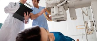
Advantages of the First Surgical Department of the Russian Research Center for Surgery
The department carries out scientific and practical work on the study and development of methods for treating diseases of the gastrointestinal tract. Much attention is paid to the use of minimally invasive surgical methods.
At the Center you can complete the full course: examination, treatment, postoperative recovery. Comfortable rooms accommodate up to 25 people in total. Each room is equipped with a TV, refrigerator, remote control for calling medical personnel and everything necessary for a quick recovery.
Esophagoplasty methods
The creation of an artificial esophagus for cicatricial obstruction and esophageal cancer was started by H. Bircher (1894), who proposed forming the esophagus from a skin flap cut out in the sternum area. This operation was not widely used due to the occurrence of peptic ulcers of the newly formed esophagus, severe dermatitis, multiple fistulas, malignant neoplasms, etc.
L. Wullstein in 1904 proposed using the small intestine in combination with skin grafting to create an artificial esophagus.
Rice. 1. Schematic representation of some methods of creating an artificial esophagus: a - using the small intestine (according to Roux-Herzen-Yudin); b - using a flap cut from the wall of the greater curvature of the stomach (according to Gavriliu); c — using the stomach, crossed in the area of the cardia (according to Kirchner); d — using the right half of the transverse colon (according to Royt); d - using the left half of the transverse colon (according to Orsoni and Tyne); 1 - proximal esophagus, 2 - esophageal-intestinal anastomosis, 3 - small intestine, 4 - stomach, 5 - transverse colon, 6 - interintestinal anastomosis, 7 - esophageal stricture, 8 - esophageal-gastric anastomosis, 9 - graft cut from walls of the greater curvature of the stomach, 10 - cardia of the stomach, 11 - anastomosis between the esophagus and the colon, 12 - anastomosis between the transverse colon and the stomach, 13 - lleotransverse anastomosis, 14 - anastomosis between the stomach and the descending part of the transverse colon, 15 - colonic anastomosis.
Ts. Ru (1906) was the first to create an artificial esophagus in humans from the small intestine, isolating a graft of such length that it was moved under the skin to the manubrium of the sternum. The second stage of this operation was the creation of an anastomosis of the graft with the esophagus. P. A. Herzen performed esophagoplasty in three stages: mobilization of the intestine, creation of an anastomosis of the intestine with the stomach, and anastomosis of the intestine with the esophagus. S.S. Yudin, improved the method and technique of graft mobilization, proposed special tools for creating a subcutaneous canal. He did not include the stomach in the passage to prevent the development of peptic ulcers of the jejunum and anastomosed the small intestine only with the esophagus (Fig. 1, a). This type of esophagoplasty is called the Roux-Herzen-Yudin method. Garlock (JH Garlock), B.V. Petrovsky, Sweet (HR Sweet), Ohshawa, and others used intrathoracic anastomosis of the Esophagus with the stomach after resection of the Esophagus.
Beck (S. Beck) in 1905, I.O. Halpern and A. Jianu in 1912. In 1957, D. Gavriliu proposed the method of esophagoplasty with a flap formed from the greater curvature of the stomach (Fig. 1.6). With this method, the abdominal cavity is opened with an oblique incision in the left hypochondrium. A stalk-like sleeve is cut out from the greater curvature of the stomach and moved under the skin of the chest to the neck. After a few days, the graft is anastomosed with the cervical esophagus.
Kirschner (1920) (Fig. 1c) after crossing the cardia, moved the entire stomach under the skin to the neck and anastomosed it with the esophagus. He connected the cardia remaining in the abdominal cavity to the jejunum with an additional Roux-en-Y enteroenteroanastomosis. Principle: Kirchner operations were successfully used by the Japanese surgeon Nakayama (K. Nakayama, 1954), and in the Soviet Union by A. A. Rusanov and others.
Kelling (G. Kelling, 1911) used esophagoplasty from the transverse colon. However, a transverse colon graft can rarely be extended to the level of the cervical esophagus, and therefore additional skin grafting is often required. Roith (1923) used the right half of the colon (blind, ascending and part of the transverse colon) for esophagoplasty (Fig. 1d), Orsoni and Toupet (P. Orsoni, R. Toupet, 1950) - the left half of the colon (Fig. 1.5), and Lafargue (P. Lafargue) et al. (1951) - the right half of the colon with the terminal segment of the ileum. When mobilizing the cecum and ileum, the technique of B. A. Petrov and G. R. Khundadze is used, which consists in peeling off the root of the mesentery of the small intestine from the retroperitoneal tissue and moving upward the entire small intestine along with the mesentery. This technique allows you to move the graft upward an additional 15-20 cm.
The most widespread in wedge practice are the methods of creating an artificial esophagus from the small intestine according to Yudin and from the right half of the colon.
Esophagoplasty according to Yudin consists of the following stages. An upper median laparotomy is performed. Isolation of the intestine and dissection of the mesentery of the small intestine begin 8-10 cm below the ligament of Treitz, ligating and crossing 3-5 radial vessels. The intestine is transected, its distal end is sutured and peritonized. Intestinal continuity is restored using end-to-side enteroenteroanastomosis. The selected part of the intestine is removed from the abdominal cavity and carried presternally through a subcutaneous tunnel created with the help of special instruments to the level of the thyroid cartilage or the angle of the lower jaw, where it is fixed to the skin. The laparotomy wound is sutured. After 5-6 days, after making sure that the intestine is viable, it is anastomosed with the cervical esophagus.
Esophagoplasty of the right half of the colon includes the following stages. An upper median laparotomy is performed, bypassing the navel on the right and 6-8 cm below it. The parietal peritoneum is dissected above the cecum and along the outer edge of the ascending colon; the greater omentum is cut off from the transverse colon, and the right diaphragmatic-colic ligament is crossed. The structural features of the ileocolic, right colic and middle colic arteries and veins are studied, after which a graft is cut out, the length of which depends on its location. The ileum is crossed 10-15 cm from the cecum, an appendectomy is performed, and the transverse colon is crossed at the level of the branching of the middle colon artery. The distal end of the transverse colon is sutured.
The mobilized intestine is passed through the hole in the lesser omentum behind the stomach. The lower end of the graft is anastomosed with the antrum of the stomach. An anastomosis is formed between the ileum and the distal colon. The graft is inserted into the neck and isoperistaltically anastomosed to the esophagus.
Until the early fifties, all surgeons created the newly created P. and. was carried out on the neck antethoracically; later, along with this, they began to use the intrathoracic route of transplantation. A.G. Savinykh used a sagittal diaphragmotomy to place a small intestinal graft onto the neck through the posterior mediastinum.
N. I. Eremeev, Robertson and Sarjeant (R. Robertson, T. R. Sarjeant, 1950-1951) described the technique of retrosternal plasty of the esophagus.
Intrapleural esophagoplasty was performed by S. S. Yudin (1947), Rienhoff (WF Rienhoff, 1946).
The question of choosing one or another method of esophagoplasty and the route of transplantation should be decided purely individually, depending on the general condition of the patient, the anatomical features of the abdominal organs, as well as the nature of the architectonics of the intestinal vessels. Thus, it is more rational to perform esophagoplasty after extirpation of the esophagus for cancer antethoracically, because in such patients, retrosternal or intrapleural placement of the graft is often complicated by the occurrence of a violation of its blood supply.
In case of cicatricial stricture of the esophagus, the location of the graft is chosen depending on the level of the stricture. If its location is low, the intrapleural location of the graft is most rational, since when using pre- or retrosternal esophagoplasty in the remaining blind sac of the esophagus, above the stricture, inflammation, cancer and other complications may develop.
Rice. 2. Schematic representation of segmental plastic surgery using the colon: 1 - proximal esophagus, 2 - upper esophageal-colic anastomosis, 3 - esophageal stricture, 4 - lower esophageal-colic anastomosis, 5 - stomach.
In the case of an extended stricture in the middle third of the thoracic esophagus, when the lumen of the esophagus is preserved above and below the stricture, segmental plasty is indicated. To resolve the issue of the advisability of using segmental plasty, it is necessary to study the distal segment of the esophagus, for which retrograde esophagoscopy and esophagography are used. A rigid endoscope (eg, rectoscope) is inserted into the gastrostomy tube. When the stomach is inflated, an endoscope tube is inserted into the cardia and the mucous membrane of the esophagus is examined. Then a gastric tube is inserted through the tube and a suspension of barium sulfate is injected with a Janet syringe. At the same time, the patient is given a sip of contrast agent through the mouth and X-rays are taken. This technique allows you to contrast the esophagus above and below the stricture, clearly determining the extent of the narrowing. For segmental plasty, the small or large intestine is used, a segment of which is inserted into the chest cavity and anastomosed with the esophagus above and below the stricture (Fig. 2).
If the stricture extends to the cardia of the stomach, then a lower anastomosis is performed with the stomach.
In the case of stricture or cancer of the cervical esophagus, it is resected, followed by esophagoplasty with a free segment of the small intestine, the blood supply of which is provided by an anastomosis between the graft vessels and the branches of the cervical vessels.
Alloplastic replacement of the esophagus has not been used in clinical practice.
Who may be at risk
Until now, scientists have not been able to definitively establish for what reason tissue cells transform into malignant ones. However, they found a relationship between the formation of cancer and such negative factors affecting the body as smoking and alcohol abuse, swallowing too hot food and drinks, harmful production and chemical damage to the skin, as well as the presence of esophageal ulcers, polyps and papillomas. Experts do not exclude heredity, so it is important to periodically observe those who have already had such a problem in their family.
Postoperative management
In the first two days, patients are fed parenterally. On the 3-4th day after surgery, additional feeding through a gastrostomy tube begins. It is necessary to monitor the viability of the graft. If the graft is located under the skin, monitoring it is easier. Usually on the second day after surgery, when lightly tapping with your fingers in the area where the graft is located, you can see its peristalsis or tonic contraction, which indicates its viability. With intestinal necrosis, swelling of the graft occurs along its entire length, then redness of the skin over the intestine and signs of intoxication appear. In doubtful cases, a small skin incision should be used to expose the upper portion of the graft in order to monitor its viability. When the graft is located in the retrosternal space or in the pleural cavity, the only objective method of control is to examine the graft in the neck. If necrosis is detected, all or part of the graft must be removed. After removal of necrotic intestine, the mediastinum should be drained from the neck and abdominal cavity.
After esophagoplasty, diseases such as reflux esophagitis (see Esophagitis), associated with the absence of the cardia or a violation of its sphincter-valve function, may develop; disruption of the patency of the artificial esophagus as a result of cicatricial changes in the anastomotic area or due to reflux esophagitis; artificial esophagus diverticula; graft ulcers; fistulas, polyposis and rarely cancer of the artificial esophagus.
Our prices for chemotherapy
If you choose our high-quality treatment option costing from three hundred thousand, then you will no longer have the problem of choice. The option in Moscow or Israel is premium, high-quality therapy for esophageal cancer. Why is the cost of treatment so high? What do you get from us:
- high-quality drugs, original and newest,
- every drop of your drug goes to you,
- the preparation of the IV is done using strict Israeli technologies, and in your presence,
- the procedure itself is precise, that is, it is carried out strictly according to the specialist’s protocol,
- Negative side effects are carefully managed,
- our doctor really makes sure that such therapy helps, that is, you have a return from it,
- you are in a favorable atmosphere, in spiritual comfort.
Chemotherapy for esophageal cancer will cost from 10,000 rubles per procedure; medications are paid separately, which can be very expensive, but extremely effective. We use only original drugs of the latest generation, which give the highest chance of a positive outcome of therapy. The patient orders such drugs abroad and they are delivered to him personally. We trust the quality of our suppliers' products.
If you have already been diagnosed and are planning treatment, or doctors are unsure which treatment option to choose, we recommend getting a second opinion from our specialist. Send us by mail all the documents on the disease (extracts, results of tests and examinations), the doctor will look at them and give his opinion on the correctness of the diagnosis, the effectiveness of the therapy already carried out and possible options for further treatment according to international standards. This step may be decisive in choosing a treatment strategy and prognosis for recovery in general!
Pathological anatomy of the artificial esophagus
Rice.
3. Macropreparation of the artificial esophagus (16 years after surgery): papillomatosis of the inner surface of the graft (indicated by arrows). Rice. 4. Macropreparation of the artificial esophagus (29 years after surgery): multiple papillomatosis of the inner surface of the graft; in a large papilloma (indicated by an arrow), squamous cell carcinoma was detected Fig. 5. Macro-preparation of an antethoracic artificial esophagus created from the small intestine (37 years after surgery): a conglomerate of loops of the small intestine is located on the sternum. Macroscopic changes in the artificial esophagus depend on its location and the organ from which it is created. During antethoracic esophagoplasty, a large number of adhesions occur around the graft, sometimes compressing the lumen of the subcutaneous esophagus. With the long-term existence of the graft, the subcutaneous tissue around it sharply becomes denser. Clearance P. and. it can be unevenly narrowed with single or multiple papillomatous growths on its inner surface or extensive scarring around the graft (Fig. 3, 4). The loops of an antethoracically located graft from the small intestine have the same width; the thickness of its walls can increase due to the surrounding adhesions (Fig. 5). The relief of the mucous membrane of the graft is preserved even after many years of its existence.
Rice. 6. Microscopic specimens of the mucous membrane of an artificial esophagus created from the small intestine (17 years after surgery): a - shortening and sclerosis of the villi (indicated by arrows); hematoxylin-eosin staining, x 140; b — hyperplasia of goblet cells (indicated by arrows) in the villi of the small intestinal tract; carmine staining, X 280. Fig. 7. Microslide of the mucous membrane of an artificial esophagus created from a colonic transplant (10 years after surgery): hyperproduction of mucus by goblet cells (indicated by arrows); Hematoxylin-eosin staining, x 280.
In functioning intestinal transplants, in the coming months after esophagoplasty, mucus accumulates on the surface of the mucous membrane due to the proliferation of goblet cells and the lumen of the crypts of the mucous membrane gradually expands. With the long-term existence of the transplant, atrophy of the villi of the intestinal mucosa develops, which acquire different sizes, but the proliferation of goblet cells remains in them (Fig. 6, 7). Restructuring of the walls of the vessels of the submucosal layer of the intestine is revealed by hypertrophy of muscle and elastic fibers. Hypertrophy of the muscle fibers of the intestinal wall develops slowly and is more pronounced in the small intestinal transplant. After two to three decades of operation of the small intestinal transplant, the muscle fibers of the intestinal wall atrophy, which is not observed in the transplant from the colon. Differences in structural changes of P. and. from the right or left half of the colon is not observed.
In the stomach used for esophagoplasty, inflammatory changes in the mucous membrane are detected in the early stages, gradually giving way to its atrophy during the first year of its existence.
Rice. 8. Microscopic specimen of the bottom of a peptic ulcer of the stomach wall in the area of anastomosis of the artificial esophagus created from the small intestine: hypertrophy of the arterial walls with drying of their lumen; hematoxylin-eosin staining, x 56.
Changes in the stomach during esophagoplasty are determined by the type of transplant and the participation of the stomach in digestion. Ulcerative dermatitis often occurs near skin-gastric anastomoses due to gastric contents being thrown into the lumen of the skin graft. Scarring of the anastomotic ulcer leads to stenosis of the anastomosis, one of the common late complications of esophagoplasty. In the stomach cut off from digestion, atrophy of the mucous membrane and the muscular layer of the wall develops, against which, after a few decades, typical stomach or duodenal ulcers and even stomach cancer can develop. The inclusion of the stomach, even if it has undergone atrophy, in the digestive process during small and large intestinal esophagoplasty after a few years can lead to the formation of peptic ulcers of the gastrointestinal anastomoses (in 2-5% of cases). Peptic ulcers of anastomoses are small in size and are usually located at the edges or near the anastomosis. At the bottom of such ulcers, under a layer of granulations, there are large arteries, the erosion of which can lead to profuse gastric bleeding (Fig. 8). Around anastomoses and peptic ulcers in the gastric mucosa, proliferation of parietal cells is detected, which occurs in response to the entry into the stomach of large quantities of alkaline intestinal contents.
Patient reviews
Valery Myazina: My dad underwent surgery for a discovered tumor of the esophagus, Dr. Greenberg did it, and now such surgical problems are behind him. This is how we hope and this is how we understand the situation. Now we just have to make a decision about chemotherapy, which we will do at the Medis clinic in Moscow. Many thanks to Dr. Greenberg and his team for the successful operation.
Alexey Nefedov: When the cancer relapsed, we came to the Israeli medical clinic in Moscow, took a PET scan, and it turned out that a second operation could not be done, but that we could only fight it with chemotherapy and radiation therapy. Dr. Aronov, who is now always with us, guides the entire treatment process. The situation is serious, but we are grateful to all the doctors at the clinic who are helping us fight the disease.