Article for the “Bio/Mol/Text” competition: Helicobacter pylori is a pathogen that in humans is associated with gastritis, ulcers and other harmful effects on the gastric mucosa, including cancer. The molecular pathogenesis of this infection is described, but at the same time confusing. In addition, the course of the disease is associated with other pathologies. For example, bronchial asthma and thrombocytopenic purpura. Scientists recently uncovered a mechanism that the bacterium uses to regulate its pathogenicity. We will try to analyze all these links in this review. True, instead of fingerprints and an accidentally left button, we will have experimental data at our disposal.
Competition “Bio/Mol/Text”-2021/2022
This work was published in the “Free Topic” category of the “Bio/Mol/Text” competition - 2021/2022.
The nomination partner is SkyGen: a leading distributor of life science products on the Russian market.
The general partner of the competition is the international innovative biotechnology company BIOCAD.
The general partner of the competition is: the largest supplier of equipment, reagents and consumables for biological research and production.
"Book" sponsor of the competition - "Alpina Non-Fiction"
Every criminal inevitably makes some mistake and gives himself away. Oscar Wilde. The Picture of Dorian Grey
Criminal record
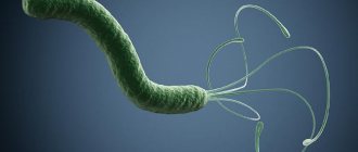
Helicobacter pylori is a gram-negative spiral-shaped bacterium. It belongs to the Helicobacteraceae family and has several flagella that ensure cell movement through the gastric mucosa. Based on its physiological properties, H. pylori is a microaerophile - a microorganism that grows at low oxygen concentrations in the environment, which partly explains the place of its colonization. An important biochemical property of this bacterium is urease activity, that is, the ability to enzymatically break down urea. This is necessary to alkalize the environment.
Urease test for diagnosing Helicobacter infection
Urease activity is primarily of diagnostic value: a quick urease test allows you to detect a shift in the pH of the stomach to the alkaline side. To carry it out, the patient drinks a solution of urea with the carbon isotope 13 C. Since urease breaks down urea into ammonia and carbon dioxide, the carbon isotope will be contained in the latter in the presence of infection. After taking exhaled air, they look at the ratio of carbon dioxide with an isotope to normal.
According to a 2022 meta-analysis [1], more than half of the world's population are carriers of Helicobacter pylori infection: among 14,006 publications from 1970 to 2016 on H. pylori, a careful selection of all cases was made. The distribution pattern can be seen in Figure 1.
Figure 1. Prevalence of H. pylori infection across continents. The most infected regions and parts of the world are indicated in burgundy color - most of Eurasia, Brazil, and the countries of Central Africa.
[1]
Blood tests
If the patient complains of pain in the stomach, any discomfort, with functional digestive disorders, with gastritis and peptic ulcers, a blood test is taken for Helicobacter pylori. Antibodies to helicobacter pylori in the blood are an indicator of human infection with the bacterium helicobacteriosis.
It is recommended to donate blood for Helicobacter bacteria if:
- weak immunity
- hereditary predisposition to stomach cancer
- as a preventive diagnosis
- to evaluate the treatment received for the infection.
How to prepare for the analysis?
Before the analysis, you must not eat food, coffee, tea, alcoholic drinks, or smoke 8 hours before the test. This test for Helicobacter pylori determines the amount of immunoglobulins. When a certain toxin enters the human body, a virus, microbe, immunoglobulins interact with them and neutralize these harmful substances. The analysis shows the interaction of the pathogen and immunoglobulins, which makes it possible to determine whether Helicobacter pylori is in the stomach or duodenum.
Advantages and disadvantages
Advantages of this test:
- the diagnostic procedure is not expensive;
- high accuracy of the result;
- analysis is available to everyone.
The test for Helicobacter pylori has its drawbacks:
- there are factors that influence the result of the analysis;
- some patients experience fear when taking blood;
- thorough results are obtained in 4-6 days.
Analysis transcript
Deciphering the result of a blood test for Helicobacter pylori can be done without medical skills. Antibodies are divided into categories A, G, M. Opposite each category there is a result on the form. If all blood test parameters for Helicobacter pylori do not exceed the norm, then there is no bacteria in the body:
- immunoglabulin LgG is absent or significantly lower than normal: bacteria are absent in the body;
- LgG detected: helicobacteriasis is present or has been previously experienced;
- immunoglabulin LgM was not detected or below the norm: conditional norm for Helicobacter pylori;
- LgM class detected: initial stage of the disease;
- immunoglabulin LgA is not detected in the blood: this may indicate an early stage of the disease, recent antibiotic therapy, or the patient is in the recovery stage.
Contraindications
A blood test for Helicobacter is not performed:
- for convulsions
- with increased excitability of the patient
- if there is skin damage at the injection site.
A blood test for antibodies is also not prescribed for venous phlebitis.
CagA is a dangerous weapon
Why can many people calmly coexist with the bacteria and not experience any discomfort, while other infected people know by heart the way to the gastroenterologist’s office? The fact is that our criminal has a weapon - a pathogenicity factor that causes structural and functional changes in the cells of the stomach - cagA (cytotoxin-associated gene A). The bacterial genome contains so-called pathogenicity islands ( PAIs ) - regions where proteins of the type IV secretion system are encoded. These are protein complexes that facilitate the transport of various substrate proteins from a bacterial cell to a eukaryotic cell. In particular, the gene product - CagA - into the host cell. However, cagA does not have a similar sequence in all bacterial strains: depending on the amino acid composition, the Western cagA specific sequence (WSS) and its East Asian cagA specific sequence (EASS) are distinguished [2]. In addition, in the region that directly encodes the pathogenicity factor, differences in the nucleotide sequence at the 3′ end can be observed [3]. In other cases, a piece of PAI may be completely absent, on the basis of which several sites of 3′-end polymorphism are identified (A, B, C, D), presented in Figure 2.
Figure 2. Pattern of PAI in H. pylori. The inner and outer membranes of the microbe with the periplasmic space between them are indicated in green. The protein complex of the type IV secretion system is built into the membranes, thanks to which CagA enters the gastric epithelial cell.
[3]
How is the 13C Hp breath test performed?
The study is carried out at an appointment with a pediatric gastroenterologist at SM-Doctor.
Its duration is about 40 minutes. The test is carried out in several stages. Before passing it, the child drinks a glass of orange, apple or grapefruit juice. After 5 minutes, he should exhale deeply into a special sealed bag. After this, he is given a special medical solution to drink, after which he must wait another half hour. After this, exhaled air is taken again for analysis. Both hermetically sealed containers are labeled and sent to the clinic laboratory for testing.
No man is an island
Conventionally, the process of human infection with Helicobacter can be divided into three main stages - the active phase, the stationary phase and the colonization phase. During the active phase, the bacterium quickly moves with the help of its flagella to the pyloric section of the stomach. Urease, mentioned above, shifts the pH towards alkaline. Then it binds to specific receptors, including the fucose-binding receptor. CagA enters the cell, and the microbe begins colonization, after which the cycle repeats again and again, inducing inflammation and other degenerative processes [4].
It is precisely because of the fucose-binding receptor that the microbe poses a greater danger to people with the first blood group according to the ABO system [5]. The thing is that the Lewis antigen of erythrocytes is one of the receptors that promotes the adhesion of Helicobacter. The results of another meta-analysis show that H. pylori has such an affinity only in this blood group and is not associated with others and the Rh factor [6].
Cytological analysis
Cytology is one of the most accurate methods for detecting Helicobacter pylori in the gastric mucosa. With this method, testing for Helicobacter pylori practically does not give false positive results.
For research, a biopsy material is taken during fibrogastroduadenoscopy (fgds), a smear is an imprint of the mucous membrane. The result can be positive or negative. According to the degree of contamination:
- weak - with a magnification of 360 times, up to 20 bacteria are detected in the field of view;
- moderate – 20-40;
- high – more than 40 bacteria.
Evidence - new and not so new
There are a large number of publications that describe the role of matrix metalloproteinases in the occurrence of inflammation during human infection with various microorganisms, for example, Pseudomonas aeruginosa [7] or Borrelia burgdorferi [8]. Matrix metalloproteinases ( MMPs ) are enzymes that degrade the extracellular matrix. They are classified according to their substrate specificity. For example, collagenases decompose collagen, elastases decompose elastin. A study on the AGS cell line (human gastric adenocarcinoma cells) and other cell lines showed that the synthesis of MMP-7, MMP-1, MMP-25 and, in particular, MMP-10, increases in response to the presence of the microbe [9]. In AGS cells, the mRNA level was measured using RT-qPCR (reverse transcription quantitative polymerase chain reaction) - PCR using reverse transcriptase. The level of MMP-10 was also determined by Western blotting. If you are not yet familiar with these methods, you can read about them here [35]. However, I will focus on a study where experiments were carried out on mice and biological material from patients with H. pylori infection [10].
Figure 3. Induction of MMP-10 in response to H. pylori is detected by various immunological methods. a — The samples circled by rectangles represent matrix metalloproteinases, the level of which was detected by antibodies. The naked eye can see that the samples with MMP-10 are the brightest of those presented. b — The level of MMP-10 production according to qPCR results in the group of infected patients (n = 65) is higher than in the control group (n = 40). c — Correlation between the level of MMP-10 synthesis and colonization of the microorganism, measured in the logarithm of the amount of 16S rDNA per 1 ng of host DNA. d - The level of MMP-10 production is high in the wild-type H. pylori group (n = 34), but the difference between the control group (n = 40) and the group with the CagA-free strain (n = 31) is very small. d — In mice infected with a pathogenic strain, the synthesis of MMP-10 first increases and then decreases after the ninth week, and when infected with △cagA, that is, a strain with a knockout of this gene, (5 mice per group) it fluctuates slightly relative to the control. f and g — MMP-10 levels were compared in these groups also by Western blotting and immunohistochemistry, respectively. The results speak for themselves!
[10]
In Figure 3D, it can be seen that the level of MMP-10 synthesis increases in response to infection with H. pylori associated with CagA, and in the “non-pathogenic” strain this indicator is slightly increased. This effect is also observed in Western blotting (Figure 3e), and is also clearly visible in immunohistochemical staining (Figure 3g) when comparing all three groups among mice and patients.
You can read more about the immunohistochemistry method in the article “Working with cells: automate and conquer” [36].
Figure 4. Just look at this beauty! Immunohistochemistry of the gastric epithelium (antibody staining and identification under a fluorescence microscope). a - Human gastric epithelium on the left and mouse on the right with nuclear staining (DAPI) - blue, H+/K+ATPase - green (marker of parietal cells), and in red - our main evidence, MMP-10. b - Similar staining, but with a nuance - the main cell marker - pepsinogen - was used, on the basis of which we can conclude that the content of MMP-10 is high in both types of cells.
[10]
Into the heat of the moment
Let's imagine this situation: in the state of K. you have a store and it is robbed, the alarm goes off, the SOS signal immediately reaches the relevant authorities. At the same time, the state of K. has very strict legislation, so the latter are allowed to open fire to kill. The result is dilapidated expensive property in your store, but the robbers left with nothing (or rather, they didn’t leave at all). The cell also sends approximately the same SOS signal in unfavorable conditions to attract the immune system. The signaling role is played by chemokines - protein molecules that provide chemotaxis (movement caused by a chemical irritant) of cells to the site of inflammation. But unfortunately, something always has to be sacrificed, and such immune “attacks” do not end well for the cell itself. Accordingly, the next hypothesis that needed to be tested was whether the production of MMP-10 is associated with the production of the chemokine CXCL 16? The answer is yes. To understand which cells respond to the signal, flow cytometry was performed. It turned out that killer T-cells are the most active in flocking to the crime scene compared to other cells. Using an in vitro transwell assay (a method in which cells are separated by a membrane permeable only to molecules), the researchers found that the fewest cells migrated in wells supplemented with siRNA against MMP-10.
Figure 5: A new player enters the field, the chemokine CXCL16, followed by T cells. AGS cells were incubated with Helicobacter, then the supernatant was collected and transferred to a well with a semi-permeable membrane. T cell migration was assessed using the transwell assay: the least number of cells migrated in wells supplemented with siRNA against MMP-10.
[10], figure adapted
And who is to blame?
We are gradually getting closer to the answer - how does MMP-10 interact with the gastric epithelium and how does this lead to gastritis and ulcers? Let's return to the study [9]. The article examines the role of the EGFR signaling pathway - epidermal growth factor receptor. The main task of this pathway is to stimulate cell growth and differentiation. It turned out that it is also associated with an increase in the synthesis of MMP-10. As we have learned well, the most effective way to test whether one molecule affects the activity of another is to turn something off in the middle and see what happens at the end. Therefore, by inhibiting individual proteins in the cascade, the researchers were able to build an axis from EGF to MMP-10. Another early study [11] showed that the EGF-dependent cascade is inhibited by the p38 protein, and as a result, the production of MMP-1 is reduced.
Figure 6. EGFR signaling pathway. After CagA enters the cell, it is phosphorylated by kinases and, together with the activation of Src kinase through the EGF-dependent pathway, increases the synthesis of MMP-10.
[9]
It turns out that the induction of MMP-10 is influenced by at least two factors - IL-22 from the immune system and CagA from Helicobacter. And then comes what we’ve been waiting for: MMP-10 interacts with the body’s own defense proteins - β-defensins and Reg3. The classical role of Reg3 is to exhibit antimicrobial activity, although it is now even considered as a hormone [12]. In the presence of H. pylori, the content of Reg3 decreases. However, in mice that cannot produce MMP-10 and IL-22, the opposite effect is observed.
Finally, having collected all the evidence, we can with a clear conscience present the picture depicted in Figure 7. However, do not rush to rejoice, because this is only one crime.
Figure 7. Scheme of the solved crime. The cagA gene product enters the eukaryotic cell and is phosphorylated. In this form, CagA and IL-22 activate the EGPR signaling pathway, resulting in increased production of MMP-10 in the cell with the help of ERK kinase. The latter disrupts the natural defense of the cell in the form of Reg3a, which enhances Helicobacter colonization. The epithelial cell sends a signal in the form of the chemokine CXCL 16, which causes the migration of killer T cells and the progression of inflammation.
[10]
Indications for the study
The appearance of individual symptoms or their combination is a direct indication for performing a breath test for Helicobacter Pylori. These symptoms include:
- emerging stool disorders in the form of chronic constipation or diarrhea;
- recurring heartburn;
- belching with a sour taste;
- discomfort and pain in the epigastric region, increasing after eating or at night (so-called hunger pains);
- nausea and vomiting;
- partial or complete loss of appetite;
- difficulty swallowing food.
As a rule, with chronic gastritis or gastric ulcer, the listed symptoms appear in combination.
Other guns
In fact, H. pylori, along with CagA, has another very dangerous weapon - VacA (vacuolating cytotoxin) [13]. More than 20 years ago, it caused confusion among specialists due to conflicting data [14]. Some studies showed that VacA has high affinity only for specific target cells [15], [16], others argued the opposite - the adhesion and pinocytosis it causes are nonspecific [17]. The mechanism of action of VacA is that it forms channels in the membrane, after which it becomes permeable to low molecular weight substances. Subsequent transport is organized by binding the cytoskeleton, namely F-actin [18], and penetration into the so-called GEEC (glycosylphosphatidylinositol-anchored protein-enriched endosomal compartments - cell areas rich in proteins anchored by glycosylphosphatidylinositol). These areas are also called VCV - VacA containing vacioles (VacA-containing vacuoles) [19]. Interestingly, the toxin penetrates mitochondria in an unknown way. The article [14] suggested several options. The first idea is that Vac-containing vacuoles (VCVs) fuse with the mitochondrial membrane and help transport VacA inside them. However, it is contradicted by the assumption that VCVs do not fuse with mitochondria, but only come into contact with them. Another hypothesis is that VCVs are not involved in the movement of the VacA toxin at all. Instead of vacuoles, transport is carried out by a carrier protein located directly in the mitochondrial membrane.
Figure 8. VacA is as mischievous as CagA. Mysterious intracellular transport of VacA with three possible routes of entry into mitochondria: 1) Transport of VacA using VCVs (VacA containing vacuoles). 2) VacA enters mitochondria using a transporter. 3) VacA is located between VCVs and mitochondria.
[14]
It often happens that if some substance is going to go into the mitochondria, it is clearly not with good intentions. VacA is no exception: it triggers one of the mitochondrial mechanisms of apoptosis, when cytochrome C “flies out” of the electron transport chain. As you might already guess, H. pylori with vacA knockout colonizes the gastric mucosa much worse than the wild type [14].
IgG antibodies to Helicobacter pylori
Detection of immunoglobulins to Helicobacter pylori is carried out using an enzyme-linked immunosorbent assay, or “sandwich” method. Venous blood is used for the study; you must not smoke for 30 minutes before donating it.
The method is based on the fact that after the Helicobacter bacteria enters the body, a local and systemic response develops. It consists of an increase in the titer of immunoglobulin IgM, IgG, and IgA antibodies in the blood serum. In 100% of cases of infection, IgG immunoglobulin is detected, in 65-80% of cases - IgA, in 17-20% - IgM, therefore, accurate diagnosis is carried out by determining the concentration of IgG in the blood serum.
Advantages of the method:
- Does not require endoscopy, so the method is safe;
- Highly sensitive;
- Used to prevent primary infection.
The method may not be accurate when determining the immunoglobulin titer in older people, since they have reduced antibody production, as well as in those patients who take cytostatics. After treatment of heliobacteriosis, this method is also not effective, because the level of the hormone in the blood after treatment with antibiotics remains quite high for another 6 months.
Indications for use:
- Patient complaints of dyspepsia when it is impossible to perform endoscopy;
- Primary diagnosis of heliobacteriosis;
- Monitoring the effectiveness of treatment with antibacterial drugs.
Reference values for the examination: IgG immunoglobulin concentration from 0 to 0.9, the result is negative. This may mean the absence of infection or high effectiveness of therapy.
He's involved here too...
It's time to talk in more detail about the role of Helicobacter in carcinogenesis. Various hypotheses were put forward in this regard, but it was still unclear how the bacterium could be involved in the occurrence of cancer. One of the mechanisms is a sad classic - inhibition of the p53 protein, known for its ability to send a cell into apoptosis, by activating certain signaling cascades [27].
Figure 9. CagA-induced signaling pathways that regulate p53 protein production. Consecutive activation of protein kinases by CagA leads to a decrease in the activity of the p53 protein. Another way is to influence the expression of the p53 gene. The result is the same - a decrease in surveillance of the occurrence of tumor processes in the cell.
[27]
However, just recently an article was published in which researchers described an RNA molecule that regulates the production of oncoproteins by H. pylori [28]. Clawed gerbils and mice were infected with the pathogenic strain of H. pylori ATCC 43504, and then Helicobacter was isolated from their stomachs. Then the researchers looked at which RNA molecules were synthesized in bacteria more than others. One of them - HPnc4160 - aroused genuine interest. It is this non-coding RNA that regulates the adaptation of the microbe to the host environment. Moreover, the degree of carcinogenicity of the bacterium increases with a decrease in the level of HPnc4160. This happens due to the fact that RNA reduces the production of CagA, which, as we have already found out, plays a key role in the pathogenesis of infection. What can this find tell us? On the one hand, there is no data yet on the possible use of this molecule, for example, in treatment, since the research is recent. On the other hand, finding such an “Achilles heel” provides a whole layer for new research. We can only wait for them with anticipation.
Figure 10. Experimental design. a — Removal of biological material and subsequent NGS analysis. b — Rate of mutations in various RNA molecules.
[28]
There is quite interesting evidence that diffuse large B-cell lymphomas (DBCL) in patients with H. pylori infection are more sensitive to chemotherapy compared with lymphomas in uninfected patients [29]. To do this, we analyzed the cases of 95 patients, 46 of whom were HP-positive and 49 HP-negative, and then their survival indices were assessed - five-year (event-free survival, EFS) and overall survival (OS) . Moreover, the situation was better in infected patients with high cagA expression in tumor cells: both survival indices were higher than in uninfected patients. A reasonable question arises - how does it work? It is possible that Helicobacter acts so aggressively on all components of the cellular component of the stomach that the tumor can no longer actively continue to grow and becomes vulnerable to treatment.
How to deal with this?
All the antics of Helicobacter described above are “tempting”, but the main question arises - what to do? The most trivial approach to treating an infection is eradication therapy, which was mentioned above, which includes taking a standard course of antibiotics according to a specific regimen [30], [31]. In addition, therapy using bismuth has also achieved success [32]. Against the backdrop of a gradual increase in antibiotic resistance [33], it is especially relevant. And then we remember one of our clues - MMP-10: it can be inhibited. Not long ago, the effects of using antibodies against this protein, obtained and constructed by immunizing llamas, were described [34]. The peculiarity of these antibodies is that they lack a heavy chain constant domain and completely lack a light chain (Figure 11). This structural detail allows these antibodies to be more selective compared to canonical ones.
Camels, for example, have similar antibodies - “A camel will cure cancer!” [38].
Figure 11. Unusual antibodies against MMP-10 - a new step towards treatment. a — Comparison of the structure of canonical antibodies and those used in the work. b — Antibodies tested demonstrated selective inhibition of MMP-10.
[34]
The researchers believe that this approach could be used as a targeted therapy in many diseases in which MMP-10 is implicated, not just H. pylori. You may ask: why do we need to know about some kind of llamas? The fact is that the work is experimental. It is possible that such antibodies can be synthesized through human immunization. The necessary amount of research must be carried out to show that all this works as it should. It just takes time.
Preparing for the test
Obtaining a reliable and most informative result of a breath test for Pylori helps by following simple but effective rules. These include:
- 3 days before the test, it is strictly forbidden to drink alcohol and any alcohol-containing drinks and foods.
- Stop using antibiotics for one month.
- 1 hour before the test, it is permissible to drink no more than 10 ml of water.
- 6 hours before the test, it is strictly forbidden to eat, smoke or chew chewing gum.
- 1-2 hours before the test, you should thoroughly brush your teeth and use mouthwash.
In addition, 5 days before a breath test for Helicobacter, it is strictly forbidden to take antacids and anti-inflammatory medications. For 3 days, you should exclude from your diet foods that stimulate gas formation in the intestines (legumes, white cabbage, fresh baked goods, confectionery and foods with a high sugar content).