What is Treatment of adhesions after surgery?
In practice, it looks like this: in places where the serous membrane is damaged, collagen and elastic fibers and connective tissue cells are intensively produced. If at this time any internal organ (for example, a loop of intestine) touches the area of the damaged serosa, it is involuntarily involved in this process. A cord of connective tissue is formed, which leads from the wall of the internal organs to the inner surface of the abdominal wall. This is called adhesions.
Adhesions can also connect internal organs to each other. Each of them is also covered by a serous membrane. During the operation, micro-tears are not excluded. And these places of microtrauma can also subsequently become a source of the formation of adhesions between this organ and the organs adjacent to it.
Also, at the site of contact and healing of tissues after their dissection or rupture, a scar may form, in which the normal tissue is replaced by a more rigid and inelastic connective tissue. Scars can be on the skin, or they can be on internal organs.
What are adhesions?
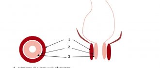
The organs of the abdominal cavity and small pelvis (uterus, fallopian tubes, ovaries, bladder, rectum) are externally covered with a thin shiny membrane - the peritoneum. The smoothness of the peritoneum, combined with a small amount of fluid in the abdominal cavity, ensures good displacement of the intestinal loops, uterus, and fallopian tubes. Therefore, normally, the functioning of the intestines does not interfere with the capture of the egg by the fallopian tube [1], and the growth of the uterus during pregnancy does not interfere with the normal functioning of the intestines and bladder.
Inflammation of the peritoneum - peritonitis - is a very dangerous disease. And the more dangerous it is, the more space it occupies in the abdominal cavity or small pelvis. But there is a mechanism in the body that limits the spread of peritonitis - the formation of adhesions.
With the development of the inflammatory process in the pelvis, the tissues at the site of inflammation become swollen, and the surface of the peritoneum is covered with a sticky coating containing fibrin (the protein that forms the basis of the blood clot). The fibrin film on the surface of the peritoneum at the site of inflammation glues adjacent surfaces to each other, resulting in a mechanical obstacle to the spread of the inflammatory process. After the end of the acute inflammatory process, adhesions in the form of transparent whitish films can form in places where internal organs stick together. These adhesions are called adhesions. The function of adhesions is to protect the body from the spread of a purulent-inflammatory process throughout the abdominal cavity.
The inflammatory process in the abdominal cavity does not always lead to the formation of adhesions. If treatment of adhesions
started on time and carried out correctly, the likelihood of adhesions is reduced. Adhesions form when an acute process becomes chronic and the healing process extends over time.
Adhesions can interfere with the normal functioning of internal organs. Impaired mobility of intestinal loops can lead to intestinal obstruction. Adhesions affecting the fallopian tubes, uterus, and ovaries disrupt the entry of the egg into the fallopian tube, the movement of sperm along the fallopian tube, the meeting of sperm and egg, and the advancement of the embryo after conception to the place of attachment in the uterine cavity. In gynecology, adhesions can cause infertility and pelvic pain.
Symptoms
Symptoms in the presence of adhesions in a woman are very different, however, several common signs can be identified:
- Chronic pelvic pain syndrome is a constant, sometimes almost imperceptible, dull pain in the area of adhesion formation. It occurs due to tension and deformation of organs. In some cases, it can radiate to surrounding organs, the lower back and legs. The pain may intensify during sexual intercourse, physical exercise and prolonged exercise, hypothermia, and when the bladder is full.
- Impaired organ function - reduction or disappearance of menstruation, lack of ovulation, inability to become pregnant.
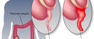
- In cases of formation of adhesions with intestinal loops - constipation, feeling of bloating, intestinal obstruction.
It is also worth noting that if a woman has adhesions, it is quite possible for her to experience increased pain during menstruation and ovulation. But it is worth considering that in half of the cases, adhesions do not cause any symptoms and are detected only during an ultrasound of the pelvic organs or a visual examination.
Adhesions formed as a result of inflammation
The fallopian tubes, uterus and ovaries can be involved in the adhesive process that occurs during inflammation of neighboring organs (appendicitis - inflammation of the appendix), as well as with lesions of the small and large intestines. In this case, the genital organs themselves suffer little: the adhesive process almost does not disturb their internal structure. If inflammation occurs inside the genital organs, not only the formation of adhesions occurs, but also damage to the genital organs themselves.
The most unprotected in this regard is the fallopian tube - one of the most delicate and finely structured smooth muscle organs. It plays a key role in facilitating conception and supporting early pregnancy.
Sperm entering the vagina are filtered through the mucus of the cervix, pass through the uterine cavity and enter the fallopian tube. Peristalsis - the movement of the fallopian tube - helps sperm enter the outer third (ampull) of the fallopian tube, where the process of conception occurs. During ovulation—the release of an egg from the ovary—the fallopian tube “sucks in” the mature egg. If at the time the egg enters the fallopian tube there are sperm there, fertilization occurs, and the resulting embryo moves to the uterine cavity within a few days, where it is immersed in the uterine mucosa (implantation). Delivery of the embryo into the uterine cavity is ensured by the movements of the fallopian tube and the active work of the microcilia of the fallopian tube.
Thus, the fallopian tube not only ensures the transportation of germ cells and the embryo, but also creates an environment for fertilization and development of the embryo during the first 5-6 days of intrauterine development. A change in the composition of the fluid that is produced in the fallopian tube can destroy the embryo. Local immunity inside the fallopian tube is minimal, that is, there are almost no mechanisms that ensure the rejection of foreign substances, because the embryo is half foreign, the fallopian tube does not reject it, and excessive activity of the immune system is unfavorable for the development of pregnancy. This is why the fallopian tubes so easily become victims of the so-called ascending infection (coming from the vagina and uterine cavity). Surgical and diagnostic interventions in the uterine cavity (abortion, diagnostic curettage, hysteroscopy[2], echohysterosalpingography[3]) facilitate the entry of infection into the fallopian tubes.
Once in the fallopian tubes, the infection first affects the mucous membrane of the fallopian tube (endosalpinx), then the muscular layer (myosalpinx), and only at the last stage the outermost layer of the fallopian tube (perisalpinx) is involved in the inflammatory process and conditions arise for the formation of adhesions. If treatment of adhesions
belated or not effective enough, after recovery not only adhesions remain, but also irreversible damage to the mucous membrane of the tube and its muscle layer. The cilia disappear, and scar tissue forms in place of the smooth muscle fibers. The fallopian tube can turn into a connective tissue sac (sactosalpinx), i.e. it loses the ability to promote a fertilized egg. With such disorders, elimination of adhesions cannot restore the function of the fallopian tube, and the presence of a focus of the inflammatory process leads to a decrease in the likelihood of pregnancy even in the tube from the opposite side or with the help of in vitro fertilization. In such cases, to increase the chances of pregnancy using IVF, which can be performed after recovery, the entire tube has to be removed. As a result of inflammation, gluing and fusion of the walls of the fallopian tube can occur, which leads to obstruction of the tube for the egg and is also an indication for separation of adhesions or removal of the tube.
Adhesion formation after hysterectomy
The risk of adhesions forming after removal of the uterus is high. This problem occurs in almost every operated patient. But the negative consequences of adhesive disease manifest themselves individually. In severe cases, complications such as intestinal obstruction and dysfunction of nearby organs occur.
After removal of the uterus, physiological adhesions form in the form of scars. They do not harm organs if they stop growing and do not turn into fibrous cords. The latter are prone to sprouting into other organs, disrupting their blood circulation, and therefore their function. The only way to get rid of the strands is through repeated surgery.
Adhesion formation in the postoperative period occurs with symptoms, the first of which is nagging pain in the lower abdomen. They are most often periodic in nature, but are so intense that they force you to take painkillers. The patient may develop a high temperature (up to 40C). Painful palpation of the suture also indicates the onset of adhesive processes. There is difficulty urinating and constipation. But none of these symptoms can indicate with absolute certainty an adhesive disease. The final diagnosis is determined only after an instrumental examination and a series of tests.
The first 2-3 weeks of abdominal discomfort and fever may be due to physiological factors of the postoperative period. But if they do not go further, then you need to see a gynecologist.
Postoperative adhesions
During surgical interventions, adhesions are formed due to:
- tissue hypoxia or ischemia - insufficient supply of tissues with blood and oxygen;
- drying tissue during surgery;
- rough manipulation of fabric;
- presence of foreign bodies;
- presence of blood;
- separation of former adhesions.
Foreign bodies that cause adhesions often include talc particles from doctor's gloves, small cotton fibers from gauze or tampons, and suture material. Adhesions also form with endometriosis. During menstruation, a small amount of menstrual blood containing living cells from the lining of the uterus (endometrium) may enter the abdominal cavity through the fallopian tubes. Normally, these cells are removed by the body's own immune system, but if there are any problems, they take root and form functioning endometrial islands that menstruate into the abdominal cavity. Adhesions form around these foci.
Diagnostics
The presence of adhesions in the abdominal cavity can be suspected in patients who have previously undergone pelvic inflammatory diseases, surgical operations on the pelvic and abdominal organs, and in women suffering from endometriosis. However, only in half of the patients with more than two risk factors for the development of adhesions in the anamnesis, adhesions are detected during laparoscopy (an operation during which small holes are made in the anterior abdominal wall through which an optical device is inserted to examine the cavity and special surgical instruments) .
A gynecological examination suggests the presence of adhesions in the abdominal cavity with a probability of 75%. Obstruction of the fallopian tubes according to hysterosalpingography (a contrast agent is injected into the uterus, X-ray images are taken) and ultrasound examination with a high degree of certainty indicate the presence of adhesions, but the patency of the fallopian tubes does not exclude the presence of adhesions that seriously impede pregnancy. Conventional ultrasound does not reliably detect the presence of pelvic adhesions. Today, the method of nuclear magnetic resonance (NMR, or magnetic resonance imaging, MRI) seems to be very promising in diagnosing the adhesive process. Using this method, images are obtained that reflect the “state of affairs” at different levels.
The main method for diagnosing adhesions is the laparoscopy method. It allows not only to detect the presence of adhesions and assess the severity of the adhesions, but also to treat adhesions
.
There are 3 stages of the adhesive process according to laparoscopy: Stage I: adhesions are located around the fallopian tube, ovary or in another area, but do not interfere with the capture of the egg; Stage II: adhesions are located between the fallopian tube and the ovary or between these organs and other structures and can interfere with the capture of the egg; Stage III: either torsion of the fallopian tube occurs, or it is blocked by adhesions, or a complete blockade of egg capture.
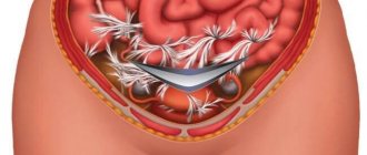
Treatment of adhesions
The main method of treating adhesions is laparoscopy . Using special micromanipulators, adhesiolysis - cutting and removing adhesions. Techniques for separating adhesions include laser therapy (cutting adhesions with a laser), aquadissection (cutting adhesions with pressurized water), and electrosurgery (cutting adhesions with an electric knife).
To prevent the formation of new postoperative adhesions during laparoscopy, the following methods can be used:
- introduction into the spaces between anatomical structures of various barrier liquids (dextran, povidin, mineral oils, etc.);
- wrapping the fallopian tubes and ovaries with special polymer absorbable films.
In addition, after laparoscopy, control diagnostic laparoscopy several months after the first laparoscopy has become increasingly common in recent years.
How to prevent adhesions from occurring?
For the prevention and treatment of adhesions, it is possible to use enzyme preparations based on hyaluronidase. This enzyme acts on the “skeleton” of connective tissue fibers. Unfortunately, natural hyaluronidase, introduced into the body, is quickly inactivated by enzymes and blood plasma inhibitors, without having time to exert its therapeutic effect.^(3)
Longidase is a new generation enzyme preparation based on hyaluronidase—Unlike the previous generation enzyme preparations, hyaluronidase in its composition is stabilized by a high-molecular carrier, which allows it to remain resistant to the action of enzymes and unimpededly exert its therapeutic effect. Longidase facilitates the movement of fluid in the intercellular space, which leads to a reduction in edema, resorption of hematomas and increases the availability of antibacterial drugs to the site of infection. In addition, the decrease in the viscosity of the connective tissue base under the influence of the drug increases the elasticity of adhesions, which reduces pain (instructions for the drug Longidaza).
Prevention
The cellular and molecular mechanisms of adhesion formation are now quite well understood. Therefore, the process of adhesions after surgical operations, including after laparoscopy, can be sharply slowed down using so-called adjuvant (auxiliary) therapy. This therapy should begin as early as possible after the operation (in the first days and hours) and continue for several weeks. Treatment is aimed at suppressing the inflammatory reaction, suppressing fibrin deposition in the abdominal cavity, and activating fibrin dissolution.
Adjuvant therapy includes the use of the following drugs:
- Fibrinolytic agents are substances that dissolve fibrin, around which adhesions are formed: FIBRINOLYSIN, STREPTOKINASE, UROCINASE, HYALURONIDASE, CHEMOTRYPSIN, TRYPSIN, TISSUE ACTIVATORS OF PLASMINOGEN.
- Anticoagulants - drugs that prevent blood clotting: drugs HEPARIN, OXALATES, CITRATES.
- Antibiotics : TETRACYCLINES, CEPHALOSPORINS, SULPHANAMIDES.
- Anti-inflammatory drugs : corticosteroids, antihistamines, non-steroidal anti-inflammatory drugs, progesterone, calcium channel blockers.
The choice of drugs and treatment regimens depends on each specific case and can only be made by the attending physician.
Numerous experimental, clinical and laboratory studies have been devoted to the prevention of postoperative adhesions. Taking into account the etiological factors and mechanisms of the formation of postoperative adhesions, a wide variety of methods have been proposed to prevent them. In this case, the mechanism of action of drugs or measures used to reduce the adhesive process is aimed primarily at reducing inflammation and exudation from wound areas, temporary delimitation of wound surfaces, preventing the accumulation of fibrin in the abdominal cavity, inhibition of fibroblast proliferation, as well as enzymatic protection of tissue from damage during hypoxia [1].
Among the variety of proposed methods for preventing the development of peritoneal adhesions, the highest preventive effect is provided by means for disconnecting wound surfaces. This group of methods consists of dividing the wound surfaces of the peritoneum for the period necessary for mesothelization of the defects (3-8 days). Anti-adhesion barriers operate due to hydroflotation, sliding, and mechanical separation of surfaces. The use of anti-adhesion barriers is an etiopathogenetic method for preventing the formation of adhesions, as it protects the wound from mechanical damage and adhesion to neighboring organs, and has a beneficial effect on reparative tissue regeneration [2].
In practice, a fairly large number of different anti-adhesion drugs have been tested to date. The most widely studied solution is dextran. The drug dextran-70 was tested as an anti-adhesion agent with good experimental and clinical results back in the 80s of the 20th century. However, with its practical use, side effects such as interstitial edema, ascites and coagulopathy have been reported. For these reasons, the use of dextran as an anti-adhesion agent has not been approved and its use is currently very limited [3].
For anti-adhesive purposes, saline solutions were used, which also create hydroflotation, but are absorbed through the surface of the peritoneum at a rate of 35 ml/h, i.e. quickly, and act during the critical period for the formation of adhesions only in the first 36 hours [4].
It should be noted that even honey has been tested as an anti-adhesive agent with good results from experimental studies [5].
The glucose polymer best known as an anti-adhesion drug is icodextrin, which is a topical anti-adhesive agent (liquid barrier) with the ability to separate damaged peritoneal surfaces and be absorbed within 3-4 days, which is sufficient to prevent the early formation of adhesions. Its 7.5% solution is used for peritoneal dialysis in 4000 patients per year. Known as the drug Adept, a 4% solution of icodextrin is widely used as an anti-adhesive agent in general surgery and gynecology and, according to several authors, can reduce adhesions by 32-52% [6-8].
Polysaccharides isolated from fungi have a well-known anti-inflammatory effect and reduce adhesions in experiments, which is explained by the modulation of tissue plasminogen activator and urokinase-type plasminogen activator [9].
Based on polytetrafluoroethylene, which is used in vascular grafts and is characterized by a low thrombus-forming factor, an anti-adhesion “barrier” Preclude (Gore-Tex Surgical Membrane, WL Gore & Associates, Flagstaff, AZ, USA) was created. This drug was positively evaluated in one of the studies and is recommended for gynecological operations (The Myomectomy Adhesion Study Group., 1995). However, Preclude has some disadvantages that limit its use. They are associated primarily with its hydrophobicity, which leads to insufficient adhesion to tissues. In addition, the material is not subject to biodegradation. For these reasons, this “barrier” must be fixed with threads and remains as a foreign body in the abdominal cavity forever, which naturally increases the risk of adhesions and infection in the long-term postoperative period [10].
Hyaluronidase compounds are widely used as anti-adhesive agents. Hyaluronidase is a glycosaminoglycan that is normally found in various tissues, synovial and abdominal fluids. As a natural biological compound of the body with immunological compatibility, it is most convenient as an anti-adhesive agent. Clinical studies have proven the effectiveness of using only hyaluronidase, as well as its compounds with iron and carboxymethylcellulose (CMC). Barriers created on the basis of hyaluronidase, in addition to anti-adhesion, also have an anti-inflammatory effect and increase the possibility of proliferation of mesothelial cells [11, 12]. Of the products created on the basis of hyaluronidase, its combination with CMC and two anionic polysaccharides, known as Seprafilm (Genzym Corporation, USA), is more common. It is a bioabsorbable membrane that is non-toxic, non-immunogenic, and biocompatible. It is used in the form of a film and covers injured surfaces. The membrane turns into a gel within 24-48 hours, but remains in place for up to 7 days. Completely resolves by day 28, does not require fixation with sutures, and is effective in the presence of blood. Seprafilm significantly reduced the extent and severity of postoperative adhesions in a variety of experiments and randomized clinical trials in gynecology and general surgery. The drug has received approval for clinical use in European countries and North America [13]. Its effectiveness has been proven in both experimental and clinical studies. However, Seprafilm is quite expensive, and exposure to hyaluronidase can lead to anastomotic leakage, abscess formation, and inflammatory reactions [14]. Claims that Seprafilm can reduce the incidence of adhesive intestinal obstruction remain controversial [15].
Another type of hyaluronidase, a compound with iron in the form of a paste, the drug Intergel (Life Core Biomedical Corporation), is not currently used, and there is fragmentary information about it in the literature [16].
Oxidized regenerated cellulose, known as Interceed Adhesion Barrier (Ethicon Inc., Somerville, NJ, USA), is a membrane that is completely resorbed within 28 days. The drug began to be used in the late 80s of the 20th century during gynecological operations; experimental and clinical use showed a decrease in the number and severity of postoperative adhesions. Widespread use of the drug is limited by decreased effectiveness in the presence of blood or excess peritoneal fluid. The need to achieve thorough hemostasis before using Interceed is believed to be due to the deposition of fibrin between tissue fibers [17].
In experimental studies, CMC showed anti-adhesive properties when used both as monotherapy and in combination with low molecular weight heparin and tissue plasminogen activator. However, CMC in its pure form is not used in the clinic, i.e. there are no commercial preparations of CMC for the treatment of adhesions in the abdominal cavity. Despite the positive properties of CMC gel, in practice it is used as an excipient. At the Congress of Surgeons in Atlanta in 1999, it was reported that the anti-adhesive effectiveness of CMC is higher when used in combination with other anti-adhesive drugs [10, 18].
Recently, a new membrane, Oxiplex (FzioMed, Inc., San Luis Obispo, CA), which consists of CMC and polyethylene oxide, has been reported. The drug reduces postoperative adhesions in experiments on rabbits, as well as in conditions of peritonitis in rats [13].
When the peritoneal surfactant layer is damaged during surgery, phospholipids can be added to maintain a lubrication effect between the organs and the abdominal wall. Phospholipids have not yet been used in clinical work to prevent adhesive disease, but in experimental work, the use of phosphatidylcholine led to good results both with and without peritonitis, without a negative effect on wound healing or anastomoses [7].
Spraygel, created on the basis of polyethylene glycol, is a hydrogel that is formed by mixing two liquids; the resulting foam adheres to organs, creates a viscous membrane, prevents contact of damaged surfaces and the formation of adhesions. Spraygel turned out to be effective in experimental work and was successfully used in gynecological practice [19].
To prevent the formation of adhesions, different types of polytetrafluoroethylene are also used - Preclude peritoneal film (WI Core corporation), Gore-Tex surgical sheath, Dual-Mesh mesh. However, at present, these films and networks do not have wide practical applications [20].
Despite the lack of results from clinical studies, experiments have yielded quite encouraging results from the use of bioactive polypeptides, such as poly-L-lysine. The combination of poly-L-lysine and poly-L-glutamate in experiments prevents the formation of adhesions with the peritoneum without affecting macrophages, the strength and tightness of anastomoses. Experiments also revealed the ability of bioactive polypeptides to reduce bleeding [4, 21].
In experimental work, collagen film, thrombin-based gelatin film, and polylactic acid film are also used as an anti-adhesion barrier [22]. For experimental and clinical research, new horizons are opened by the introduction of mesothelial membrane transplantation and the use of stem cells in areas of damaged mesothelium [23].
According to some authors, allogeneic peritoneum with preserved anti-adhesive and antimicrobial properties can serve as an effective remedy against adhesions. The allogeneic peritoneum can be attached to the abdominal wall using Sulfacrylat medical adhesive. In this case, laparoscopic adhesiolysis and film attachment using endovideosurgical techniques significantly reduce tissue damage and provide good results [24].
An original approach was demonstrated by the authors who proposed creating an alkaline environment in the abdominal cavity using a phosphate-citrate buffer with a pH of 5.6. They were based on the fact that simultaneously with a decrease in the pH of the environment, thrombin activity also sharply decreases, and at pH 5.6 the latter tends to zero, therefore, this prevents the transition of fibrinogen to fibrin [24].
Thus, the separate use of local and general anti-adhesive agents in most clinical studies did not lead to satisfactory results, and some drugs never left the experimental stage. Many unresolved questions remain in the pathogenesis of adhesive disease and in the field of treatment methods that are pathogenetically based [25].
In experimental studies, we found a significant, but inconclusive, decrease in the volume of adhesions under the influence of methylene blue, allopurinol, and vitamin E [25].
Thus, anti-adhesion “barriers” can be divided according to their state of aggregation into five groups: 1) gases (air, oxygen, helium, etc.); 2) aerosols (medicinal suspensions); 3) liquids (dextrans, hemodez, saline, etc.); 4) gels (hyaluronic acid, CMC, phosphotidylcholine, fibrin glue, etc.); 5) solids: A - films (Seprafilm, Interceed, Sepracoat); B - membranes (Gora-Teflon) [13].
A promising method for preventing the formation of postoperative adhesions is the use of barrier methods made of non-reactive, biodegradable materials. Available "barriers" are solid, semi-solid or liquid agents. Many “barriers” liquefy in the body after some time. Biocompatible, absorbable gel barriers are safe and effective. They are easy to use and quickly adapt to damaged tissues without fixation. The use of barriers differs from other methods in that they do not themselves interfere with the healing process, but rather potentially separate opposing surfaces during healing [26].
Based on extensive clinical and experimental experience in the development and use of various means to prevent the formation of postoperative adhesions, surgeons and researchers have formulated requirements for substances that can be used for this purpose. The ideal barrier, characterized by high safety and effectiveness, should not cause inflammation, immune response, should persist throughout the critical phase of remesothelialization, be held in place without sutures or staples, remain active in the presence of blood and be completely resorbable. In addition, it should not interfere with healing, provoke infection, oncological processes or cause adhesions. Barrier agents should be used intraperitoneally during or at the end of surgery [27].
The following barrier agents are currently in circulation:
— Interceed (“Johnson a. Johnson/Ethicon corporation”) — regenerated cellulose oxide. It has the form of a knitted fabric with small porosity, does not require suturing to damaged tissue and turns into a gel in approximately 8 hours. The material is usually absorbed within 4 days; if it is placed in several layers, absorption occurs within 4 weeks. Interceed must completely cover the injured area, requiring careful hemostasis and removal of irrigating fluid, otherwise it is no longer effective. It is currently approved for use only in gynecological surgical procedures [28];
— Fibrin glue is a combination of highly concentrated fibrinogen, thrombin, calcium and factor VIII. Due to its sealing effect, fibrin glue separates exposed surfaces. However, the use of human blood to produce the glue limits its use as a surgical treatment;
— Seprafilm (“Gemnzyme corporation”) is a modified hyaluronic acid and CMC, which is a bioresorbable transparent flexible membrane barrier and is applied to potentially adhesive tissue during surgery before closing the abdominal cavity. It adheres well to moist tissues, so complete hemostasis is not necessary. Seprafilm turns into a gel approximately 24–48 hours after placement, is absorbed within 28 days and does not require further surgery for removal [28];
— Preclude (“Wl Gore corporation”) is made of Gore-Tex - polytetrafluoroethylene. It does not liquefy and requires removal by repeated surgery.
- Gore-Tex, according to C. Farquhar et al. [17], is superior to Interceed in preventing the formation of adhesions, but its usefulness is limited by the need for suturing and further removal of the membrane;
- Flo-Gel (Alliance Pharmaceutical corporation) - a gel consisting mainly of Poloxamer-407, at temperatures below body temperature it is in the form of a liquid, and at body temperature it turns into a gel;
— Hyskon (“Medison Pharmaceuticalis corporation”) — 32% solution of dextran-70, a water-soluble glucose polymer, absorbed from 5 to 7 days;
— Adcon P (“Gliatech”) — a gel absorbed into the body within 4 weeks;
— Repel and Resolve (“Life Medical Sciences company”) — bioresorbable polymer film and viscous gel;
— Intergel (“Life Core Biomedical corporation”) - a combination of iron and hyaluronic acid, used only for gynecological operations, like Interceed;
— Incert (“Anika Therapeutick Inc Woburn MA”) — a derivative of hyaluronic acid;
— Seprocoat (“Hal-C Grenzyme corporation”) — a solution used after opening the abdominal cavity, during surgery by irrigation every 30 minutes;
- Oxiplex ("FzioMed") - polyethylene oxide and CMC in the form of a film, can be used in laparoscopic operations, easily twisted and inserted through a 5-mm trocar, easily spread on tissues [15];
— Adhibit (“Angiotech Pharmaceuticals”) is a self-polymerizing liquid sprayable hydrogel, ideal for use during endovideosurgical operations. The hydrogel is metabolized by the body in less than 30 days and is most often used in gynecological practice during laparoscopic operations;
— Adept (Shire Pharmaceuticals Ltd) is a non-viscose solution of 4% icodextrin, a new polymer of glucose solution, used during and after intervention for irrigation and infusion in laparoscopic and laparotomic operations. Non-viscous liquid does not reduce visibility during operation. Effective consumption - within 500 ml for irrigation and 1000 ml for infusions;
— Mesogel (“Lintex”, Russia) is an anti-adhesion absorbable gel. Acts as an artificial temporary “barrier” between damaged serous surfaces, ensuring their effective separation during healing, and then resolves. Reducing the adhesion of the surfaces of organs and tissues helps maintain their mobility and prevents the formation of adhesions. At the end of the action, the gel is completely removed from the body. When applied to healthy tissue areas, the gel helps reduce their drying due to the formation of a protective hydrophilic layer [26].
The existing “barriers” are not a panacea; the search for new means continues. K. Treutner et al. [26] suggest directing scientific efforts to the search for liquid substances for single intraperitoneal use that will significantly reduce the incidence of postoperative adhesions at a reasonable cost, without adverse effects on the blood coagulation system and wound healing processes.
From the above positions, the drug Guardabrida (Hangzhou Singclean Medical Products Co. Ltd., China) is promising. Clinical verification involving 30 patients demonstrated that Guardabrida has a pronounced preventive effect against postoperative adhesions, does not cause clinical adverse reactions and can be recommended for widespread use. The mechanism of action lies in its “barrier” properties. The gel acts as a temporary artificial barrier between damaged surfaces, ensuring their effective separation during healing. Reducing the adhesion of the surfaces of organs and tissues helps maintain their mobility and prevents the formation of adhesions. For use before the end of the operation, subject to asepsis, the outer packaging is opened and the syringe is removed. The gel is distributed in a thin, uniform layer on the surface of the treated organs in areas at risk of postoperative adhesions. The dosage and amount of administered gel depend on the area of application and are determined individually by the attending physician [29].
In conclusion, it should be noted that the effectiveness of measures to prevent postoperative adhesions depends on the level of trauma of the surgical procedure, the presence of a concomitant inflammatory process, extragenital foci of infection, and the type of anti-adhesive drug used. The search for more effective anti-adhesion components and agents at the level of molecular biology of cells continues [26].
If treatment for adhesions does not help
Unfortunately, laparoscopy cannot solve all problems associated with adhesions. It is possible to free the internal genital organs from adhesions, but it is impossible to restore the structure and function of the fallopian tubes. Therefore, if pregnancy does not occur within several months after laparoscopy, you should consider switching to more radical methods of treating infertility. In vitro fertilization[4]
(IVF) is one of the most famous methods of in vitro fertilization.
To increase the effectiveness of in vitro fertilization (IVF), the following is taken into account: 1. The presence of sactosalpinxes (limited accumulation of fluid in the fallopian tube) sharply reduces the effectiveness of the IVF method. Sactosalpinxes are removed using laparoscopy. 2. Before the in vitro fertilization (IVF) procedure, it is advisable to undergo a special immunological examination to increase the chances of embryo engraftment.
[1]
The fallopian tube is a thin, hollow tube that extends from the uterus, connecting the uterine cavity to the abdominal cavity. After the egg is released from the ovary into the abdominal cavity, it travels through the fallopian tube into the uterine cavity. [2] Hysteroscopy is the insertion of a special optical device into the uterus, which allows you to examine the uterus from the inside. [3] Hysterosalpingography – injection of an X-ray contrast agent into the uterus and taking a series of X-rays. [4] In vitro fertilization (IVF) is a relatively new method of treating infertility. It was first used in England in 1978. The essence of in vitro fertilization is that the egg is fertilized and develops outside the uterus, and an embryo (sometimes consisting of only a few cells) is transferred into it to bear a child. Tags: infertility