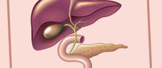
- Signs and symptoms of liver tumors
- Diagnosis of liver tumors
- Determining the stage of malignant liver tumors
- Treatment of liver tumors
- Results of treatment of malignant liver tumors
- What happens after treatment ends?
Malignant liver tumors account for 1-2% of all childhood tumors.
Boys get sick almost 2 times more often than girls.
Among liver tumors in children, hepatoblastoma and hepatocellular cancer are most often diagnosed. Other very rare malignant tumors include embryonal sarcoma, rhabdoid tumor and angiosarcoma.
In Western countries, hepatoblastoma occurs 2 times more often than hepatocellular cancer. The average age of children at the time of diagnosis of hepatoblastoma is 1 year. After 5 years, this form of liver tumor is detected very rarely. The average age of detection of hepatocellular cancer is 12 years.
Benign liver tumors are even rarer and include hemangiomas, hamartomas and nodular hyperplasia.
Risk factors that increase the likelihood of malignant liver tumors include congenital malformations, liver cirrhosis, colon polyposis, helminthic infestations, viral hepatitis B, metabolic disorders, aflatoxins, nitrosamines and some others.
Signs and symptoms of liver tumors
The most common manifestation of liver tumors in children is an increase in the size of the abdomen and a detectable thickening. Other symptoms include loss of appetite and weight loss. Loose stools or constipation are rare.
Anemia (anemia), fever, vomiting and jaundice usually appear with an advanced tumor process, and more often in patients with hepatocellular cancer.
In rare cases, patients with hepatoblastoma experience premature puberty due to the tumor's production of the hormone human chorionic gonadotropin.
1.General information
In comparison with such dangerous, common and widely discussed liver diseases as cirrhosis, cancer or viral hepatitis, the problem of benign tumors does not seem so acute. At the same time, benign neoplasia in hepatology is still not a sporadic rarity - and for a number of reasons (nonspecificity and weak severity of symptoms, reasonable caution of doctors regarding any tumor processes, inaccessibility of objective research due to localization, etc.) often become source of diagnostic doubts, difficulties or errors.
The names of benign tumors are traditionally derived from the name of the tissue in which the neoplastic process begins. Among benign liver tumors, the absolute leader are tumors growing from the tissue of the vascular walls, i.e. hemangiomas (up to 90%); in second place in terms of frequency of occurrence are tumors of glandular tissue, or adenomas (hepatocellular adenoma grows from parenchymal liver cells, or hepatocytes). Other types (lipomas, fibromas, hamartomas, etc.) are much less common.
A must read! Help with treatment and hospitalization!
Diagnosis of liver tumors
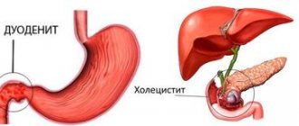
When palpating the abdomen, one or more tumor nodes can be identified in the right hypochondrium. An enlarged venous network on the anterior abdominal wall and ascites (fluid) in the abdomen may indicate damage to the hepatic veins and inferior vena cava.
The most necessary laboratory method for diagnosing malignant liver tumors is to determine the level of a special protein in the blood - alpha-fetoprotein (AFP). Its level is elevated in most patients with hepatoblastoma and in 50% of children with hepatocellular cancer.
An increase in the number of platelets (thrombocytosis) in peripheral blood is one of the signs of hepatoblastoma.
A biopsy (taking a piece of tumor for microscopic examination) using a puncture or during surgery is performed in cases where the data from a comprehensive examination are doubtful.
Chest X-ray makes it possible to detect metastatic lesions of the lung tissue, which occurs in 20% of cases at the time of diagnosis of the main tumor.
X-ray of the abdominal cavity can detect calcifications (calcifications) in liver tumors and exclude other tumor diseases, for example, metastases of neuroblastoma.
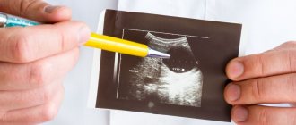
Ultrasound examination (ultrasound) helps confirm the origin of the tumor from the liver, clarify its location and distribution in the organ, and identify damage to the hepatic vessels.
Computed tomography (CT) with contrast, magnetic resonance imaging (MRI), radioisotope scanning of the liver, angiography (contrast study of blood vessels) are used to obtain additional information necessary when planning the extent of the operation.
3. Symptoms and diagnosis
The vast majority of benign liver neoplasias are asymptomatic—more precisely, they remain so for a long time, until a slow increase in size begins to affect the condition of surrounding tissues and structures. In the latent, subclinical period, such neoplasms are discovered only by chance. At the stage of clinical severity, most often the patient feels heaviness in the hypochondrium, characteristic of almost all liver diseases, and aching or nagging pain, independent of food intake and caused by the mechanical pressure of the tumor on adjacent tissues. Sometimes symptoms of dyspepsia occur, very rarely symptom complexes of portal hypertension, bile outflow disorders, cardiac complications, etc. are formed.
Differential diagnosis in such cases is facilitated by the benign nature of the tumor, i.e. absence of metastases, germination to other organs, symptoms of intoxication, tumor markers in laboratory tests.
A puncture biopsy performed under ultrasound guidance has decisive diagnostic information. To clarify the size, location, morphological features and other characteristics of the tumor, CT or MRI is prescribed; X-ray contrast angiography and/or Dopplerography are used to study hemodynamic changes caused by large liver hemangioma.
About our clinic Chistye Prudy metro station Medintercom page!
Determination of the stage (prevalence) of malignant liver tumors
After a comprehensive examination, the stage of the disease is determined, which is important for planning treatment and assessing the prognosis (outcome) of the disease.
Stage I - the tumor is completely removable
Stage II - after removal of the tumor, individual tumor cells remain.
Stage III - tumor rupture during surgery, the presence of affected lymph nodes, or only partial removal of the tumor.
Stage IV - presence of distant metastases
Liver cancer symptoms
Liver cancer can be asymptomatic for a long time. In many patients it is discovered during follow-up examinations or accidentally during examination for another pathology. Cases have been described where a patient had a tumor measuring 15 cm, but there were no clinical signs.
Already at advanced stages the following symptoms appear:
- Heaviness and dull pain in the right side.
- Increasing weakness, rapid fatigue.
- Loss of appetite, nausea, vomiting.
- Increased abdominal volume due to fluid accumulating in the abdominal cavity (ascites).
- Dilated veins in the abdominal wall.
- Yellowing of the skin and sclera.
- Pain. The more the liver capsule is stretched, the more intense the pain. In some cases it may be absent.
Clinical signs include the following:
- Increased liver size.
- Increased bilirubin levels. It can also be elevated against the background of cirrhosis.
- Enlargement of the spleen.
- Fever resistant to antibiotics and anti-inflammatory therapy.
- Gastroesophageal bleeding from varicose veins of the esophagus.
Stages of liver cancer
About 10-15% of patients have signs of paraneoplastic syndrome:
- Hypoglycemia.
- Skin itching without jaundice.
- Osteoporosis.
- Disruption of the thyroid and parathyroid glands.
In some cases, there are no primary signs from the liver, and symptoms appear with the development of distant metastases, for example, in the lungs, bones or brain.
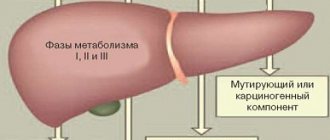
Treatment of liver tumors
The main treatment for liver tumors is surgery. The prognosis (outcome) of the disease mainly depends on how radically (completely) the tumor is removed.
Surgery in children with hepatocellular cancer is difficult due to the fact that there are usually multiple tumor nodes inside the liver tissue.
Preoperative chemotherapy using vincristine, platinum, Adriamycin (doxorubicin), Vepesid allows in some cases to reduce the size of hepatoblastoma, which makes it possible to further completely remove the tumor.
In the postoperative period, chemotherapy is prescribed to destroy the remaining tumor cells. In patients with hepatocellular cancer, chemotherapy is ineffective.
Radiation therapy can only be used for palliative purposes to relieve pain and reduce the manifestations of jaundice.
Types of liver adenomas
Liver adenoma, as a rule, is localized in the right lobe of the liver, has a heterogeneous structure and is located away from blood vessels. The diameter of the adenoma can range from a few mm to 20 centimeters. During palpation, this formation is an insensitive or painful node with a uniform surface. Liver adenomas can be either solitary (single) or multiple. Scientists identify several types of liver adenomas:
- cholangioadenoma. This benign neoplasm consists of cells of the bile ducts or the epithelium of the bile ducts. More common in women than in men;
- cystadenoma. It is a subtype of cholangioadenoma. It is malignant in nature, it is a bubble filled with a clear liquid with impurities of cholesterol, bilirubin, etc.;
- Hepatocellular adenoma is a tumor, the main component of which is liver cells - hepatocytes.
Complications
Despite the fact that hemangioma does not turn into cancer, its complications are very serious. The most common of them:
- A rupture occurs and internal bleeding begins;
- Thrombosis develops and the tumor thickens;
- When the tumor size is significant, compression of nearby organs and blood vessels occurs.
The development of the tumor is largely influenced by the content of estrogen in the blood, so it is necessary to monitor its development during pregnancy or during the use of hormonal contraception.
Introduction
According to the definition of pathologists, focal nodular hyperplasia of the liver is a benign formation related to hamartomas, which is manifested by focal hyperplasia of liver cells forming parenchymal nodules surrounded by fibrous layers [1]. According to the International Classification of Diseases (ICD-10), this disease is included in the heading “Other specified liver diseases”, K 76.8. In the international histogenetic classification of tumors of the liver and intrahepatic bile ducts, it appears as “focal nodular hyperplasia.” In the literature there are synonyms for the name of this disease: “fibronodular hyperplasia” and “focal cirrhosis of the liver.” In our opinion, the terms “focal nodular hyperplasia” and “focal cirrhosis” are not entirely appropriate. The phrase “focal nodular” is essentially a tautology, since it practically means the same thing (from the Latin focus - hearth, nodulus - node). The term “focal cirrhosis” accurately reflects the morphological essence of the disease, but in the minds of most clinicians, cirrhosis is strongly associated with diffuse liver damage. From our point of view, it is “fibronodular hyperplasia” (FNH) that is a completely acceptable term from a semantic point of view, since it most clearly corresponds to the morphological substrate of the disease - a node of hepatocyte hyperplasia with fibrosis.
Until relatively recently, FNH was classified as a rare (3%) space-occupying liver lesion (LLI) [2, 5, 6]. Currently, due to the active use of modern liver imaging methods, the number of identified patients with FNH (as well as focal liver diseases in general) has increased [3, 12]. According to the literature, FNH in the structure of focal liver diseases ranks third among benign AKIs after hemangioma and adenoma [4, 8]. The etiology and pathogenesis of FNH remain unclear [11]. The theory about the participation of hormonal contraceptives in the development of FNH has not been confirmed, and currently most researchers believe that FNH is the result of the progression of congenital or acquired vascular malformations, leading to hepatocyte hyperplasia [8, 9, 14]. Macroscopically, FNH is most often defined as a well-demarcated soft-elastic formation from the liver tissue, sometimes dense, yellow, cherry, whitish, light brown or pinkish in color, a lobulated structure with the presence of fibrous layers, usually extending from a centrally located scar.
FNH is a formation that is visually and palpably often indistinguishable from a malignant tumor. The macroscopic picture is determined by the ratio of parenchymal, fibrous and vascular components, which can be different, therefore, with full confidence, the diagnosis of FNH can only be established after a histological examination of the entire removed specimen. FNH can be observed in the form of one or multiple foci.
The microscopic picture is similar to that of true large-nodular cirrhosis of the liver. There is hyperplasia of the liver parenchyma, divided into nodes by fibrous layers; the latter form star-shaped scars. Hepatocytes contain excess amounts of glycogen and fat. In the fibrous layers there are small bile ducts, sometimes inflammatory infiltration is detected. Abnormal vessels with thickened walls are observed. The process is focal in nature, the liver tissue outside the focus appears unchanged [8, 11].
The disease is often asymptomatic and is an incidental finding during an ultrasound of the liver performed for another reason. Clinical manifestations (discomfort, feeling of heaviness) are caused by the “mass effect” and differ little from those with other AKI. There are no specific laboratory symptoms. FNH does not become malignant and extremely rarely causes complications such as intra-abdominal bleeding [13]. Cases of tumor regression up to its complete disappearance have been described [10]. When the diagnosis of FNH is confirmed and there are no complications, a number of authors recommend dynamic observation, although such tactics are debatable [7, 9, 10]. Therapeutic tactics for FNH remain controversial and, in addition, in the available literature we have not found any reports of attempts to trace the “natural” course of the disease when surgery is refused. This prompted us to share our own experience in treating this disease.
The purpose of the study was to retrospectively analyze the frequency of FNH among AKI and the treatment and diagnostic tactics for FNH over two time periods: from 1996 to 2005 and from 2006 to the present.
Material and methods
Over the past 17 years, 1425 patients with AKI have been treated in the hospital. 53 patients with FNH were observed, which accounted for 3.6% of all patients with AKI and 11.06% of patients with benign tumors. The age of patients with FNH ranged from 12 to 77 years, women predominated - 44 (83.2%). 47% of patients presented with complaints characteristic of AKI at an early stage of development - a feeling of heaviness, abdominal discomfort, which were based on stretching of the Glissonian capsule of the liver by a growing tumor; in other cases, the formation was accidentally discovered during ultrasound, less often during CT (“incidentaloma”). The final diagnosis of FNH was the result of the application of a comprehensive diagnostic algorithm for the management of patients with AKI of a solid structure [6], including ultrasound as a method of primary detection and monitoring, SCT and MRI as methods of clarifying diagnosis, and, if indicated, laparoscopy, biopsy, angiography, and laparotomy as the final stage of diagnosis. In our observations, tumor nodes were more often single, and only one patient had two lesions in both lobes of the liver. The diameter of the formations in the greatest dimension varied from 3.5 to 20 cm. 40 of 53 patients with FNH were operated on. The extent of the operation was determined by the size and location of the tumor. Extended left-sided hemihepatectomy was performed in 1 case, left-side - in 2, right-side - in 1, bisegmentectomy - in 8, atypical liver resection - in 16, tumor enucleation - in 8 cases. Exploratory laparotomy was performed in 3 patients, laparoscopic cholecystectomy for calculous cholecystitis with a biopsy of the detected tumor was performed in 1 patient. There were no deaths or complications directly related to liver surgery. Patient U.
, 18 years old, with congenital diabetes mellitus, on the 10th day after left hemihepatectomy, decompensation of diabetes was noted with the subsequent development of multiple organ failure syndrome. In another 24-year-old patient, latent porphyria manifested itself on the 8th day after right hemihepatectomy with the development of severe polyneuropathy. Death was avoided in both patients, but long-term intensive treatment was required in the intensive care unit. 12 patients were not operated on and are under observation with regular ultrasound. The follow-up period for patients with FNH ranged from 1 year to 13 years.
Results and discussion
A retrospective analysis of case histories since 1996 made it possible to identify two periods during which tactical approaches to the management of patients with FNH underwent some changes. The period from 1996 to 2005 was characterized by the predominant use of ultrasound as the main means of diagnosing focal liver disease, with FNH listed as a “rare” liver disease. The echosemiotics of FNH was poorly developed; there were no “conscious” diagnostic and treatment tactics. Under these conditions, the very fact of detection of AKI of unknown etiology determined the indications for surgical treatment, and the exact morphological diagnosis of FNH was established after surgery. During this period, 22 patients were operated on, and in 3 cases exploratory laparotomy was performed. Indications for surgical treatment were set out quite widely, but they refrained from removing the tumor if its benign nature was confirmed in the event of a large-scale intervention. This made it possible to trace the “natural” course of FNH. It turned out that the thesis about the “harmlessness” of the course of FNH is doubtful. This is confirmed by two of our observations published in the journal Surgery in 2005 [7].
As experience accumulated during the second period, from 2006 to 2013, it became apparent that FNH was more common than expected. In the diagnosis of FNH, the main place was occupied by SCT and MRI. Indications for dynamic monitoring of patients with FNH have become more frequent (with confidence in the absence of a malignant tumor). We have experience in observing the “natural” course of FNH, which did not require surgical treatment, in 12 patients. Moreover, in 11 cases, the diagnosis of FNH was established according to non-invasive diagnostic methods (ultrasound, SCT, MRI), in 1 - after laparoscopic cholecystectomy with tumor biopsy. The size of the formation in these patients was small (up to 5 cm in diameter), and there were no clinical symptoms. Monitoring with regular ultrasound over a period of 1 to 7 years revealed a slight increase in formation in only 3 patients, which so far excluded the need for surgery. In addition, analysis of the course of the disease in patients undergoing radical surgery in both periods made it possible to verify that after surgical treatment, during a control examination that included ultrasound, SCT, no signs of relapse of FNH were detected in any observation. In this regard, the main task of the second period (from 2006 to the present) is to determine clear indications for both surgical treatment and dynamic monitoring of people with FNH. An example of successful active treatment tactics is the following observation.
Patient L.
, 18 years old, ultrasound and MRI revealed a space-occupying formation with a diameter of up to 15 cm in the IV and V segments of the liver. There were no specific complaints, however, given the young age of the patient, the size of the formation, the impossibility of excluding its malignant nature, also the previous “negative” observation experience, we decided to operate.
On 08/12/10 the patient was operated on. Upon inspection in the IV (anterior sector) and V segments of the liver, there is a large tuberous formation with a diameter of 15 cm, protruding above the diaphragmatic and visceral surfaces, soft-elastic consistency, dark cherry color, with whitish veins, spreading to the bed of the gallbladder, most of the tumor is located in left lobe (Fig. 1)
.
Figure 1. Intraoperative photograph.
Fibronodular hyperplasia of the IV, V segments of the liver. a — diaphragmatic surface of the liver; b — visceral surface of the liver. A left-side extended hemihepatectomy and cholecystectomy were performed. Histological conclusion: fibronodular hyperplasia of the liver (Fig. 2)
.
Figure 2. Micrograph.
Fibrinodular hyperplasia. Hematoxylin and eosin staining. Uv. 100. The postoperative period was without complications. During a 3-year follow-up, there were no signs of disease recurrence. Full social adaptation. In the practice of hepatological surgeons, FNH is not so rare. It should be noted that surgeons of other specialties, as well as “visualization” doctors, are not well acquainted with this disease. At the stage of outpatient examination, the diagnosis of FNH was established only in 16 (30.7%) cases, which reflects both the lack of awareness of visualization doctors about this disease and the objective difficulties of diagnosis caused by the variability in the ratio of stromal, parenchymal and vascular components in the node . Most often, a formation in the liver at the preoperative stage was interpreted as a hemangioma, liver cancer, or “massive liver lesion of unknown origin.” It is not always possible to exclude the malignant nature of a pathological focus without surgery, therefore the advisability of laparotomy as the final stage of diagnosis in a number of cases is beyond doubt. The advisability of performing preoperative puncture biopsy under ultrasound or CT guidance in all patients seems controversial. It is often necessary to differentiate FNH from a malignant liver tumor, in particular hepatocellular cancer. A puncture biopsy of a lesion is not always informative (the biopsy sample is representative) and the finality of the histological conclusion in such situations is questionable. In addition, a biopsy for cancer is fraught with the risk of dissemination or implantation metastasis. We agree with the opinion of the authors who call not to perform a preoperative biopsy if cancer is suspected, since this factor worsens the prognosis of the disease [15]. If a hemangioma is suspected, such manipulation is in principle contraindicated due to the risk of intra-abdominal bleeding.
Based on our experience, we are currently cautious about long-term follow-up of patients, even with an established and histologically confirmed diagnosis of FNH. Mandatory conditions for this should be the possibility of regular ultrasound monitoring (once every 6-8 months) and sufficient compliance on the part of the patient. As with most AKI, “exit” of the lesion beyond the limits of easy operability is fraught with intraoperative problems and a severe course of the postoperative period [6].
Thus, FNH is a disease with unclear etiopathogenesis, uncontrolled course and a very uncertain individual prognosis. Tumor growth can lead to fatal complications. At present, the following tactics seem to be optimal for this disease: surgical intervention is indicated for “symptomatic” and rapidly growing FNH, the size of the lesion is over 5 cm, and it is impossible to exclude its malignant nature during a full examination. Dynamic observation is advisable if there is confidence in the diagnosis, small (less than 5 cm) size of the lesion, as well as well-known surgical risk factors (age, concomitant diseases), since the danger of the operation should not exceed the danger of the disease itself.
Comment from the editorial board
The widespread and availability of modern non-invasive diagnostic methods (CT, MRI), as well as an increase in their resolution, have led to an increase in the frequency of detection of asymptomatic focal liver tumors. The vast majority of them are benign, however, the doctor is often faced with the question of tactics for managing such patients. An article devoted to focal nodular hyperplasia, one of the most common benign liver tumors, will certainly be of interest to hepatologist surgeons, especially since Russian-language publications on this topic are rare.
First you need to decide on the terminology. At the beginning of the article, the authors suggest using the term “fibronodular hyperplasia.” You can agree with this or not, but in order to avoid confusion and misunderstanding, it is better to adhere to the international terminology proposed by the research group in 1995 (Terminology of Nodular Hepatocellular Lesions. International Working Party. Hepatology, 1995), in which this formation is defined as focal nodular hyperplasia and proposed criteria by which it can be distinguished from other regenerative formations.
Non-invasive methods play a dominant role in the diagnosis of FNH. MRI and CT have high sensitivity and specificity, however, in some patients the diagnosis may remain questionable, in which case a biopsy is justified, especially since “major” and “minor” criteria have been proposed for interpreting the material obtained from a biopsy (A. Fabre and al., 2002). The authors are wary of biopsy, fearing complications and implantation metastasis in the case of a malignant tumor, however, we believe that to establish a diagnosis it is necessary to use the entire arsenal of available means, and only if doubts remain, resort to laparotomy and resection of the formation. We consider the risks of a biopsy, if a number of rules are followed, to be exaggerated.
We adhere to a restrained tactics for managing FNH, since the prognosis is favorable: no cases of malignancy have been recorded, complications are rare (only a few cases of tumor rupture with bleeding have been described), most tumors are asymptomatic, and their sizes remain stable for long periods.
In this regard, we support the indications for surgical treatment given in the article: symptomatic tumor, its growth, the inability to exclude malignancy after a comprehensive examination. It is also important to note that the symptoms of FNH are nonspecific, so the exclusion of concomitant pathology of the abdominal organs, which may cause the clinical picture, is necessary. As for the recommendation to remove formations whose size exceeds 5 cm, one may disagree with this, since the presence of symptoms is influenced not only by the size of the formation, but also by its location and the proximity of surrounding organs. In addition, the size of the patient and his organs matters. For example, an FNH measuring 10 cm in a large person, located intraparenchymal, may not manifest itself in any way, and therefore the need for liver resection in him will be doubtful.
Thus, when choosing management tactics for a particular patient, one should focus on a set of factors, without exposing him to an unreasonable risk of surgical intervention, since mortality in the group of patients with benign liver tumors is unacceptable.
Modern methods of treatment
Some hemangiomas are diagnosed at birth or in early childhood (up to 5 - 10% of one-year-old children).
Hemangioma usually shrinks over time and in some cases may disappear. If it is small, stable and not causing any symptoms, it can be monitored with imaging studies every 6 to 12 months. There are no medications to treat liver hemangioma. Surgery may be necessary to remove the tumor if it is growing quickly or causing significant discomfort or pain. A technique called vascular embolization, in which the blood vessels supplying the hemangioma are closed, can slow or reverse its growth.
Treatment of benign neoplasms
Because of their benign nature and lack of known malignant potential, hepatic hemangiomas do not usually require treatment. For large, symptomatic hemangiomas that affect the patient's quality of life or when the diagnosis is in doubt, surgical resection is used.
Once diagnosed, patients with hepatic adenoma should discontinue use of COCs or any anabolic steroids. A ruptured adenoma may require emergency intervention to control internal bleeding. Surgical resection is classically indicated for symptomatic adenomas or lesions larger than 5 cm.
For nodular hyperplasia, in most cases there is no indication for surgery, and treatment includes conservative clinical observation in asymptomatic patients. Surgery is usually reserved for large, symptomatic tumors or when the diagnosis is in doubt. Some authors recommend surgical resection of tumors that show progressive growth or tumors larger than 10 cm.
Complications, prognosis and prevention
In order to prevent pathology, it is necessary to lead a healthy lifestyle.
The prognosis is generally favorable with timely treatment.
With late diagnosis, massive bleeding with the development of anemia, tumor rupture, and transition to a malignant form is possible.
For the purpose of prevention, it is necessary to lead a healthy lifestyle: quit bad habits, eat right, exercise (walking, swimming, running, fitness). If possible, avoid using hormonal medications unless their use is necessary to treat the underlying disease.
Prevention of liver cancer
Considering that liver cancer mainly develops against the background of cirrhosis, regular examination is recommended for all such patients. An analysis is carried out to determine the level of the tumor marker alpha-fetoprotein and an ultrasound scan of the liver is performed.
For patients not at risk, i.e. who do not have cirrhosis and concomitant diseases, the following measures can be offered:
- Vaccination against hepatitis B.
- Regular examination to determine markers of viral hepatitis, and upon diagnosis, timely antiviral therapy.
- Stop drinking alcohol.
- Compliance with the principles of rational nutrition.
- Weight control and diabetes.
- Control of metabolic syndrome.
Risk factors for liver cancer
The main risk factor for the development of liver cancer is cirrhosis of various etiologies. Essentially, cirrhosis is the outcome of chronic liver disease, which in some cases can last for years. The main reasons for the development of cirrhosis are:
- Chronic hepatitis B and C.
- Alcoholism. With prolonged and heavy consumption of alcohol, acute hepatitis can develop with transformation into chronic.
- Some parasitic infestations.
- Hemochromatosis.
- Autoimmune hepatitis.
- Toxic poisoning with aflatoxins or vinyl chloride.
- Insulin-resistant diabetes mellitus.
In only 10% of patients with liver cancer, the tumor develops against the background of healthy liver tissue.
Popular questions and answers
We asked X-ray endovascular surgeon Alexander Shiryaev to answer questions about liver hemangioma.
What complications can occur with liver hemangioma?
Liver hemangioma can lead to tissue rupture, internal bleeding, and hemorrhagic shock. There is a possibility that due to the large size of the formation, nearby organs, vessels and nerves may be compressed.
When is surgery needed for liver hemangioma?
The choice of treatment tactics for hemangioma largely depends on its size and shape.
Tumors measuring 4 - 6 cm (in volume) do not require surgical action. In this case, the patient’s condition is simply monitored; after 3 months from the moment of detection, ultrasound control is performed, and then it is repeated every 6 to 12 months. In a more complex situation, the specialist will select hormonal therapy, radiation therapy or surgery.