What is pyelectasia?
Pyeloectasia is an expansion of the renal pelvis (pyelos (Greek) - pelvis; ectasia - expansion). In children, as a rule, pyeelectasis is congenital. If the calyxes are dilated along with the pelvis, then they speak of pyelocalicoectasia or hydronephrotic transformation of the kidneys. If the ureter is dilated along with the pelvis, this condition is called ureteropyeloectasia (ureter), megaureter or ureterohydronephrosis. Pyeelectasis is 3-5 times more common in boys than in girls. There is both unilateral and bilateral pathology. Mild forms of pyelectasis often go away on their own, while severe forms often require surgical treatment.
How is calicoectasia detected on the left and right?
Symptomatic diagnosis of calicoectasia presents a certain difficulty for doctors, since its symptoms described above are similar to the symptoms of many other common diseases of the gastrointestinal tract: cholecystitis, appendicitis, pancreatitis. However, with the help of laboratory tests, calicoectasia easily reveals itself through increased levels of leukocytes, red blood cells and certain types of epithelium. As a rule, with the laboratory diagnostic method, the patient is prescribed a biochemical blood test.
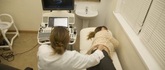
Another effective method to detect this disease is an ultrasound of the problem area, which will clearly show an enlargement of the renal pelvis, precipitation of salts in the urine (microlites), and an increased size of the cups. This diagnostic method is necessary to correctly determine the causes of difficulty in passing urine, as well as the affected area (left, right, or both kidneys).
Instrumental diagnostics are quite effective in the following ways:
- Urography is the injection of a contrast agent into large blood vessels, with the help of which the functioning of the kidneys and urinary tract is monitored.
- Angiography is a procedure similar to urography. The only difference is that the substance is injected directly into the renal artery without involving other blood vessels.
- Retrograde pyelography - a contrast agent is injected into the kidney through a ureteral catheter.
The final stage of examining the patient is to conduct various types of radiation diagnostics, for example, magnetic resonance imaging. This diagnostic method can provide information:
- About the functional activity of organs and the presence of complications and anomalies.
- About the presence or absence of malignant or benign neoplasms.
- About the presence or absence of fistula connections with neighboring organs.
What can be an obstacle to the outflow of urine?
Often, an obstacle to the outflow of urine from the kidney is a narrowing of the ureter at the junction of the pelvis and the ureter, or when the ureter flows into the bladder. Narrowing of the ureter may be a consequence of its underdevelopment or compression from the outside by an additional formation (vessel, adhesions, tumor). Less commonly, the cause of impaired urine outflow from the pelvis is the formation of a valve in the area of the ureteropelvic junction (high ureteric outlet). Increased pressure in the bladder, resulting from a disruption of the nerve supply to the bladder (neurogenic bladder) or from the formation of a urethral valve, can also impede the flow of urine from the renal pelvis.
Types of disease
Highlight:
- Unilateral hydronephrosis
- Bilateral hydronephrosis
In the first case, the pyelocaliceal complex is affected only on one side, in the second - on both sides. Unilateral damage usually occurs against the background of congenital narrowing, conflict with abnormal renal vessels, as well as urolithiasis. The cause of a bilateral process can be stones, tumors or prostate adenoma.
The doctor knows how to treat kidney hydronephrosis at one stage or another. It is only important for the patient to contact a specialist as soon as possible.
What diagnostic methods are used for pyeelectasis in a newborn?
For mild pyelectasis, it may be sufficient to conduct regular ultrasound examinations (ultrasounds) every three months. If a urinary infection occurs or the degree of pyelectasia increases, a complete urological examination is indicated, including radiological research methods: cystography, excretory (intravenous) urography, radioisotope study of the kidneys. These methods allow you to establish a diagnosis - determine the level, degree and cause of the disturbance in the outflow of urine, as well as prescribe reasonable treatment. The research results themselves do not constitute a verdict that unambiguously determines the fate of the child. The decision to manage the patient is made by an experienced urologist, usually based on observation of the child, analysis of the causes and severity of the disease.
Treatment of the disease
Depending on the results obtained during a comprehensive diagnosis, treatment may be conservative or require surgical intervention.
Regardless of the location and presence of complications, surgical treatment is preferable and most effective.
When carrying out conservative treatment, the patient is prescribed drugs that eliminate the root cause of the disease, painkillers and antibiotics.
Surgery can be performed in one of the following ways:
- Endoscopy. In this case, the operation is performed without an incision. Surgical intervention takes place within one, or less often two, punctures.
- Strip. This operation is used in the presence of contraindications to endoscopy, as well as in the presence of tumors located at a considerable distance from each other.
An important stage in treating the disease is following a diet and avoiding certain foods, such as:
- Salty and fatty foods.
- Alcoholic drinks and carbonated drinks with a large number of additives and colorings.
- Hot and spicy foods, smoked meats and marinades.
In severe cases of the disease, a more strict diet may be prescribed with a strict caloric restriction (3500 kcal) due to an increase in carbohydrates and proteins, and the complete exclusion of fats.
In the absence of contraindications, treatment can be supplemented by the use of medicinal plants, such as:
- Burdock.
- Bearberry.
- Rose hip.
- St. John's wort.
- Other plants.
How to cure mastitis in women with folk remedies, and is it worth self-medicating?
The following publication will reveal the signs and causes of uterine endometriosis.
About the symptoms and treatment of parametritis: https://venerolog-ginekolog.ru/gynecology/diseases/parametrit-ego-priznaki-kak-lechit.html.
What diagnoses are made based on the examination?
Some examples of common diseases accompanied by pyelectasis:
- Hydronephrosis caused by an obstruction (obstruction) in the area of the ureteropelvic junction. It manifests itself as a sharp dilation of the pelvis without dilatation of the ureter.
- Vesicoureteral reflux is the backflow of urine from the bladder into the kidney. It manifests itself as significant changes in the size of the pelvis during ultrasound examinations and even during one examination.
- Megaureter - a sharp dilation of the ureter can accompany pyelectasis. Causes: severe vesicoureteral reflux, narrowing of the ureter in the lower section, high pressure in the bladder, etc.
- Posterior urethral valves in boys . Ultrasound reveals bilateral pyelectasis and dilatation of the ureters.
- Ectopic ureter - The flow of the ureter not into the bladder, but into the urethra in boys or the vagina in girls. Often occurs with double kidneys and is accompanied by pyeelectasis of the upper segment of the double kidney
- Ureterocele - the ureter, when it enters the bladder, is inflated in the form of a bubble, and its outlet is narrowed. Ultrasound reveals an additional cavity in the lumen of the bladder and often pyelectasis on the same side.
Does renal calicoectasia affect reproductive function?
The presence of this disease does not in any way affect a woman’s ability to become pregnant. But, during pregnancy, problems may arise associated with overload of the kidneys due to the release of a large amount of nitrogenous compounds by the fetus and compression of the urinary tract by the female reproductive organs, which leads to complications caused by too much undischarged urine.
To ensure that the kidneys function well, special preventive exercises will help. About them in the video:
Consequences
In the absence of timely diagnosis and treatment, caliectasis can provoke very serious complications. One of the dangerous conditions is considered to be a bacterial infection, which can cause death. In addition, with this pathology, the kidney loses its functions, shrinks, and its parenchyma is replaced by connective tissue.
With calicoectasia, stagnation of urine occurs, increasing the risk of stone formation and the development of urolithiasis. Hydronephrosis or renal failure can also be a consequence of the disease. In order to reduce the risk of complications, it is important to diagnose the disease in time and take all necessary measures to treat it.
Stages
There are 3 stages of renal hydronephrosis:
- Initial.
At this stage, the pelvis gradually expands. Kidney function is not affected - Stage of pronounced manifestations of the disease.
At this stage, the pelvis and calyces are involved in the pathological process, the functioning of the kidneys is disrupted due to compression of their tissues - Terminal stage.
It is characterized by atrophy of kidney tissue. Both chronic renal failure and complete loss of kidney functionality may occur.
The doctor knows how to treat kidney hydronephrosis at one stage or another. It is only important for the patient to contact a specialist as soon as possible. With early diagnosis, the chances of a full recovery are the highest!
Impact with traditional methods
Let's consider the methods offered by traditional medicine:
- We take equal amounts of ginseng and eleutherococcus roots, fill them with forty-proof alcohol and let them brew in a dark, dry room for several weeks.
- Sick people need to drink milk until the swelling disappears.
- A good remedy is stewed pumpkin.
- Watermelon, red currant and raspberry juice have a beneficial effect on the organ.
- We take dried rosehip and remove ten shavings from it, the length of which is ten centimeters. Fill with a liter of liquid and leave overnight. In the morning, strain the broth and consume throughout the day. The course is a month.
- Take blackcurrant leaves and pour 500 ml of boiling water over them. After fifteen minutes, the broth should be strained and the leaves should be squeezed out. Place the resulting broth on the fire until it boils. Add two tablespoons of dried or fresh blackcurrants. This course should be carried out four times a day: drink the liquid and eat the remaining fruits.
- We take knotweed, parsley, birch buds, cranberry fruits, carrot seeds and rose hips in equal parts and chop them. Pour the resulting powder into 250 ml of water. After an hour you need to strain. This decoction should be consumed three times a day before meals. The course is three months.
Do not self-medicate under any circumstances, so as not to aggravate the situation. Before using folk remedies, be sure to consult a specialist. Observation by a nephrologist or urologist, as well as conservative or surgical treatment for patients with renal caliectasis are necessary throughout their lives.
Main symptoms
Most patients complain of the following manifestations of hydronephrosis:
- severe pain in the lower back and abdomen, which can radiate to other parts of the body
- increase in body temperature
- weakness
- chills
- the appearance of blood clots in the urine
- headaches and increased blood pressure
- there may be a completely asymptomatic course when the pathology is present from birth and the body has adapted to it
The more advanced the disease, the more pronounced its symptoms become.
The more advanced the disease, the more pronounced its symptoms become.
Important! Body temperature rises when the disease is accompanied by an infection. In this case, it is very important to carry out immediate treatment.
Symptoms of hydronephrosis may also include bloating, constant nausea and severe vomiting, increased fatigue with decreased performance.
In children, signs of the disease include:
- cloudy urine
- reducing the amount of daily urine
- pain
Like adults, children complain of weakness, headaches, and nausea. Parents should pay close attention to the child’s health if he refuses his favorite food, games, walks, becomes lethargic and sleeps a lot.
Competent treatment of kidney hydronephrosis in an adult or child is impossible without a high-quality comprehensive examination.
Advantages of contacting MEDSI
- Experienced specialists.
Our doctors have all the skills and knowledge to treat renal hydronephrosis in adults and children. Specialists work in accordance with international standards and use organ-preserving techniques - Opportunities for comprehensive diagnostics.
Before starting treatment for kidney hydronephrosis in adults and children, an examination is carried out to assess the cause of symptoms, determine the degree of damage and other features of the course of the disease. We have modern expert-class equipment. It allows you to detect pathologies in the early stages - Individual therapy methods.
They are selected in accordance with the results of the patient’s examination, after analyzing laboratory and instrumental data - Gentle methods of surgical treatment.
The therapy uses the capabilities of endoscopy, microsurgery and laparoscopy. If possible, operations are performed through small punctures, including using robotic technologies - Modern hospital.
We have high-tech operating rooms with innovative equipment from leading manufacturers. In hospitals, patients are provided with balanced nutrition and round-the-clock care - Fast recovery after interventions.
Particular attention is paid to the new philosophy of patient management after surgery (fast track), which involves the fastest possible return to active life. - Comfort of visiting clinics.
They are located near the metro. This allows residents of any district of Moscow to visit MEDSI clinics. We made sure there are no queues so you don't have to wait long for an appointment
To undergo treatment for renal hydronephrosis in adults and children in our clinics, call (495) 7-800-500. Our specialist will answer all questions and offer a convenient time for consultation with a doctor. You can also make an appointment through the SmartMed app.
Specialized centers
- Standards of the International Association of Urology
- High-precision diagnostics using cutting-edge equipment
- Modern high-tech operating rooms and hospitals
More details


