Author of the material
Professor Kapranov S.A. — Doctor of Medical Sciences, twice Laureate of State Prizes of the Government of the Russian Federation in the field of science and technology, Laureate of the Lenin Komsomol Prize, author of more than 350 scientific works on medicine, 7 monographs, and 10 patents for inventions in medicine, with more than 30 years of personal experience 10,000 different endovascular operations
At the Center for Endovascular Surgery of Professor Kapranov, controlled embolization for varicose veins is performed. For intervention, special detachable spirals are used. Thanks to them, the dilation of the veins of the esophagus is eliminated in the shortest possible time. You will be able to independently choose a clinic for the operation.
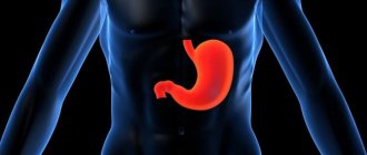
Varicose veins of the esophagus: features of pathology
Varicose veins of the esophagus are a rather dangerous pathology. It represents a deformation of blood vessels. As a result of the development of the disease, the lumen of the veins increases, their walls stand out and nodes form. Deformed vessels become very tortuous and quite loose. The mucous membrane, which is located above the vessels, is subject to damage and inflammation.
The disease is also dangerous due to the absence of symptoms at an early stage of development. In the initial stages, varicose veins do not show any signs. For this reason, a person has no idea that pathological processes are occurring in his body. Meanwhile, varicose veins are very dangerous!
With the development of varicose veins, the patient complains of:
- belching and heartburn,
- from time to time difficulties when swallowing food (gradually discomfort becomes a constant companion),
- increased heart rate,
- discomfort and heaviness in the chest area.
Esophagitis is often associated with varicose veins of the esophagus. This process is inflammatory.
Bleeding becomes a dangerous complication of this pathology. The result is weakness and shortness of breath. General health deteriorates. The patient may suffer from weight loss.
Indications for the procedure
Ligation is carried out as primary and secondary prevention of bleeding in esophageal varices. Despite the fact that in medicine there are still no uniform criteria for prescribing the procedure, specialists tend to use it when the following signs appear:
- progression of esophageal varicose veins, in which regular examinations reveal a steady increase in the diameter of the affected veins;
- the appearance of venules in the esophagus - darkly colored stripes or spots in the area where varicose nodes are located, which indicate the onset of the ischemic process;
- a progressive underlying disease against which esophageal varices developed - liver cirrhosis, portal hypertension;
- pronounced bulging of veins into the lumen of the esophagus, in which there is a high probability of mechanical damage to varicose nodes.
Ligation is mandatory for patients who have previously experienced internal bleeding from varicose veins in the esophagus. As a preventive measure, the procedure is carried out when the diameter of the vessels increases to 5 mm or more.
Prophylactic ligation can be performed before varices in the esophagus reach critical sizes. A signal for surgery can be a rapid change in the veins connecting the abdominal vein with the vessels of the esophagus.
How is the disease diagnosed?
Esophageal varicose veins are diagnosed by:
- Studying anamnesis.
- Medical examination. During it, you can note such signs of varicose veins as swelling, changes in the color of the skin and mucous membranes.
- Laboratory research. To study varicose pathology, the patient undergoes general and biochemical blood tests. Specialists count the number of platelets, examine liver enzymes, serum iron, protein, and lipid spectrum.
- Instrumental research. If varicose veins of the esophagus are suspected, ultrasound, esophagoscopy and x-ray diagnostics are performed. These studies are aimed at studying a specific area of the esophagus. The remaining organs of the peritoneum are also studied.
How is esophageal varicose veins treated?
The main goal of therapy for esophageal varices is to prevent bleeding. In some cases, specialists have to eliminate the consequences of bleeding that has already been suffered.
Vein enlargement is treated by prescribing:
- Vitamins, antacids and astringents.
- Hemostatic agents.
- Drugs that constrict blood vessels.
In some cases, special colloidal solutions are introduced. Transfusions of plasma, blood and red blood cells are also carried out.
Surgeries on the esophagus are resorted to when drug therapy does not give the desired result.
All interventions are divided into 2 groups:
- Endoscopic.
- Surgical.
Endoscopic interventions on the esophagus are carried out by:
- Electrocoagulation. With this intervention, damaged vein tissue is effectively removed under the influence of electric current.
- Doping of the esophageal veins. This technique boils down to careful ligation of the veins of the esophagus. To carry out the intervention, specialists use small rings. 1, 2 or 3 rings are placed on each vein. Alloying is also possible using nylon loops.
- Bandaging. This technique is used to fix dilated veins. Thanks to this, you can quickly stop bleeding that has already begun due to vasodilation.
- Insertion of a probe that compresses the veins. Vein probes are made of rubber. They apply pressure to the bleeding vessel.
- Application of a special adhesive film or a substance such as thrombin to the affected areas of the veins.
Surgical operations to prevent bleeding as a result of dilation of the veins of the esophagus are carried out by:
- Sclerosis. A special solution is injected into the dilated veins. The solution is supplied by injection. They are injected into the lumen of the veins of the esophagus. The technique is quite effective. There is one significant drawback. The procedure must be carried out constantly and regularly. Typically, sclerotherapy is repeated every 5 days. Then the procedure is carried out once a month. Subsequently, sclerotherapy is carried out once every 3 months. At least 4-5 procedures must be performed per year. Only in this case can the method be sufficiently effective.
- Portosystemic stent shunting. This technique for eliminating varicose pathologies involves introducing a special stent into the middle part of the liver. It connects the functionality of the hepatic and portal veins.
- Anostomosis. The objects of intervention are the splenic vein and the left kidney.
- Vessel lining.
- Removal of esophageal veins that cannot be restored.
All operations on the veins of the esophagus are quite dangerous. In addition, varicose pathologies cannot be completely eliminated with their help. Relapses often occur, and operations provoke the risk of a number of complications.
That is why varicose pathologies are now being fought using another method. It is innovative and allows you to forget about the problem!
Embolization for dilated veins is popular for good reason!
Material and methods
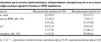
Since 2015, 48 patients with ongoing bleeding from the esophageal varices have been admitted to our clinic. In 23 such patients, we performed endoscopic ligation of the esophageal varices of the esophagus with ongoing bleeding from the esophagus, which served as the basis for assessing the results of the treatment.
Endoscopic ligation of esophageal varices was performed in patients with ongoing bleeding with liver cirrhosis of various etiologies, with functional reserve of Child-Pugh classes A, B and C, with concomitant complications of the disease (sequestration thrombocytopenia of varying severity, hepatic encephalopathy, hypoalbuminemia, posthemorrhagic anemia), and also with various forms of portal hypertension. We strictly adhered to the treatment algorithm for acute bleeding from the esophageal varices in accordance with the National Clinical Guidelines [5].
The criteria for inclusion of patients in this study were: grade III esophageal varices (but not more than 10 mm in diameter), ongoing bleeding from esophageal varices, clear visualization of the source of bleeding, and the absence of contraindications to surgical intervention. The exclusion criteria were: gastric varices of types II, III and IV and bleeding from them, the patient’s refusal to perform this intervention.
All patients with a clinical picture of ongoing bleeding from the esophagus (vomiting blood, weakness, pallor of the skin, melena) were hospitalized in the intensive care unit for intensive care and hemodynamic stabilization (vasoactive drugs, non-selective beta-blockers, transfusion of fresh frozen plasma, red blood cells were prescribed for necessary, as well as antibiotic therapy as prevention of spontaneous bacterial peritonitis). After hemodynamic stabilization, EGD was used to identify the location of the source of bleeding to determine further treatment tactics. In 25 patients in whom the source of bleeding was gastric varices or the varices could not be visualized due to poor surgical visibility, a Blackmore obturator probe was installed and endoscopic ligation of esophageal varices was performed in the “cold” period as secondary prevention of recurrent bleeding. In 23 patients, endoscopic ligation was performed for ongoing bleeding from the esophageal esophagus with successful visualization of the source of bleeding.
The average age of patients (23 patients; 100%) operated on during the acute period of bleeding was 56 years (from 28 to 77 years). The etiology of portal hypertension and, as a consequence, varicose veins of the esophagus included: liver cirrhosis of HCV etiology (14 cases; 60.9%), portal vein thrombosis (5 cases; 21.7%), liver cirrhosis of toxic etiology (4 cases; 17. 4%).
According to the functional class of liver cirrhosis, patients were distributed as follows: with compensated liver cirrhosis (functional class A according to Child-Pugh) - 1 (4.3%) patient, with subcompensated liver cirrhosis (functional class B according to Child-Pugh) - 8 ( 34.8%) patients with decompensated liver cirrhosis (functional class C according to Child-Pugh) - 14 (60.9%) patients. The MELD index (Model of End Stage Liver Diseases—a scale for predicting the mortality index in patients with cirrhosis of the liver) ranged from 8 to 23.
Sequestration thrombocytopenia of varying severity was detected in 23 (100%) patients. Posthemorrhagic anemia during hospitalization was noted in 23 (100%) patients, of which 3 (13.0%) had mild severity, 16 (69.6%) had moderate severity, and 4 (17.4%) - severe anemia.
In order to determine the degree of esophageal varicose veins, we used A.G.’s three-grade classification. Scherzinger (1986). Endoscopic ligation was performed in patients with grade III esophageal varices (from 5 to 10 mm in diameter).
Endoscopic ligation with ongoing bleeding was performed in the intensive care unit under intravenous anesthesia to avoid excessive regurgitation of blood and a high risk of rupture of varicose veins, as well as for technical convenience during manipulation.
This intervention was performed using a multi-charged endoscopic ligator (). A set of 10 latex rings was used.
Surgical technique for ligation of esophageal varices with ongoing bleeding
After performing endoscopy and visualizing the source of bleeding, the endoscopic ligator and endoscope were immediately assembled into a single structure, and the operation began. The intervention was carried out in the intensive care unit in compliance with all surgical rules; the patient was placed in a position on the left side.
After bringing the endoscope to the source of bleeding (most often it was located just above the dentate line), aspiration of this varicose node into the cap began at more than half its height and the first ligature was dropped onto it by turning the screw-handle clockwise. In this case, the endoscope itself was positioned “at 12 o’clock” in relation to the selected varicose vein.
Next, endoscopic ligation of the esophageal varices was continued in a spiral pattern in a checkerboard pattern, dropping 1 latex ring onto 1 varicose trunk, without affecting the upper third of the esophagus. In this case, a total of 4 or 5 latex rings were used.
Key features of the intervention!
The advantages of vein embolization include:
- High speed of intervention. The operation takes 30-60 minutes. Complex procedures may take longer.
- No significant discomfort. Pain and any discomfort during vein embolization are relieved with special modern medications.
- Speed of recovery after intervention. The patient will have to stay in the clinic for only 1-2 days. Note! In some cases, the length of hospitalization after vein embolization increases. In any case, the patient can return to normal life in the near future.
What recommendations do doctors give to patients?
- Strengthening the drinking regime. In the first 7 days after surgery, you need to drink plenty of clean water.
- Elimination of water procedures for 3-5 days. After this, you can take both a shower and a bath.
- Physical peace. It is usually prescribed for 2-3 weeks. Patients are prohibited from lifting heavy objects or playing sports. Bed rest is not prescribed for this period. Slow walks are even beneficial.
What lifestyle should I lead after embolization of esophageal vessels?
Patients should:
- Follow a specially prescribed diet that includes a large number of foods that help reduce total cholesterol levels.
- Avoid heavy exertion.
- Take various medications. They will be prescribed by your attending physician.
- Eat food often enough, but in as small portions as possible. The daily dose is best divided into 5-6 doses. Eating food is undesirable 2-3 hours before bedtime.
- Give preference to boiled dishes. Food can also be steamed. This way it retains a lot of useful substances, is easily digestible and does not become a source of additional harmful cholesterol.
It is forbidden to eat too hot or cold food. This can damage the esophagus and its veins.
The main advantages of the vein embolization method used in the center of Professor Kapranov
Endovascular embolization in our center is a low-traumatic procedure. It is fundamentally different from standard surgical interventions in the absence of large incisions in the vein and tissue. In addition, embolization does not require the patient to be put under general anesthesia. This reduces numerous risks and makes intervention possible in the presence of general contraindications to standard surgery.
The advantages of carrying out the intervention at the center of Professor Kapranov also include:
- Experience of professionals. All doctors performed a lot of interventions to restore the functionality of the esophagus. They are ready to help you too.
- Comfortable conditions of stay in any clinic. Embolization of the esophageal vessels will not cause pain or severe discomfort.
- Reducing the likelihood of relapse. The technique used to block the vessel allows you to quickly and reliably stop the blood flow of the vein (vessel) of the esophagus.
- No discomfort when restoring the functionality of the esophagus. During vein embolization, the patient does not experience pain. All unpleasant sensations are eliminated with the help of special medications.
- Short recovery period after intervention for varicose veins of the esophagus. For minor vascular interventions, the patient may be discharged on the day of the procedure. In this case, special care, complex treatment, and regular dressings are not required.
- Minimal number of contraindications and complications. The success of an operation for embolization of esophageal vessels largely depends on the professionalism of the doctor. That is why it is important to choose a surgeon wisely.
- Opportunities for organ preservation. Esophageal embolization, for example, avoids a number of serious problems.
Endoscopic ligation of esophageal varices
The last decade has been marked by an active search for an alternative endoscopic method that would be as effective as injection sclerotherapy, but would be simpler to perform and lead to a lower percentage of complications. The search led to the invention of the endoscopic ligation method. The method was first developed in the 70s for the treatment of internal hemorrhoids, but was not widely used. The idea of using a similar technique to stop bleeding from a varicose vein and achieve temporary hemostasis belongs to Stiegmann et al., who proposed it in 1986. use a device of original design, which is basically still used today.
A standard set of instruments consists of a cylinder with rubber bands attached to it, which is attached to the front end of the endoscope. The cylinder is connected through the biopsy channel of the endoscope to a handle, which directly carries out the alternate release of the fixed elastic bands.
The manipulation technique is that the varicose node is sucked into a cylinder located at the end of the endoscope, then an elastic band is released using a handle, which is thrown onto the neck of the varicose node.
The usual ligation technique is that the varicose veins should be treated as low as possible to the cardia. The procedure is performed in several sessions, and the number of ligated nodes in one session is not limited; cases of ligation of 17 nodes at once in one session are described]. The effect of ligation on tissue has been studied quite well]: on the second day after the procedure there are no changes in the endoscopic picture; in the period from 4 to 7 days, the node becomes necrotic and is rejected, forming a superficial ulcer, which usually epithelializes by the end of the third week. At the histological level, by the 1st day a polypoid formation with ischemic necrosis is determined, which affects only the mucous and submucosal layers; on the 4th - 7th day a superficial ulcer with granulation tissue and an active inflammatory reaction around is determined; complete epithelization occurs by 21-28 days, and by 50-60 days the submucosal layer is mixed with scar tissue, leaving the muscle layer intact.
Indications for use
Currently, the main indications for using the endoscopic ligation method are:
- Stopping ongoing bleeding from the urinary tract
- Prevention of recurrent bleeding from varicose veins
- Primary prevention of bleeding from varicose veins
Contraindications and complications
Most researchers note that the risk of developing systemic complications with endoscopic ligation of varicose veins is practically absent, since the method is mechanical and is not associated with the introduction of any chemicals into the vascular bed. Some authors describe complications associated with changes in hemodynamics in the portal vein (formation of varicose veins in the fundus of the stomach), or local complications in the form of isolated cases of transient episodes of dysphagia that were self-limited]. The described rare episodes of bleeding from ulcers formed after rejection of a necrotic node, as a rule, stopped on their own, were not profuse and did not pose a threat to the patient’s life.
results
The immediate and long-term results of using the method of endoscopic ligation of varicose veins are quite encouraging. Thus, many researchers have noted a higher efficiency of the endoscopic ligation method in comparison with the injection sclerotherapy method, which manifested itself in a reduction in the number of recurrent bleedings by an average of 20% and a reduction in mortality by an average of 10-15%. It should be emphasized that to achieve complete elimination of varicose veins using this method requires 2 - 3 sessions less than with injection sclerotherapy, the percentage of eradication of varicose veins with ligation (70 - 80%) is higher than with injection sclerotherapy (40 - 60% ). Complications such as aspiration pneumonia, perforation and strictures of the esophagus are practically absent. Regular endoscopic monitoring in the long term makes it easy to perform repeated ligations of newly appeared veins.
What determines the cost of esophageal vein embolization?
The final cost of embolization of esophageal vessels is largely determined by factors such as:
- Comfort of hospitalization and its speed.
- Convenience of examination before intervention.
- Speed of diagnosis and determination of an accurate diagnosis.
Thus, the cost of the procedure depends on a number of factors that are not directly related to the quality of medical services. Thanks to this, even at relatively low costs, you can count on receiving qualified professional support.
Approximate prices are presented on our center's website.
Any questions?
Call the clinics where esophageal treatment is carried out by Professor Kapranov and ask your questions.
You can also contact a professional using his personal phone numbers:
- +,
- +.
You can discuss all the features of treatment of the esophagus with Professor Kapranov. You will be able to discuss all the details of the intervention and schedule the first appointment and examination. The operation can be carried out in the near future.
List of used literature:
- Angiography and transcatheter endovascular interventions for bleeding from esophageal varices. All-Union Conference “Radiation diagnostics and X-ray diagnostics of liver and kidney diseases”, Leningrad, 1984, pp. 22-24 (co-authors V.I. Prokubovsky, M.N. Ovchininsky, S.A. Kapranov).
- Possibilities and prospects for the development of X-ray endovascular interventions for bleeding from esophageal varices. VII All-Union Symposium “X-ray Endovascular Surgery”, 1985, pp. 133-134 (co-authors M.N. Ovchininsky, V.A. Cherkasov, S.A. Kapranov, S.B. Bakhvalov).
- Methods and tactics of endovascular hemostasis for profuse bleeding from esophageal varices. Republican conference of surgeons “Surgical treatment of portal hypertension, diseases and injuries of the liver”, Kharkov, 1986, pp. 60-62 (co-authors V.I. Prokubovsky, M.N. Ovchininsky, V.A. Cherkasov, S.A. Kapranov ).
- Ways to increase the effectiveness of endovascular interventions for bleeding from esophageal varices. Republican scientific and practical conference “X-ray contrast research methods and endovascular surgery”, Alma-Ata, 1986, pp. 167-169 (co-authors V.A. Cherkasov, M.N. Ovchininsky, S.A. Kapranov).
- A method for complex embolization of varicose veins of the esophagus and stomach. Rationalization proposal of industry significance 0-2976 dated October 2, 1987 (co-authors M.N. Ovchininsky, S.A. Kapranov, A.S. Belenky).
Karmanov
Author: Kapranov S.A.
Doctor of Medical Sciences, Professor, twice Laureate of State Prizes of the Government of the Russian Federation in the field of science and technology, Laureate of the Lenin Komsomol Prize, author of more than 350 scientific papers on medicine, 7 monographs, and 10 patents for inventions in medicine
Prevention of complications
During the first week after ligation of the esophageal veins, the nodes will die. At this stage, the patient may experience a sensation of a foreign object in the throat and nausea. During this period, it is important to exclude the possibility of injury to the mucous membranes of the esophageal tube with food: if part of the vein, tightened by a latex ring, detaches before scar tissue forms, internal bleeding will open.
Nutrition
The first step in preventing complications after ligation is a diet that prescribes:
- eating slightly warm food and completely avoiding hot foods and drinks;
- eating small portions up to 5-7 times a day;
- thorough grinding of food - dishes should not contain solid particles;
- exclusion from the diet of spices, seasonings, alcohol, which irritate the mucous membranes of the esophagus.
The nutritional and energy value of food must satisfy the body's needs. The diet includes enough proteins (meat, eggs, fish, dairy products, legumes), fats (cream, vegetable and butter), carbohydrates (cereals, vegetables). On the first day, it is advisable to eat liquid food:
- clear broths and decoctions;
- liquid mashed porridges;
- fermented milk drinks.
From the second day you can include cream soup, pureed boiled vegetables and pureed fresh fruits and berries in the menu. On the 3-4th day, you can introduce into the diet a soufflé of lean meat, fish cutlets, meatballs with a side dish of pureed porridge, vegetable purees. Every day you need to drink at least 2 liters of liquid - water, herbal tea, juices diluted with water, fermented milk drinks or milk.
Until the wounds heal at the site of the rejected nodules, it is not recommended to take alcohol, even if it is part of the medication.
Other preventive measures
To prevent infection of wounds, maintaining oral hygiene will be sufficient. It is recommended to brush your teeth at least 2 times a day. After each meal, you should rinse your mouth with water. You cannot use rinses from the pharmacy - even a small amount of the product that gets into the esophagus can cause irritation of the mucous membrane.
Since the risk of bleeding remains elevated for 2 weeks after ligation, the patient's condition must be carefully monitored throughout this period. Weakness, fever, vomiting streaked with blood or completely colored vomit should be a reason to urgently consult a doctor. Visible traces of blood in the stool should also alert you.
To prevent re-development of varicose veins in the esophagus, it is recommended to pay attention to the treatment of the underlying disease against which the disease developed. Giving up bad habits and moderate physical activity will help reduce the likelihood of relapse. Swimming, measured walks in the fresh air, and yoga are ideal. You should forget about the loads associated with jumping, sudden changes in body position, bending and lifting weights.
Regular examinations by a gastroenterologist and phlebologist, systematic instrumental studies (EGD and others) will help to detect residual problems with the veins of the esophagus in a timely manner and avoid possible complications. Strict adherence to the recommendations of your doctor, taking medications and following a diet will help maintain quality of life and extend the relapse-free period to 5-7 years.