Barrett's esophagus - what is it?
The process of replacing one epithelium with another is metaplasia.
It is always preceded by dysplasia - a change in the structure, functions and shape of organ cells. According to statistics, men over 45 years of age are most likely to experience Barrett's esophagus disease. In medicine, there are two degrees of severity of esophageal cell dysplasia:
- moderate (minor changes in the structure of organ cells);
- severe (high risk of developing Barrett's esophagus).
In turn, cylindrical metaplasia can also occur in different scenarios - the epithelium of the esophagus is capable of being replaced:
- cardiac gastric;
- fundic gastric;
- specialized intestinal epithelium with goblet cells.
The latter option is considered the most dangerous, as it has the greatest malignant potential and often causes esophageal cancer.
Causes of Barrett's esophagus
Barrett's esophagus disease occurs for the following reasons:
- Genetic predisposition (the problem is inherited).
- Duodenogastric reflux disease (a condition in which the contents of the duodenum reflux into the stomach and damage its walls).
- Smoking, drinking alcoholic beverages in large quantities.
- Abdominal obesity (fat is deposited in the abdominal area).
- Double reflux (a combination of gastroesophageal reflux disease and duodenogastric reflux disease).
- Work activity associated with constant bending (more than 20 years).
- Overeating, excessive consumption of spicy and fried foods.
- Peptic ulcer of the stomach and duodenum.
- Diseases of the operated stomach (occur after gastric surgery).
- Gastroesophageal reflux disease (stomach contents enter the esophagus and irritate its walls).
- Zollinger-Elisson syndrome (a disease caused by a tumor of the pancreas that secretes gastrin).
Factors predisposing to the development of Barrett's esophagus include:
- long-term use of certain medications (Baralgin, No-spa, analgesics);
- damage to the vagus nerve;
- diabetes;
- consumption of caffeine-containing products;
- excessive consumption of flour products, fatty and spicy foods;
- bloating.
Esophageal spasm
The clinical picture of esophageal spasm depends on the location and form of the pathological process. The most typical signs of any form of the disease are chest pain and difficulty swallowing. Patients most often associate pain with swallowing food and saliva, although it can also occur spontaneously. Stress leads to worsening pain. Pain may radiate to the shoulder blades, shoulders, lower jaw, and back. Most often, the attack lasts no more than an hour, although a longer duration is not excluded. Typically, patients describe their sensations as a feeling of pressure behind the sternum. While taking antispasmodics, the pain weakens or disappears.
Dysphagia can develop while eating both solid and liquid foods. Most often it is not constant and occurs simultaneously with pain. Heartburn bothers every fifth patient, and regurgitation of food is observed only against the background of very strong spasms or a significant accumulation of food masses in the esophagus.
Spasms of the upper narrowing of the esophagus (the most common form of pathology) most often occur in patients prone to hysteria, neuroses, and mood swings. Clinically manifested by pressing pain behind the sternum, nausea, cough, redness of the face, fear and excitement. Esophageal spasm can occur acutely, or its manifestations increase gradually, interspersed with periods of remission. The intermittent nature of the pathology leads to the patient becoming restless, eating irregularly, and fearing a return of symptoms, and this further aggravates the pathological manifestations of esophageal spasm.
Chronic spasm in the area of the upper narrowing most often develops in elderly patients with defects in the dentition, chewing disorders, and a tendency to swallow large unchewed pieces of food; especially if there is a history of acute esophageal spasm. The clinical picture is dominated by unpleasant sensations in the chest, difficulty passing solid food, and the need to wash down every sip with water. Permanent obstruction of the esophagus leads to the formation of its compensatory expansion over the spasmodic area.
Spasm of the lower parts of the esophagus and cardia can also be acute and chronic. Acute spasm is manifested by pain in the epigastrium and behind the sternum, a feeling of retention of food masses above the stomach; Washing down food with water does not bring relief. Non-sphincteric spasm of the esophagus (Barshon-Teschendorff syndrome), in which several parts of the organ are simultaneously affected along its entire length, is considered separately. Patients complain of occasional episodes of dysphagia (from a couple of minutes to several weeks), accompanied by chest pain and regurgitation of mucus. Pathology often occurs against the background of peptic ulcer, cholelithiasis; it is characterized by an increase in appetite.
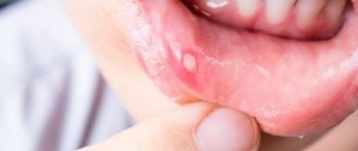
Symptoms of Barrett's Esophagus Disease
The disease has no specific symptoms. Very often it is generally asymptomatic (especially in elderly patients). Doctors include the main clinical signs:
- sore throat that appears half an hour after eating and gets worse in a horizontal position when bending over;
- heartburn resulting from overeating, after eating smoked, fried and spicy foods, strong tea, coffee, soda;
- sour belching that occurs after eating;
- regurgitation (reflux of semi-digested food from the stomach into the esophagus);
- dysphagia (impaired swallowing function).
May also be present:
- abdominal pain that gets worse after eating;
- nausea that occurs from time to time and goes away on its own;
- single vomiting after eating;
- tooth erosion, thinning of enamel.
If you notice similar symptoms, consult a doctor immediately. It is easier to prevent a disease than to deal with the consequences.
Diagnosis of Barrett's esophagus
Diff. Diagnosis of Barrett's esophagus involves various medical procedures. First, the doctor listens to the patient’s complaints and finds out how long ago heartburn began to bother him, whether the discomfort that arises is related to food intake, and what other complaints there are related to the gastrointestinal tract (pain, diarrhea, vomiting, nausea).
Then may follow:
- Analysis of the patient’s life history, during which it is determined whether there are other diseases associated with the gastrointestinal tract (gastroesophageal reflux disease, pancreatitis, cholecystitis, gastritis). It is being clarified whether anyone in the family had Barrett's disease.
- Palpation of the abdomen (most often the patient feels discomfort when pressing in the area above the navel).
- Laboratory tests (complete blood count, urinalysis, blood biochemistry, stool occult blood test).
- Stomach acid production test. If it is found that the acidity of the stomach is below 2, the doctor can assume the pathological nature of heartburn.
- Contrast X-ray examination of the esophagus to study the structure and patency of the organ.
- Esophagogastroduodenoscopy. The doctor examines the condition of the inner surface of the esophagus, duodenum and stomach using an endoscope. In this case, a fragment of an organ is taken (biopsy) to determine the structure of its cells and tissues. Also, during esophagogastroduodenoscopy, chromoscopy is performed - to identify small and hidden damage to the mucous membrane of the esophagus and stomach, their tissues are stained.
- Esophageal manometry. Allows you to examine the motor function of the esophagus and the condition of the sphincters.
- Impedance pH-metry. The alternating resistance current is measured between electrodes inserted into the esophagus. As a result, the acidity of the medium is determined.
- Ultrasound of the abdominal organs. Makes it possible to exclude the presence of a tumor of the pancreas, duodenum and stomach wall.
- Collection of stomach material and a breath test for Helicobacter pylori (bacteria that damage the walls of the duodenum and stomach).
Full diagnostics and treatment can be completed in any modern gastroenterology clinic
.
Renev E.N. Modern treatment of gastroesophageal reflux disease.
Modern treatment of gastroesophageal reflux disease
E.N.
Renev We would like to preface the discussion of therapeutic options for
gastroesophageal reflux disease
(
GERD
) with brief information on the mechanisms of development and diagnosis of this pathology. The possibilities of surgical treatment of GERD will not be discussed in this article.
Definition
So, A.S.
Trukhmanov defines GERD
as the occurrence of characteristic symptoms and (or) inflammatory damage to the distal parts of the esophagus due to repeated reflux of gastric contents into the esophagus [1].
According to the definition of the International Working Group, the term " gastroesophageal reflux disease"
» should be applied to all individuals at risk of physical complications of gastroesophageal reflux, or experiencing a significant deterioration in health-related well-being (quality of life), as a result of reflux symptoms, after adequate reassurance of the benign nature of the symptoms [6].
The term “endoscopically negative reflux disease” should be used in individuals who meet the definition of gastroesophageal reflux disease, but who do not have either Barrett's esophagus or visible mucosal defects (erosions or ulcers) on endoscopic examination [6].
Development mechanisms
Without dwelling in detail on the pathogenetic mechanisms of development of this disease, we will only say that it is based on the effect of acid and pepsin on the esophageal mucosa due to the combination (in varying proportions) of pathological reflux of gastric contents into the esophagus with a violation of its clearance. Pathological reflux of contents, in turn, is caused by dysfunction of the lower esophageal sphincter (either as a result of a decrease in its tone or an increase in the frequency of spontaneous relaxation, or due to its anatomical defect, for example, with a hernia of the esophagus). Impaired esophageal clearance may be caused by decreased saliva production or impaired esophageal motility. As a result of all of the above, there is an imbalance between aggressive factors and protective factors, which leads, but not necessarily, to the occurrence of reflux esophagitis.
Epidemiology
According to S.I.
Pimanova, symptoms of GERD
observed in half of the adult population, and the endoscopic picture of esophagitis is observed in 2-10% of examined people [1]. It must be remembered that GERD is not always accompanied by esophagitis. Up to 50-70% of patients with heartburn at the time of seeking medical help have endoscopically negative GERD [8]. The attitude of a number of practitioners towards endoscopically negative GERD as the mildest degree of this disease that does not require intensive drug therapy is fundamentally incorrect. A number of studies have demonstrated that the quality of life in patients with endoscopically positive and negative GERD is impaired to almost the same extent [29]. Studies have shown that endoscopically negative GERD very rarely develops into reflux esophagitis, which in turn rarely progresses to more severe forms over time [7,8].
Diagnostics
Since
the diagnosis of GERD
is widely described in many manuals, we will dwell only on some of its points.
The main symptom of GERD, observed in at least 75% of patients, is heartburn
[6].
There may also be pain or a burning sensation behind the sternum, belching, etc. Most often, GERD symptoms
occur after eating.
Diagnosis of erosive esophagitis is based on endoscopic examination. Barium radiography has a fairly high sensitivity for severe (98.7%) and moderate (81.6%) esophagitis, but low sensitivity (24.6%) for mild esophagitis [12, 22, 23]. Endoscopy with biopsy is the only reliable method for diagnosing Barrett's esophagus. The severity of erosive reflux esophagitis on the endoscopic picture is divided into 4 degrees A, B, C and D (according to the Los Angeles classification).
pH monitoring
is a sensitive and specific diagnostic test and is especially important for identifying
endoscopically negative GERD
. More than 50 episodes of decrease below 4 are considered as a diagnostic criterion for GERD [1]. In a number of patients, a less significant decrease in the pH of the esophagus occurs, but when most episodes of such a decrease coincide with the onset of symptoms, it allows us to speak of a “hypersensitive esophagus.”
Among provocative tests, the Bernstein test plays a certain role (the appearance of typical symptoms after the introduction of a weak solution of hydrochloric acid into the esophagus and their disappearance after the introduction of saline). Determining the pressure of the lower esophageal sphincter is useful when deciding on surgical treatment.
Treatment
Before moving on to consideration of individual aspects
of the treatment of GERD,
it is necessary to emphasize the fact that its main goal is to quickly relieve patients from the symptoms that bother them. The disappearance of symptoms usually correlates well with the healing of mucosal defects in erosive esophagitis [6].
Lifestyle change
Although according to the GERD Working Group, lifestyle factors do not play a determining role in the development of GERD [6], recommendations aimed at eliminating factors that promote reflux or impair esophageal clearance should be made.
Diet.
It is necessary to stop taking reflux-inducing foods (fatty foods, chocolate and excessive amounts of alcohol, onions and garlic, coffee, carbonated drinks, especially various types of colas) and drugs with low pH (orange and pineapple juices, red wine). However, an attempt to sharply limit a patient’s diet (especially a young one) is rarely possible in practice; your recommendations simply will not be followed. It makes more sense to identify which products cause the appearance or exacerbation of symptoms in a given patient and try to at least give them up. The patient should be informed that overeating should be avoided. After eating, it is advisable not to take a horizontal position or work in an inclined position. The last meal should be 3 hours before bedtime.
Weight control.
Losing weight does not always resolve symptoms, but losing weight may reduce the risk of developing a hiatal hernia. However, giving advice to lose weight is much easier than implementing it. Overweight people sometimes try to hide their lack of a waist by over-tightening the waist belt, which leads to increased intra-abdominal pressure and the development of reflux (as does wearing clothes that are too tight).
Smoking
is a contributing factor to GERD as a result of both sphincter relaxation and decreased salivation and should be discontinued accordingly [7]. Although, according to some researchers, smoking cessation has a minimal positive effect on GERD [6].
Raising the head of the bed is important for patients with nocturnal or laryngeal symptoms (which constitute a small proportion of patients with GERD), but its necessity in other cases is questionable.
A number of medications such as antispasmodics, beta blockers, hypnotics and sedatives, nitrates and calcium antagonists can contribute to the development of reflux.
Antacids
Discussing the use of antacids, of which there are a great many in our time (almagel, phosphalugel, maalox, rutacid, etc.), I would like to emphasize that, in our opinion, antacids do not play an independent role in the treatment of GERD and can only be used as a short-term remedy symptom control. The low effectiveness of antacids is based on the short duration of pH control achieved by their use. Data from many authors confirm the minimal effect of antacids (even in combination with lifestyle changes) in reflux esophagitis, although it is superior to the placebo effect [27]. We suggest that patients (being treated for GERD) use antacids as a method of quickly controlling symptoms that occur, usually after a violation of diet or exercise, and in those with rare (no more than 4 per month) episodes of heartburn without endoscopic signs of esophagitis.
Antisecretory drugs
The most effective way to treat GERD is to reduce acid production in the stomach using H2 blockers or proton pump inhibitors. The goal of this therapy is to increase the pH of gastric juice to 4 and during the period of greatest likelihood of reflux occurring, i.e. not the prevention of reflux as such, but the elimination of the pathological effects of gastric juice components on the esophagus. H2 blockers. Before the advent of proton pump inhibitors, H2 blockers were the drug of choice for the treatment of GERD. There are currently 4 H2-histamine receptor blockers used in practice (cimetidine, ranitidine, famotidine and nizatidine). The mechanism of action of the drugs is to block gastric secretion stimulated by histamine. However, two other stimulation pathways, acetylcholine and gastrin, remain open. It is this fact that is associated with a lower degree of suppression of secretion than with proton pump inhibitors (PPI) and a gradual decrease in the degree of inhibition of gastric secretion with long-term use of H2 blockers, when stimulation of acid production begins to increasingly occur through other mediators (mainly gastrin).
Cimetidine
(First generation H2 blocker). Use 200 mg 3-4 times a day and 400 mg at night. The maximum daily dose is 12 grams.
Ranitidine
(second generation) is used in a dosage of 150 mg 2 times a day, which can, if necessary, reach 300 mg 2 times a day (maximum dose 9 grams per day).
For nighttime symptoms - 150-300 mg at night. Maintenance therapy - 150 mg at night. Famotidine
(third generation) is used at a dose of 20 mg twice daily, with a maximum daily dose of 480 mg. For nocturnal symptoms, 20-40 mg at night, maintenance therapy 20 mg at night.
Nizatiditis
(fourth generation) take 150 mg twice a day or 300 mg at bedtime.
Due to a very wide range of side effects (from androgenic effects to blockade of respiratory enzymes) and inconvenient dosage, cimetidine is not currently used in practice. Of all the other H2 blockers, we prefer famotidine (as the drug with the least common side effects). It must be remembered that all H2 blockers are discontinued gradually in order to prevent “recoil” syndrome - a sharp increase in acidity after stopping treatment.
Based on 33 randomized trials (involving 3000 people), the following data were obtained: the use of placebo led to relief of GERD symptoms in 27% of patients, H2-blockers in 60% and PPI in 83% [7]. Esophagitis was relieved in 24%, 50% and 78% of cases, respectively. These figures suggest the effectiveness of H2 blockers in the treatment of GERD, which, however, is significantly inferior to that of PPI. H2 blockers retain a certain role in the treatment of GERD. They are effective as a treatment for nocturnal reflux [21], even with continued PPI use [25], and as an on-demand therapy.
Proton pump blockers
Their action is based on blocking the ATPase of the proton pump (due to the formation of an irreversible bond with the cystine residue of the enzyme). It must be remembered that PPI only blocks the currently active proton pump. Drugs of this group are absorbed in the form of inactive compounds, turning into the active substance directly in the tubular systems of secretory cells. All PPIs, except esomeprazole, have a short half-life (30-120 min). PPI destruction occurs in the liver, and there are two ways of their destruction - fast and slow. The destruction process is stereodependent. The dextrorotatory isomer decays along the fast path, and the left-handed isomer decays along the slow path. All PPIs, again except esomeprazole (only the levorotatory isomer), are represented by right and left-handed isomers. This fact explains the longer retention of the minimum therapeutic concentration by esomeprazole compared to other PPIs.
PPIs are prescribed before meals (usually 30 minutes before breakfast, with a single dose), so that the effect occurs when the maximum number of active proton pumps is present - 70-80% of their total number. The next dose of PPI again blocks 70-80% of the receptors (remaining and regenerated), so the peak of the antisecretory effect occurs on days 2-3 (slightly faster when using esomeprazole). PPIs are practically ineffective as an on-demand therapy (the onset of symptoms - heartburn, indicates an acid surge that has already occurred, followed, as a rule, by a decrease in the number of active pumps and, therefore, the absence of a target for PPI action).
When analyzing the comparative effectiveness of various PPIs, it can be concluded that there are no significant advantages between omeprazole, rabeprazole, lansoprazole and pantoprazole. The effectiveness of esomeprazole (Nexium) is slightly higher. When comparing the duration of maintaining intragastric pH>4 using different PPIs, data were obtained about better control of gastric secretion when using Nexium (Fig. 1).
Although it should be noted that when using 40 mg of omeprazole, the difference is not so noticeable. The benefits of Nexium are more pronounced in severe forms of esophagitis (grade D) [4]. Omeprazole is used in a dose of 20-40 mg per day (either once in the morning or twice a day). In severe cases, the dose can reach 60 mg per day. Lansoprazole is used at 30 mg/day, pantoprazole at 40 mg/day, rabeprazole at 20 mg/day and Nexium at 40 mg/day. Discontinuation of the drug should also be gradual.
Prokinetic drugs
Prokinetic drugs (domperidone, metoclopramide, and cisapride) may increase lower esophageal sphincter pressure, improve esophageal clearance, and accelerate gastric emptying. Cisapride is only available for limited use in the US due to concerns regarding cardiac arrhythmias (see below). Metoclopamide causes weakness, anxiety, tremor, parkinsonism or tardive dyskinesia in 20-50% of cases. Use 10 mg 3-4 times a day. The maximum single dose is 20 mg, daily dose is 60 mg.
Cisapride.
Although cisapride was generally considered virtually safe, its recent widespread use in the United States has been associated with the occurrence of cardiac arrhythmias. Most often they developed when taking cisapride in combination with drugs that inhibit cytochrome P-450 and increase the level of cisapride. As a result, the manufacturer has partially restricted the use of this drug in the United States. Studies comparing the effectiveness of cisapride 910 mg four times a day with H2-receptor antagonists (ranitidine 150 mg twice a day) and cimetidine (400 mg four times a day) demonstrated their superiority over placebo and similar effectiveness in relieving the symptoms of GERD and cure of esophagitis [9, 18, 24]. The combination of H2 blockers with cisapride gives a better effect than either drug alone, but is inferior to omeprazole [31].
Domperidone
(Motilium) is similar in mechanism of action to metoclopramide, but does not penetrate the blood-brain barrier and, therefore, does not cause central side effects, but increases the level of prolactin in the blood. Use 10 mg 3-4 times a day. None of the drugs gave a good therapeutic effect in severe degrees of esophagitis.
Role of HP infection
current role of HP infection in GERD
remains debatable. Although, according to the Maastrik Agreements, GERD is an indication for eradication therapy, not all authors agree with this. A number of studies have shown that Hp eradication does not cure reflux esophagitis, nor does it have a preventive role in terms of its relapse [32, 14]. The fact that Hp infection can cause either an increase or decrease in gastric secretory function makes its role in the development of GERD even more controversial. Data from some authors even indicate the protective role of HP infection in GERD [10, 28, 30], due to the alkalizing effect, and in the further development of mucosal atrophy.
Almost the only factor justifying eradication therapy for GERD is that chronic use of PPI, against the background of existing HP infection, promotes the development of atrophic gastritis and metaplasia [3, 13, 16]. According to Kuipers EJ [13], who compared the likelihood of developing atrophic gastritis in groups of patients with GERD and HP infection who received omeprazole or underwent fundoplication, it developed in 31% and 5% of patients, respectively. Although another study did not find such a pattern [20]. In turn, eradication therapy does not cause exacerbation or worsening of GERD [15].
In our practice, we test for the presence of HP and perform eradication in patients with GERD only if they have a concomitant disease of the upper gastrointestinal tract whose connection with HP infection has been established (for example, peptic ulcer) or when planning chronic (more than a year) constant use of proton pump inhibitors.
New directions of pharmacotherapy
According to Ciccaglione et al, a drug that reduces the number of spontaneous relaxations of the lower esophageal sphincter - baclofen at a dosage of 10 mg 3 times a day for a month showed significant superiority over placebo, improved pH monitoring of the esophagus and reduced the severity of GERD symptoms [5]. It was also noted to be well tolerated. The drug inhibits 34-60% of spontaneous relaxation of the lower esophageal sphincter and increases its basal pressure [11]. However, there is still insufficient data to justify the widespread use of baclofen in the treatment of GERD.
Treatment regimens
Currently, there are two main tactical approaches to the treatment of GERD, the so-called step-up and step-down. The first is the use of the weakest measures (lifestyle modification, antacids) as the first stage of treatment with the gradual use of increasingly powerful drugs if ineffective (H2-blockers, then their combination with prokinetics and only then PPI). The second treatment option involves prescribing the most effective treatment (PPI), which allows you to quickly relieve symptoms, and then reduce the dose of medications and possibly switch to weaker drugs.
In our practice, we adhere only to step-down therapy because... We believe that the patient comes to us for the fastest relief of the symptoms that bother him, which should be achieved by prescribing a group of drugs from which the best effect can be expected. You should not forget about the advice on lifestyle changes, but in combination with the administration of a standard dose of PPI. As for starting treatment with H2 blockers, and then switching, if necessary, to PPI - you won’t be judged for this, but does it make sense? H2 blockers have no less potential side effects, and their price is not significantly lower. We'll leave them for on-demand therapy and nocturnal reflux episodes. However, there is a very small group of patients with reflux esophagitis refractory to proton pump inhibitor therapy in whom sufficient pH control can be achieved using high doses of H2 blockers [17].
What to do with endoscopically negative GERD? Yes, exactly the same. As mentioned above, the degree of morphological changes in the esophagus does not correlate well with the severity of symptoms [6]. Moreover, in this group of patients there is often a less pronounced effect of antisecretory therapy with longer persistence of symptoms [8]. It must also be remembered that the effectiveness of H2 blockers for endoscopically negative GERD does not exceed that for erosive reflux esophagitis [6].
In severe reflux esophagitis (C, D), therapy with the most powerful PPI (Nexium) or the maximum dose of other proton pump inhibitors is rational.
For nocturnal episodes of heartburn, despite the use of PPI, it is rational to add a single evening dose of an H2 blocker. Antacids can be used as patient-controlled, on-demand therapy.
So, we follow a knowledgeable management strategy when a new patient with GERD appears.
- Proton pump inhibitors in a standard dose (for 2-4 weeks for endoscopically negative reflux esophagitis and erosive esophagitis grades A, B and for 8 weeks for its more severe forms).
- In case of ineffectiveness (determined by the persistence of symptoms after 7-10 days of treatment or the persistence of the endoscopic picture of esophagitis), increase the dose of PPI to the maximum or switch to a potentially more effective PPI - Nexium.
- If ineffective, pH monitoring is required during treatment. Trying to switch to high doses of H2-blockers in combination with prokinetics? Antireflux surgery?
- If effective, gradually reduce the dosage until the drug is discontinued. If symptoms recur, take the minimum effective dose of the drug (every other day therapy or weekend therapy is possible), discuss the possibility of antireflux surgery.
Maintenance therapy
Based on the chronic nature of GERD, there is a need for maintenance therapy. Reducing the dose of medication or attempting maintenance therapy with a drug less potent than the drug used for treatment often leads to a high relapse rate. Only in approximately 20% of patients after a course of treatment, lifestyle changes and periodic use of antacids are sufficient to maintain remission. H2 blockers and prokinetics are ineffective in maintaining remission in patients in whom it was achieved using PPI [2]. Low dose PPI therapy is most effective. The effectiveness of weekend therapy and every other day is controversial.
Conclusion
Drug therapy remains the mainstay of treatment for GERD. PPIs are the drugs of choice for treatment and long-term maintenance therapy. The role of HP infection in the development and natural history of GERD, as well as its effect on the outcome of treatment, are not completely clear. The development of new drugs and comparison of the effectiveness of various schemes for their use is a promising direction for further improving the quality of treatment of this pathology.
Literature
1. Pimanov I.S. Esophagitis, gastritis and peptic ulcer. N. Novgorod. - 2000. 2. Antonson CW, Robinson MG, Hawkins TM, et al. High doses of histamine antagonists do not prevent relapses of peptic esophagitis following therapy with a proton pump inhibitor. Gastroenterology 1990;98:A16. 3. Berstad AE, Hatlebakk FG, Maartmann-Moe H, et al. Helicobacter pylori, gastritis and epithelial cell proliferation in patients with reflux oesophagitis after treatment with omeprazole. Gut 1997;41:735-739 4. Castell DO, Kahrilas PJ, Richter JE, Vakil NB, Johnson DA, Zuckerman S et al. Esomeprazole (40 mg) compared with lansoprazole (30 mg) on the treatment of erosive esophagitis. Am J Gastroenterol 2002;97:575-83. 5. Ciccaglione AF, Bartolacci S, Marzio L. Effects of one month treatment with GABA agonist baclofen on gastro-esophageal reflux and symptoms in patients with gastro-esophageal reflux disease. Gastroenterology. 2002;122:A-196. 6. Dent J, et al. An evidence-based appraisal of reflux disease management the Genval Workshop Report. Gut 1998;44(Suppl 2):S1-S16 (April). 7. DeVault K, Castell D and The Practice Parameters Committee of the American College of Gastroenterology. Updated Guidelines for the Diagnosis and Treatment of Gastroesophageal Reflux Disease. Am J Gastroenterol 1999;94:1434-1442. 8. Fass R. Epidemiology and pathophysiology of symptomatic gastroesophageal reflux disease. Am J Gastroenterol. 9. Galmiche JP, Fraitag B, Filoche B, et al. Double-blind comparison of cisapride and cimetidine in treatment of reflux esophagitis. Dig Dis Sci 1990;35:649-55. 10. Haruma K, Mihara M, Kawaguchi H, et al. Low prevalence of Helicobacter pylori infection in patients with reflux oesophagitis [abstract]. Gastroenterology 1996;110:A130. 11. Holloway RH. GABA-B receptors and control of gastrointestinal motility. In: AGA Research Symposium: GABA-B receptor agonists as novel treatments of reflux disorders. Program and abstracts of Digestive Disease Week 2002; May 19-22, 2002; San Francisco, California. 12. Koehler RE, Weymean PJ, Oakley HF. Single- and double-contrast techniques in esophagitis. AJR 1980;135:15-9. 13. Kuipers EJ, Lundell L, Klinkenberg-Knoll EC, et al. Atrophic gastritis and Helicobacter pylori infection in patients with reflux oesophagitis treated with omeprazole or fundoplication. N Engl J Med 1996;334:1018-1022. 14. Labenz J, Malfertheiner P. Helicobacter pylori in gastro-oesophageal reflux disease: causal agent, independent or protective factor? Gut 1997;41:277-280. 15. Laine L; Sugg J Effect of Helicobacter pylori eradication on development of erosive esophagitis and gastroesophageal reflux disease symptoms: a post hoc analysis of eight double blind prospective studies. Am J Gastroenterol 2002 Dec;97(12):2992-7 (ISSN: 0002-9270). 16. Lamberts R, Creutzfeldt W, Struber HG, et al. Long term omeprazole therapy in peptic ulcer disease: gastrin, endocrine cell growth and gastritis. Gastroenterology 1993;104:1356-1370. 17. Leite LP, Just RJ, Castell DO, et al. Control of gastric acid with high dose H2-receptor antagonists after omeprazole failure: Report of two cases. Am J Gastroenterol 195;90:1874-7. 18. Lepoutre L, Van der Spek P, Vanderlinden I, et al. Healing of grade-II and III oesophagitis through motility stimulation with cisapride. Digestion 1990;45:109-14. 19. Lind T, Rydberg L, Kylebäck A, Jonsson A, Andersson T, Hasselgren G et al. Esomeprazole provides improved acid control vs. omeprazole in patients with symptoms of gastro-oesophageal reflux disease. Aliment Pharmacol Ther 2000;14:861-7. 20. Lundell L, Havu N, Andersson A, et al. Gastric atrophy development and acid suppression therapy revisited. Result of a randomized clinical study with long term follow up [abstract]. Gastroenterology 1997;112:A28. 21. Mann SG, Murakami A, McCarroll K, et al. Low dose famotidine in the prevention of sleep disturbance caused by heartburn after an evening meal. Aliment Pharmacol Ther 1995;9:395-401. 22. Ott DJ, Wu WC, Gelfand DW. Reflux esophagitis revisited: Prospective analysis of radiological accuracy. Gastrointest Radiol 1981;6:1-7. Koehler RE, Weymean PJ, Oakley HF. Single- and double-contrast techniques in esophagitis. AJR 1980;135:15-9. 23. Ott DJ, Chen YM, Gelfand DW, et al. Analysis of a multiphasic radiographic examination for detecting reflux esophagitis. Gastrointest Radiol 1986;11:1-6. 24. Richter JE, Long JF. Cisapride for gastroesophageal reflux disease: A placebo-controlled, double-blind study. Am J Gastroenterol 1995;90:423-30. 25. Robinson M, Yale J Biol Med 1999 Mar-Jun;72(2-3):169-72 (ISSN: 0044-0086). 26. Rohss K, Hasselgren G, Hedenström H. Effect of esomeprazole 40 mg vs omeprazole 40 mg on 24-hour intragastric pH in patients with symptoms of gastroesophageal reflux disease. Dig Dis Sci 2002;47:954-8. 27. Saco LS, Orlando RC, Levinson SL, et al. Double-blind controlled trial of bethanecol and antacid versus placebo and antacid in the treatment of erosive esophagitis. Gastroenterology 1982;82:1369-73. 28. Sehiguchi T, Shirota T, Horikoshi T, et al. Helicobacter pylori infection and severity of reflux oesophagitis [abstract]. Gastroenterology 1996;110:A755. 29. Tew S, Jamieson GG, Pilowski I, et al. The illness behavior of patients with gastroesophageal reflux disease with and without endoscopic esophagitis. Dis Esophagus 1997;10:9-15. 30. Vicari JJ, Peek RM, Falk GW, et al. The seroprevalence of cag A-positive Helicobacter pylori strains in the spectrum of gastro-oesophageal reflux disease. Gastroenterology 1998;115:50-57. 31. Vigneri S, Termini R, Leandro G, et al. A comparison of five maintenance therapies for reflux esophagitis. N Engl J Med 1995;333:1106-10. 32. Werdmuller BFM, Loffeld RJLF. Helicobacter pylori infection has no role in the pathogenesis of reflux esophagitis. Dig Dis Sci 1997;42:103-105. 135. 33. Wilder-Smith C, Röhss K, Lundin C, Rydholm H. Esomeprazole 40 mg provides more effective acid control than pantoprazole 40 mg. J Gastroenterol Hepatol 2002;17 Suppl:A784. 34. Wilder-Smith C, Rohss K, Claar-Nilsson C, Lundin C. Esomeprazole 40 mg provides faster and more effective acid control than lansoprazole 30 mg in patients with symptoms of gastroesophageal reflux disease. Gastroenterology 2002;122 4 Suppl 1:A200. 35. Wilder-Smith C, Claar-Nilsson C, Hasselgren G, Rohss K. Esomeprazole 40 mg provides faster and more effective acid control than rabeprazole 20 mg in patients with symptoms of GERD. J Gastroenterol Hepatol 2002;17 Suppl:A612.
How to cure Barrett's esophagus disease
Treatment of Barrett's esophagus can be carried out using methods:
- drug therapy;
- endoscopy;
- surgery.
Medications that can be used to treat Barrett's esophagus include:
- Proton pump inhibitors, which reduce the production of hydrochloric acid.
- Antacids that reduce the acidity of gastric juice and protect the esophageal mucosa from the irritating effects of acids.
- Selective non-steroidal anti-inflammatory drugs.
- Proton pump inhibitors in combination with prokinetics (stimulate the movement of food through the gastrointestinal tract) and ursodeoxycholic acid preparations (stimulate and accelerate digestive processes).
Endoscopic treatment of Barrett's esophagus disease involves the use of:
- photodynamic therapy (photosensitive substances are used, which, after being introduced into the patient’s body, accumulate at the site of the tumor/metaplasia/dysplasia and are irradiated with special light);
- laser therapy (changed areas of the esophagus are destroyed using a laser).
Surgical treatment of Barrett's esophagus is indicated when drug therapy is ineffective or serious complications develop. Two types of operations are possible:
- removal (resection) of the lower third of the esophagus,
- plastic surgery of the lower esophageal sphincter with part of the stomach to prevent acidic gastric contents from refluxing into the esophagus (Nissen fundoplication).
General recommendations for patients with pathology of Barrett's esophagus
Individuals undergoing treatment for Barrett's esophagus are advised to:
- sleep on a high pillow;
- do not wear belts or trousers that compress the stomach;
- after eating, walk for half an hour;
- do not eat before bedtime;
- eat a balanced diet, do not overeat;
- stop smoking and drinking alcohol;
- Avoid taking medications that reduce the tone of the quadrille sphincter (antidepressants, nitrates, calcium antagonists, birth control pills, non-steroidal anti-inflammatory drugs).
Treatment of Barrett's esophagus with folk remedies
It is important to understand that Barrett's esophagus is a chronic disease. It is completely impossible to cure it. You can only relieve the patient of pronounced symptoms and transfer the disease to the “dormant phase.”
Alternative treatment for Barrett's esophagus involves the use of recipes:
- Mix chamomile, calendula, St. John's wort, sage and elecampane in equal proportions. Add 1/2 part motherwort and 2 parts flaxseed. Mix and chop everything, put in a glass jar. Cover the top with a plastic lid. Store in a cool, dry place. Brew one spoon of the mixture with 400 ml of boiling water. Leave for 5 hours. Strain. Keep refrigerated. Drink 100 ml of decoction 3 times a day 30 minutes before meals or an hour and a half after.
- Mix 2 tbsp. small centaury herb, fennel fruits and trifoliate leaves, a spoonful of celandine and half a tablespoon of valerian roots. Pour 500 ml of boiling water over a spoonful of the mixture. Leave for 3 hours. Drink 150 ml half an hour before meals 3 times a day. The duration of the first course is 1.5 months. Then a 10-day break. Second course – 2 months. After a break of 1 month. Third course – 1.5 months.
Barrett's esophagus - what not to eat
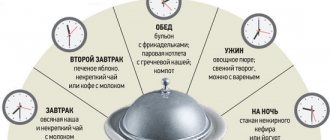
The diet for Barrett's esophagus involves avoiding snacking late in the day and overeating. It is important to avoid using:
- cream, milk with a high percentage of fat, butter, spread;
- cakes;
- beef, pork, goose, duck, lamb;
- drinks containing caffeine (Coca-Cola, tea, coffee);
- citrus fruits;
- carbonated drinks;
- alcohol.
Take food 4 times a day in small portions. After eating, walk in the fresh air for 30 minutes. It is important that the diet is rich in proteins.
The development of physiotherapeutic technologies correlates with the problems of treating patients with gastroesophageal reflux disease (GERD), which occupies a leading place in the structure of morbidity of the digestive system.
In recent decades, there has been a significant increase in the number of patients suffering from GERD [1, 2]. The prevalence of GERD among adults is up to 40%. The formation of GERD is determined by the action of many factors, but it is generally accepted that the trigger is the incompetence of the lower esophageal sphincter (LES), the genesis of which is associated with a violation of neurohumoral regulation [3-5].
Drug treatment of GERD is based on reducing the acidic effect on the esophagus and the use of drugs that correct motor disorders of the upper gastrointestinal tract. In this regard, the main direction of treatment is antisecretory therapy and the prescription of prokinetics [2, 3, 6, 7].
While the mechanisms of classical GERD are largely known, non-erosive reflux disease (NERD) has not yet been studied in many respects, so interest in it is steadily increasing. Among the numerous hypotheses with which they try to explain the nature of NERD, disturbances in the emotional and autonomic spheres play an important role [7, 8], and the extent to which the use of standard drug therapy is justified in this category of patients is a rather serious question. In addition, if we consider that in 5-21% of patients with GERD the cause of heartburn is alkaline reflux, and daily pH monitoring is not available to every patient, then antisecretory therapy in some of this category of patients may be unsuccessful. Patients with GERD pose a significant problem for healthcare in terms of the insufficient effectiveness of most of the methods used and the high economic costs of treatment.
The multiplicity of pathways for regulating acid formation gives rise to the need to develop treatment methods that have a multifaceted effect. It is known that smooth muscle tone has a certain dependence on autonomic regulation and adrenoreception [9]. Traditional methods of treatment, natural and preformed physical factors are capable of influencing functional disorders and the pathological process by influencing the regulatory systems of the body. Important in the treatment of GERD is the effect on the neurohumoral regulation of the LES, impaired motility of the esophagus and stomach, and the aggression factor - acid and alkaline reflux.
Methods of physiotherapy and balneotherapy for patients with GERD have been developed [10, 11]. However, to date, the influence of physical factors on the neurohumoral regulation of the LES, the psychological and vegetative status of patients with GERD, and their quality of life (QoL) has not been sufficiently studied.
From 2001 to the present time, the Federal State Budgetary Institution Russian Scientific Center for Medical and Radiotherapy has been conducting scientific research to study the effectiveness of using various non-drug methods in the treatment of patients with GERD. At the first stage, from 2001 to 2004, studies were devoted to studying the effect of acupuncture on the neurohumoral regulation of the LES and the acid-forming function of the stomach. The obtained research results prompted the authors in the future, together with E.B. Tishkova [12, 13], head of the gastroenterology department of the rehabilitation complex of the Russian Scientific Center for Medical and Radiological Sciences, to study the effect of structural resonance electromagnetic therapy on the clinical and functional state, vegetative, psycho-emotional status and quality of life of patients with GERD.
The goal of scientific research is to develop pathogenetically based methods of treating patients with GERD using non-drug treatment methods - acupuncture, natural and preformed physical factors.
Material and methods
197 patients with GERD aged from 18 to 65 years were observed.
The observed patients were divided into 4 (main) groups and one control group.
Group 1 - 40 patients with GERD, received treatment including exposure to an ultra-high frequency electromagnetic field (UHF therapy) and general iodine-bromine baths.
Group 2 - 67 patients with GERD who underwent a course of acupuncture treatment.
60 patients with GERD received structural resonance electromagnetic therapy (SRT): group 3 (30 patients) as monotherapy, group 4 (30 patients) in combination with iodine-bromine baths.
Group 5 - control, 30 patients with GERD who were on diet therapy.
The design of the study involved a clinical examination and diagnosis verification, randomization and formation of a group of patients, a control examination with assessment of clinical symptoms, esophagogastroduodenoscopy data, and neurohumoral regulation of the LES. Studies of intestinal hormones were carried out using the radioimmunological method at the Scientific Center for Chemistry of the Russian Academy of Medical Sciences in the laboratory of immunology and regulatory mechanisms in surgery. To verify the diagnosis, daily pH monitoring was performed using a Gastroscan-24 pH meter (Istok-Sistema, Fryazino). Assessment of the state of the autonomic nervous system based on the results of heart rate variability (HRV) - using the method of spectral analysis using the Kardiotekhnika-4000AD cardiac monitor as part of daily ECG monitoring. The assessment of psychological status was carried out using a computer version of the abbreviated multifactor personality questionnaire (SMOL). As a criterion for the patient’s subjective self-assessment, a method was used to determine the quality of life of patients based on a questionnaire using the general questionnaire SF-36 (The MOS 36-Item Shot-Form Health Survey - Russian version).
Statistical data processing was carried out using the Statistica 6.0 (Win) software package. Calculation of the significance of differences between groups was carried out by one-way analysis of variance using Student's t-test. Differences between two average values were considered significant at a p value <0.05.
Treatment methods
The complex of rehabilitation treatment was built taking into account literature data on the mechanism of action of physical factors and the clinical syndromes and disorders of the functional state of the digestive organs and neurohumoral regulation that we identified, which were discovered when patients were admitted to the clinic.
UHF therapy was prescribed using a Volna-2 device with a power of 25-30 W. The exposure was carried out on the collar area with a rectangular emitter 35x16 cm for 8-10 minutes, 8-10 procedures were prescribed every other day for the course of treatment; DMV therapy alternated with general iodine-bromine baths.
General iodine-bromine baths (on a sodium chloride basis at a concentration of 10 g/l) were prescribed at a temperature of 36-37 °C. The duration of the baths was increased from 8-10 minutes at the beginning of the treatment course to 12-15 minutes at the end of treatment. Baths were given every other day, for a course of treatment - 10-12 baths.
The course of acupuncture treatment was carried out according to traditional methods. The basic recipe was based on the following points: E36, E25, E23, GI11, MC6, RP6, VC12, VC10, VC13, E45.
Along with the basic recipe, acupuncture points were used, taking into account the individual manifestations of the disease. There are 10-12 procedures per course of treatment.
SRT was performed using the REMATERP device. The effect is non-contact, systemic (via inductors), the operating factor is an alternating electromagnetic field, mode No. 43, frequency spectrum 0.26-45100.0 Hz, duration of the procedure in the specified mode 43 minutes, course of treatment 8-10 procedures.
Results and discussion
A study of the clinical picture of GERD indicated that the main complaint forcing the patient to see a doctor—heartburn—was present in all those observed. 75% of patients complained of belching, 46% of regurgitation.
Endoscopic examination revealed cardial insufficiency; There was foamy mucus in the lumen of the esophagus, and there was swelling and hyperemia of the mucous membrane. Most often, pathological changes began in the middle or lower third of the esophagus and their intensity increased in the aboral direction. In 21% of those examined, single erosions of the lower third of the esophagus were detected. All patients with erosive esophagitis were included in the main groups.
Determination of the neurotransmitter involved in the regulation of the functional state of the LES—vasoactive intestinal polypeptide (VIP)—revealed a significant increase in its level in the blood serum of patients with GERD to 36.5±1.01 pg/ml, compared with values in healthy individuals of 29.85±2. 8 pg/ml (p<0.05). The level of gastrin in the blood serum of the observed patients also significantly exceeded the values in healthy individuals.
Spectral analysis of the wave structure of the heart rhythm in patients suffering from GERD revealed in the structure of the total power of the spectrum a significant predominance of the activity of the higher centers of autonomic regulation and sympathetic influences and a decrease in the activity of parasympathetic influences. Thus, in patients with GERD, an imbalance of autonomic regulation was revealed with a predominance of central regulatory mechanisms and a decrease in vagal influences.
The data obtained on the imbalance of autonomic regulation in patients with GERD contributes to the understanding of the pathogenesis of the disease, which can be used in the development of effective methods of treatment for this category of patients.
Analysis of the psychological characteristics of patients with GERD according to the results of the SMOL test revealed such features of psychological status as anxiety, internal tension, restlessness, decreased mood, along with an increased level of depressive-hypochondriacal and psychasthenic disorders (scales 1, 2, 7). Clinically, this was manifested by a depressed, depressed mood, apathy, pessimism, and disbelief in the possibility of a favorable outcome of the disease.
The results of a study of the quality of life of patients with GERD showed that patients generally rated their quality of life as low, most scale scores were in the range from 42.6 ± 3.4 to 55.8 ± 2.2 points. This fact indicates low differentiation of important areas of life that influence the assessment of QoL.
Thus, in patients with GERD, along with esophageal and extraesophageal clinical manifestations of the disease, there are: changes in the functional state of the esophagus, an increase in the level of VIP in the blood, an imbalance of the autonomic nervous system, characterized by a predominance of central regulatory mechanisms and a decrease in parasympathetic influences. Psychological disorders and decreased quality of life in patients with GERD determine not only the medical, but also the social significance of the problem.
As a result of the treatment, the general condition improved in the vast majority of patients, and favorable changes in the subjective and objective manifestations of the disease were noted. Complaints of heartburn disappeared in 75% of patients in group 1, 84% in group 2, 63% in group 3, and 73% in group 4. Regurgitation after treatment was not observed in 75% of patients in group 1, 97% in group 2, 67% in group 3, and 80% in group 4.
In 2 (33.3%) patients out of 6 with erosive esophagitis, after 5 procedures of DMV therapy and iodine-bromine baths, complaints of pain under the xiphoid process of the sternum remained. One patient experienced a deterioration in health after DMV therapy procedures. All 3 patients were prescribed appropriate drug therapy. After five physiotherapy procedures, 5 (83.3%) of 6 patients in group 3 and 6 (86%) of 7 patients in group 4 with erosive esophagitis still complained of pain under the xiphoid process of the sternum. The above patients were prescribed appropriate drug therapy.
Endoscopic examination of the esophagus revealed the disappearance of inflammatory phenomena in 56% of patients in group 1, 75% in group 2, 71% in group 3, and 68% in group 4. Erosions disappeared only in patients of group 2.
The study of the functional state of the LES according to pH monitoring of the esophagus showed a decrease in the number of transient relaxations of the LES with pH<4 in patients of the 1st group from 4.3 per hour to 1.3 after the DMV procedure on the collar area, in the 2nd group - with 4.8 per hour to 0.7 after the acupuncture procedure, which indicates an increase in the tone of the LES. A decrease in the intensity of acid formation was noted only in patients receiving acupuncture (from 1.42±0.03 to 1.64±0.03; p<0.05).
After the course of treatment, there was a significant decrease in the elevated level of VIP (p<0.05) in the blood of patients with GERD in all main groups. A decrease in VIP may indicate an improvement in the function of the LES and, as a consequence, a decrease and/or disappearance of reflux of gastric contents into the esophagus. There was a decrease in the initially elevated level of gastrin in the blood from 69.46±2.85 to 60.16±1.8 pg/ml (p<0.05) in patients with GERD after a course of acupuncture treatment. In patients with GERD treated with physiotherapy, no significant changes in gastrin levels were observed.
Thus, acupuncture influenced various parts of the pathogenesis of GERD, on the one hand, the functional state of the LES, and on the other, the aggression factor - the acid-peptic factor.
Assessment of autonomic regulation according to indicators of spectral analysis of HRV (carried out only in patients of groups 3 and 4, a later stage of research) revealed unidirectional positive changes after treatment in patients with GERD. Thus, after a course of SRT, there was a decrease in the power of the spectrum of slow LF waves, reflecting the activity of the sympathetic centers of the brain, in absolute values by 18% (from 1233±150 to 1010±105 ms2) and an increase in the power of the spectrum of fast HF waves, reflecting the activity of the parasympathetic center of the medulla oblongata. brain, by 19% (from 294±55.2 to 363±52.6 ms2), which caused a significant decrease in the coefficient of vagosympathetic balance from 4.3±0.3 to 3.25±0.3 (p<0.05 ). As a result of complex treatment with SRT and iodine-bromine baths, the power of the spectrum of slow LF waves decreased by 8% (from 1155±119 to 1064±114 ms2), the power of fast waves increased by 21% (from 365±43 to 442±48 ms2), which caused decrease in the coefficient of vagosympathetic balance from 3.8±0.4 to 2.6±0.3 (p<0.05).
In the control group, no dynamics of the studied parameters of HRV spectral analysis were detected.
Thus, SRT helps to increase parasympathetic influences, thereby optimizing autonomic regulation, which can have a positive effect on esophageal motor dysfunction.
The results of psychodiagnostic testing carried out over time in patients with GERD indicated a unidirectional psychocorrective effect of the proposed methods. After the course of treatment, there was a significant decrease in indicators on the scales of the “triad of anxiety” (SMOL 1; SMOL 2; SMOL 7), which is characterized by a decrease in internal tension, anxiety and worry. In patients in the control group, no significant changes in the studied parameters were observed.
It is well known that the nature of the disease, as well as the response to treatment, largely depends on the individual characteristics of the patient: his physiological, psychological and emotional state. In this regard, the main goal of GERD treatment is not only to relieve the symptoms of the disease, but also to improve the quality of life of patients.
The improvement in the general condition of the patients was characterized by a change in the quality of life indicators of patients with GERD. Thus, analysis of quality of life indicators after treatment indicated positive dynamics on the scale of role functioning, determined by the emotional state, and the scale of pain intensity in all observed patients. In patients of the main groups, significant dynamics were revealed on the general health scales; role functioning due to physical condition; social functioning; vital activity; mental health.
In the control group, no significant changes in the indicators of these scales were observed. On the scale of physical functioning, significant dynamics were observed only in patients of groups 2 and 4.
conclusions
Summarizing the above, we can conclude that the proposed treatment methods, including DMV therapy and general iodine-bromine baths, acupuncture, as well as SRT, both in the form of monotherapy and in combination with iodine-bromine baths, are pathogenetically substantiated and effective methods of treating patients with GERD. Higher therapeutic efficacy, the ability to influence various parts of the pathogenesis of GERD, the absence of adverse reactions and the ability to select points depending on the individual characteristics of the patient, taking into account concomitant pathology, allow us to give preference in the treatment of patients with GERD with esophagitis of degrees 0 and 1 (according to the Savary-Miller classification) acupuncture
CRT, either as monotherapy or in combination with iodine-bromine baths, can be recommended for patients with GERD with grade 0 esophagitis. The use of DMV therapy in combination with iodine-bromine baths is also indicated for patients with GERD with grade 0 esophagitis. For patients with GERD with grade I esophagitis, treatment with the above physical factors is recommended in combination with appropriate drug therapy. The use of the developed methods for treating patients with GERD is possible in hospitals, sanatoriums, sanatoriums, and also in clinics.
In light of modern approaches to the treatment of common diseases, the principles of personalization in medicine are becoming increasingly important [14]. From this position, continued research and study of pathogenetically based methods of treating patients with GERD, coupled with the results of previous studies, will allow us to systematize and implement the principles of personalization in the rehabilitation treatment of this category of patients.
Prevention of Barrett's esophagus
To avoid developing Barrett's esophagus, you need to:
- eat rationally and balanced;
- sleep on a high pillow;
- normalize body weight (it is important to exclude risk factors such as obesity);
- to refuse from bad habits;
- do not eat before bedtime;
- take walks and lead an active lifestyle.
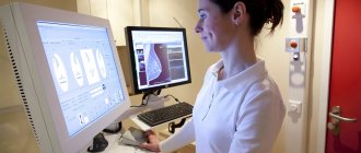
It is also important to undergo a routine esophagogastroduodenoscopy every two years - a procedure for examining the mucous membrane of the stomach and esophagus and performing a biopsy using an endoscope.
This article is posted for educational purposes only and does not constitute scientific material or professional medical advice.