A urethral stricture is a narrowing of the internal lumen of the urethra. This disorder causes problems with urination. It causes a person severe discomfort and pain, and in severe cases causes kidney problems. To avoid this, specialists strive to quickly diagnose and decide how to treat urethral stricture. The method of eliminating the defect is selected individually, based on the cause and severity of the violation.
What are the causes of urethral stricture?
Another name for the defect is stenosis, which means an abnormal narrowing of the physiological openings in the human body. Urethral stricture occurs in 1-2% of men and 0.5% of women. About 2% is allocated for cases of congenital form of the defect. In other cases, the disorder occurs during life.
Urethral stricture is more common in men. This is due to the structure of the urethra. It is long and complex, so it is more susceptible to stenosis than in women. Acquired narrowing of the urethra is more common. Its main reasons:
- In 70% of cases - blunt trauma to the perineum, penetrating wounds, fractures of the penis or pelvic bones, as well as chemical or thermal injuries.
- In 15% of cases - inflammation of the genitourinary organs: balanitis or urethritis against the background of tuberculosis, gonorrhea, chlamydia.
- In 13% of cases, there were errors by medical workers during urological manipulations.
In women, the cause may be birth trauma, amputation of the cervix or the entire uterus. A separate role in the development of stricture is given to diseases in which the blood supply to the urethral tissue is disrupted. These include coronary heart disease, atherosclerosis, hypertension and diabetes mellitus.
Prevention
To reduce the likelihood of developing urethral strictures, it is recommended:
- avoid injury to the genitourinary organs;
- promptly treat inflammatory processes;
- consult a doctor when the first suspicious signs indicating a pathology of the genitourinary system appear.
Parents should ensure that chemically aggressive substances are located at a height where the child cannot reach them. This will help prevent chemical burns to the genitals.
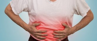
What are the symptoms of urethral stricture?
The main symptom of urethral stricture is difficulty urinating. Due to the narrowing of the canal, urine cannot be excreted normally naturally. The process of urination causes severe pain to a person. Against this background, other symptoms are observed:
- feeling of an incompletely emptied bladder;
- weakening of the stream of urine, its splashing;
- urine leakage;
- blood in urine or semen;
- decreased intensity of sperm release in men;
- the need to tense muscles to release urine.
In the most severe cases, acute urinary retention occurs, which requires immediate assistance from specialists. Symptoms of urethral stricture in women typically include pain when urinating and recurrent urinary tract infections.
Due to impaired urination, favorable conditions are created in the urethra for the development of urinary tract infections. Loss of patency of the urethra also contributes to the formation of stones, which impair the functioning of the bladder. The most dangerous complications of urethral stricture:
- cystitis,
- pyelonephritis,
- urolithiasis,
- hydronephrosis,
- bladder diverticula,
- renal failure.
The most serious complication of urethral stricture is kidney failure. This is a condition where they stop working completely, which can threaten the patient's life. If the urethra is completely obstructed, urine excretion becomes possible only through a catheter, which can also be a source of infection.
Postoperative complications
The most unpleasant postoperative complication is bleeding, when a hematoma or accumulation of blood forms in the area of surgical manipulation. Such situations require repeated surgical interventions to evacuate blood accumulations and stop bleeding. Postoperative wound infection is the second most common complication. Symptoms of such inflammation are pain in the wound, twitching in nature, and purulent discharge. Reducing the length of hospital stay, as well as antimicrobial therapy with antibiotics, helps to minimize the risks of the latter. The optimal time for a patient to stay in hospital is no more than 3-4 days.
Article information
| Last update | April 12, 2021 |
| Next update | April 12, 2022 |
| Author | Doctor Plekhanov A.Yu. |
| Reviewer | Doctor Zhivov A.V. |
| Is it possible to have plastic surgery in a year or a year and a half if the urethra is completely obliterated? He underwent three operations in 2017-2018 with long and frequent catheterization... See other questions or ask your own on the topic: urethral stricture |
Types and stages of the disease
Treatment for urethral stricture is determined based on its cause and type. The narrowing can be partial or complete, and it can be in different parts of the urethra, in the front or back. Depending on the length, stenosis is divided:
- for short – up to 2 cm long;
- long – over 2 cm long.
When the entire urethra is narrowed, we are talking about a total stricture. If it appears for the first time, it is called primary, if it occurs repeatedly, it is recurrent, and if there are complications, it is called complicated. Complete loss of the urethral opening indicates obliteration, i.e., fusion of the canal.
Diagnostic methods
Diagnosis of urethral stricture is carried out to determine the cause of the defect. These may be diseases or circumstances that preceded the development of stenosis. To do this, the complex of diagnostic measures includes:
- Smear for sexually transmitted infections with bacterial culture or PCR diagnostics.
- Uroflowmetry is a study of the dynamics of urination and urine flow rate.
- X-ray to detect false tracts, bladder stones or urethral diverticula.
- Cystoscopy or urethroscopy with tissue biopsy.
- Ultrasound of the bladder to assess the volume of residual urine and determine the severity of stenosis.
How is urethral stricture treated?
Treatment of urethral stricture in men and women can be different:
- Radical. These are urethroplasty operations that are considered the most effective.
- Non-radical. Such operations most often provide a temporary effect and are accompanied by frequent relapses.
Radical methods for removing urethral stricture include:
- Anastomotic urethroplasty – excision of the area of stenosis and subsequent connection of urethral fragments.
- Replacement urethroplasty is the replacement of part of the lumen with the patient’s own mucous membrane.
- Perineal urethroplasty – removal of the urethral opening into the perineum.
Non-radical methods of treating urethral stricture:
- bougienage (temporary restoration of the lumen of the urethra)
- urethrotomy (dissection of urethral stricture);
- stenting (placement of a stent in the lumen of the urethra).
At the State Urology Center you can get qualified help from a urologist and get rid of such a defect and its unpleasant symptoms. Here you will undergo comprehensive diagnostics and treatment, and also receive clinical recommendations for combating urethral stricture in order to avoid its complications. To receive medical services, you only need to make an appointment with a urologist at a time convenient for you.
On weekdays you can get an appointment with a urologist on the day of your visit
Introduction
The social significance of gastroesophageal reflux disease (GERD) is determined by its extremely wide prevalence among the population of economically developed countries. In Western Europe and North America, clinical manifestations of gastroesophageal reflux are noted by about 20% of the population. In various regions of Russia, the incidence of GERD ranges from 11 to 14% [6, 14].
Secondary changes in the esophagus associated with GERD are divided into inflammatory (ulcerative esophagitis, strictures) and dysplastic conditions (Barrett's esophagus, Barrett's adenocarcinoma). However, such systematization is conditional, since a combination of these processes is often noted [1, 22].
The pathogenesis of the inflammatory manifestations of GERD is due to the effect of aggressive acidic or alkaline gastric contents on the squamous epithelium of the esophagus. Chronic contact of gastric (gastroduodenal) contents with the esophageal mucosa leads to its damage - the development of esophagitis. The prevalence and depth of inflammatory and destructive changes are determined by a number of conditions: the composition (pH) of gastric contents, the frequency and duration of its impact on the esophageal epithelium, the effectiveness of physiological protective and compensatory-adaptive mechanisms [1, 2]. Damage to the growth layer of squamous esophageal epithelium and the formation of granulation tissue in areas of ulceration induce mechanisms of fibrogenesis and development of connective tissue in the wall of the esophagus. In severe cases of the disease, the muscular layer and adventitia of the esophagus and the tissue surrounding the organ are involved in the destructive-inflammatory process. The combination of these factors inevitably leads to morphological and functional changes: the lumen of the esophagus decreases, its elasticity decreases, and motility is impaired. This is exactly how the pathogenesis of peptic strictures appears from the perspective of modern knowledge [1, 2, 8, 15].
Material and methods
Between 2007 and 2010, 16 patients with peptic strictures of the esophagus that developed against the background of GERD were treated in the surgical department of the Leningrad Regional Clinical Hospital. In 7 (43.8%) cases, disturbances when swallowing solid foods were clinically noted, in 9 (56.2%) - discomfort and pain when eating semi-liquid foods. In 5 (31.2%) patients, the occurrence of dysphagia was the clinical debut of the disease.
The examination algorithm was standard and included X-ray contrast examination of the esophagus and stomach, esophagomanometry, esophagoscopy (including ZOOM and NBI modes) with biopsy and daily study of pH in the lumen of the esophagus [1, 3, 7, 9].
During X-ray examination, short (less than 3 cm) peptic strictures were diagnosed in 15 (93.8%) cases, and an extensive decrease in the lumen of the esophagus in 1 (6.2%) case (Fig. 1).
Figure 1. Esophagograms of patients with hiatal hernia. a-long peptic stricture of the esophagus.
Figure 1. Esophagograms of patients with hiatal hernia. b-short peptic stricture and esophageal ulcer.
The diameter of the esophagus in the stricture zone was about 10 mm in 3 (18.8%) patients, from 3 to 10 mm in 9 (56.2%) patients, and less than 3 mm in 4 (25.0%) patients.
The results of esophagoscopy in patients with peptic strictures of the esophagus were generally consistent with the results of radiocontrast studies.
The combination of an ulcer and cicatricial narrowing of the esophagus was identified in 3 cases. In 4 patients, the presence of columnar cell metaplasia at the edges of the stricture—Barrett's esophagus—was visually determined (Fig. 2).
Figure 2. Video esophagograms in NBI mode. a-peptic ulcer and peptic stricture of the esophagus.
Figure 2. Video esophagograms in NBI mode. b-Barrett's esophagus and peptic stricture of the esophagus.
Histological examination of the biopsy material in all cases revealed pronounced inflammatory infiltration of the esophageal mucosa. Gastrointestinal metaplasia without neoplasia was detected in 4 patients.
When analyzing the type of gastroesophageal reflux (daily pH study), in 12 (75.0%) cases a predominance of the acidic component in the composition of the gastric contents was noted, and in 4 (25.0%) - alkaline.
The study of motor function of the esophagus was carried out using multichannel esophagomanometry. Dislocation of the lower esophageal sphincter above the diaphragm was observed in all patients, its insufficiency - in 13 (81.3%), length less than 2 cm - in 5. Hypokinesia of the organ was determined in 7 (43.8%) cases, in 4 (25.0%) %) - mixed changes in motility with a predominance of spastic contractions.
The totality of information obtained from the research became the basis for determining and subsequent implementation of the treatment strategy.
Radical ways to eliminate the cause of dysphagia are various methods of surgical treatment, involving resection or plastic surgery of the altered segment of the esophagus with the performance of certain anatomical reconstructions. A serious disadvantage of this group of procedures is the high risk of life-threatening complications, which increases in direct proportion to the effectiveness of surgical interventions [2, 4, 15].
Another way to solve the problem may be mechanical dilatation of the organ lumen - bougienage with endoscopic or radiological control. The advantages of the procedure include its safety, effectiveness, and the possibility of repetition if dysphagia recurs. However, expansion of the lumen of the gastroesophageal junction leads to increased reflux of gastric contents into the esophagus and requires additional pharmacological or surgical measures [2, 4, 8, 15].
The above considerations forced us to choose the safest treatment option - endoscopic bougienage of the narrowing zone of the esophagus. Dilatation was performed strictly after healing of ulcerative defects. In all observations, the standard method of bougienage using a guide, proposed by E.N., was used. Vantsyan and R.A. Toshchakov in 1965, or hydrodilation with cylinders with a diameter of 10-15 mm from COOK. The intervals between sessions were 2-3 days. The criterion for the achieved therapeutic effect was a persistent expansion of the lumen of the esophagus to 10-11 mm (allowing the endoscope to be freely passed through the narrowing). Typically, the course of treatment took 12-16 days and consisted of 5-7 procedures. There were no complications of dilatation of the scar narrowing of the esophagus.
After endoscopic elimination of dysphagia and restoration of normal passage through the esophagus, the question arose about further tactics for managing patients in this group. The presence of an anatomical cause of the disease (hiatal hernia) and the possibility of its radical elimination by surgery determined the choice in favor of antireflux reconstruction [10, 13, 18, 20].
To heal traumatic changes in the esophageal mucosa after bougienage, before the second stage of treatment, a break was taken for 2-4 weeks. Throughout this period, patients received therapy with antisecretory drugs. Immediately before the operation, endoscopic and radiological monitoring of the condition of the esophagus was carried out.
All antireflux surgical interventions in patients with esophageal strictures were performed laparoscopically.
Restoration of the abdominal position of the stomach and correction of the size of the esophageal opening of the diaphragm were standard. The gastrohepatic and gastrophrenic ligaments and the esophageal-diaphragmatic membrane were dissected. Along the contour of the esophageal opening of the diaphragm, its legs were identified. To prevent recurrent dislocation of the stomach into the mediastinum, the lower thoracic esophagus was carefully mobilized. Compliance with this condition significantly reduced tissue tension during the reconstructive stage of the operation. In addition, pronounced fibrotic changes in the tissue surrounding the organ were an additional factor predisposing to relapse of dysphagia [5, 11].
After all of the above measures were completed, posterior crurorrhaphy was performed with single interrupted sutures. To prevent dysphagia, an esophageal probe (D=60 Fr) was used when calibrating the esophageal opening of the diaphragm.
The next stage of surgical intervention was reconstruction of the gastroesophageal junction. The choice of the optimal surgical option was determined by the task of creating an effective anti-reflux mechanism with minimal risk of disrupting passage through the esophagus. Among the wide range of fundoplication techniques developed and used today, the A. Toupet operation is considered the most suitable for correcting the manifestations of GERD in patients with esophageal dysfunction [12, 16, 21].
The valve mechanism of reconstruction of the gastroesophageal junction using the Toupet technique is implemented by restoring the acute His angle, positioning the fundus of the stomach above the distal esophageal sphincter (reproducing the inkwell-sippy cup model) and its additional compression by the fundal coupling. An increase in intragastric pressure causes a proportional increase in the compression effect of the cuff and prevents the reflux of aggressive gastric contents into the esophagus [17, 19].
The noted advantages of the Toupet procedure determined its choice as an option for surgical treatment of GERD. In all 16 cases, partial fundoplication at 240° was performed.
To freely move the fundus of the stomach behind the esophagus and create a cuff 2-2.5 cm wide, wide mobilization of the gastroesophageal junction was performed. Prevention of an extremely unpleasant complication of surgical intervention—slipping of the fundus coupling from the esophagus (the “telescope” phenomenon)—was carried out by fixing the fundus of the stomach to both legs of the diaphragm with separate sutures. The lumen of the esophagus in the area of antireflux reconstruction was controlled with a D=60 Fr probe.
Postoperative management of patients was standard and included pain relief, suppression of cough and gag reflexes, and limitation of physical activity. From the first day the intake of liquids was allowed, then - mushy food. Before discharge from the hospital, usually on the 7th or 8th day, a control radiography was performed with contrast of the esophagus with a barium suspension. 2 weeks after surgical treatment, soft food was allowed. There were no complications of antireflux reconstruction of the esophagogastric junction in the patients we observed.
results
A follow-up examination, daily pH monitoring, X-ray scanning and esophagogastroscopy 2 months after surgery were performed in all 16 patients. The main complaints were related to increased gas formation in the intestines and difficulty belching air. There were no symptoms of dysphagia or manifestations of gastroesophageal reflux. When analyzing acidity in the lumen of the esophagus, physiological pH values were recorded in all observations. There were no radiological or esophagoscopic signs of narrowing of the lumen of the esophagus within the specified time frame after combined surgical treatment.
The results of the described procedures (bougienage of the stricture and antireflux surgery) were monitored in 13 patients over a period of six months to 2.5 years. With instrumental research methods (FGDS and contrast fluoroscopy), narrowing of the distal esophagus or recurrence of a hiatal hernia was not detected within the specified time frame (Fig. 3).
Figure 3. Esophagogram of a patient after Toupet fundoplication.
Clinical manifestations of dysphagia and symptoms of gastroesophageal reflux were not identified in any observation.
Discussion
The presented experience of combined surgical correction of GERD in patients with peptic strictures of the esophagus allows us to recognize this method of treatment as simple, safe and quite effective. The obvious advantage of the described technique is the possibility of eliminating the key etiological cause of the disease - failure of the barrier function of the gastroesophageal junction caused by a hiatal hernia. Restoring the anatomical relationship between the esophagus, stomach and diaphragm, creating a reliable anti-reflux mechanism makes it possible not only to prevent further development of the pathological process, but also to achieve a pronounced regression of its clinical manifestations. Good functional results and minimal aggressiveness of the proposed treatment concept allow us to consider it promising and recommend it for wide practical use.
Hakobyan Gagik Nersesovich – professor, doctor of medical sciences, oncologist, urologist in Moscow
The appointment is conducted by a doctor of the highest category, urologist, oncologist, doctor of medical sciences, professor. Author of more than 100 scientific papers.
Urological oncology experience – more than 15 years. Helps men and women solve urological and oncourological problems.
Conducts diagnostics, treatment and complex operations for such diagnoses as:
- tumors of the kidneys and upper urinary tract;
- prostate and bladder cancer;
- urolithiasis disease;
- BPH;
- hydronephrosis, ureteral stricture, etc.
Why surgery is needed
Scar tissue changes are irreversible. Stricture disease cannot be treated with medication, and methods such as bougienage of the urethra have a temporary effect. The only way to permanently eliminate pathological changes and restore normal function of the lower urinary tract is through surgery. To effectively treat stricture disease of the urethra, a wide range of plastic and reconstructive surgeries are used, aimed at replacing the damaged area of the urinary canal with healthy tissue.
During your consultation, the urologist will answer all your questions in detail.
If you are bothered by difficulty or frequent urination, pain in the lumbar region, blood in the urine, as well as other symptoms (read about what else should alert you here), seek help from a urologist.
Admission includes:
- familiarization of the doctor with the patient’s medical history;
- inspection;
- making a preliminary diagnosis, prescribing tests and necessary procedures.
*If you plan to get tested immediately after your appointment with your doctor, go to the clinic with a full bladder.