Article for the "bio/mol/text" competition: Fatigue, loss of appetite, nausea, yellowing of the skin and even frequent headaches - all these symptoms can be indirect evidence of liver pathologies. Despite the high ability of liver cells to recover, in some severe cases they simply cannot cope with the disease. New research from a team of scientists from the Center for Regenerative Medicine in Edinburgh, the Massachusetts Institute of Technology and the Skolkovo Institute of Science and Technology shows how by blocking the main pathway of liver cell repair, they were able to open up reserve “repairers”.
A discovery foretold by myth: regeneration in reptiles
The ability of organisms to regenerate, that is, restore the structure and functions of organs, is one of the important mysteries of medicine that people have long been trying to unravel. Observations of the animal world made it possible to formulate the following pattern: the simpler an animal is, the easier it is for it to restore lost organs. And if an earthworm is able to “complete” half of its own body, and a lizard is able to grow a new tail, then in humans, regeneration abilities are represented in a narrower range [1], [2].
There are quite a lot of fairy-tale creatures that can grow a new head or tail. But both the Lernaean Hydra (Fig. 1), and the Gorgon Medusa, and even the Serpent Gorynych have a very real “relative” - triton. This representative of tailed amphibians is considered one of the oldest species of fauna on Earth. Newts successfully restore not only their tail and paws, but even damaged hearts and spinal cords. However, amphibians are far from the only creatures that have access to the “self-repair” function. For example, zebrafish are used not only in aquariums, but also to study the regeneration of heart tissue. And the first animal, thanks to which the term “regeneration” appeared, was the crayfish. The French scientist Rene Reaumur, who proposed a new term back in 1712, studied the restoration of lost legs in crayfish.
Figure 1. “The Battle of Hercules with the Lernaean Hydra” by Antonio del Pollaiuolo (1475).
"Wikipedia"
It is not surprising that scientists want to understand why a lizard, for example, can restore a lost tail, but a person cannot grow a new arm. Studying the structure and composition of tissues immediately after the lizard lost its tail made it possible to discover a model of regeneration in reptiles. During the healing period, the basal cells of the epidermis actively divide, gradually “closing” the wound. Additional aggregation of dividing cells at the distal end of the spine promotes blastema proliferation, an accumulation of unspecialized cells. At this moment, the processes of formation of new blood vessels are launched, and then new peripheral axons. New formations of bone tissue and muscles come into play most late. However, the exact mechanism of tail regeneration in lizards is not fully understood. Recent research from Arizona State University and the Institute for Genomic Research has identified microRNA molecules that promote muscle, cartilage, and spine regeneration [3]. Perhaps this work will allow the development of treatments based on the control of gene expression using microRNAs.
A Discovery Made by Accident: Regeneration in Mice
But what about the possibilities of regeneration at the level of entire organs in organisms that are more highly organized than lizards? Until recently, scientists were sure that mammals are not able to restore lost organs. But this belief was shaken by a discovery made in the laboratory of immunologist Ellen Heber-Katz at a research center in Philadelphia. There they conducted various experiments on special “patients” - genetically modified Murphy Roth Large (MRL) mice. Such individuals differed from ordinary ones in that their T-cells of the immune system did not work. One day, Dr. Heber-Katz gave her laboratory assistant a simple task: to mark the mice selected for the next experiment by making small two-millimeter holes in their ears. A few weeks later it turned out that there were no holes in the ears of the experimental subjects. The structure of blood vessels, cartilage, and tissues looked intact. However, the laboratory assistant assured the doctor that the task of “tagging” the mice was completed in a timely manner. After repeating the experiment with the auricle, the effect was the same: after four weeks, a blastema formed on the “pierced” areas of the ears (Fig. 2) [4]. The next “experimental” object was the tail - and again it was possible to demonstrate partial tissue regeneration. However, the regenerative abilities of MRL mice are not unlimited: for example, such a mouse, alas, was unable to grow a new paw. The reason lies in the different location and number of blood vessels in the organs and tissues of the animal. Without cauterization, the mouse will simply die from a large loss of blood - long before the regeneration processes start. And cauterization at the site of the amputated limb eliminates the appearance of blastema.
Figure 2. Stages of ear tissue repair in a normal laboratory mouse (bottom) and a transformed MRL strain (top).
[4]
As a result of a series of observations of transgenic mice, it was possible to show that the secret of their success lies in a certain protein. Thus, in MRL mice, the expression of the gene encoding the protein p21 (inhibitor of cyclin-dependent kinase 1A), which regulates the process of normal cell division, is blocked. Suppression of this gene in normal mice shows a similar ability to regenerate lesions [5]. But such manipulations should be carried out with great care: “disabling” the p21 gene can lead to disruption of normal cell reproduction, which can lead to the catastrophic division of all cells in the body.
Symptoms of the disease
Liver cirrhosis manifests itself already at an early stage of the disease. Drinkers experience:
- chronic fatigue, depression, loss of strength, drowsiness;
- the appearance of bruises on the body even with weak blows;
- predominance of red palms;
- nosebleeds, bleeding gums;
- the formation of telangiectasia (dilation of capillaries) on the face and body;
- arrhythmia, hypertension.
Without preventing the development of the disease, symptoms appear:
- disruptions of hormonal levels, menstrual cycle, in severe forms of cirrhosis - amenorrhea (in women);
- enlarged mammary glands, impaired estrogen production (in men);
- swelling of the limbs;
- hair loss in the armpit;
- anxiety, tremor, irritability;
- inhibited reaction, loss of attentiveness, memory problems;
- disruption of the speech apparatus;
- enlargement of the phalanges of the fingers;
- urinary tract infection, blood in urine.
Each stage is characterized by certain manifestations of cirrhosis. According to the severity of the disease, the pathology is classified into classes:
A – compensated
B – subcompensated
C – decompensated
To determine the class, the patient is diagnosed with bilirubin and serum albumin in the blood, the stage of ascites, the severity of hepatic encephalopathy, and the prothrombin index. The basis for diagnosing patients with cirrhosis is anamnesis: poor general health, shortness of breath, a noticeable change in liver size upon palpation, yellow complexion, high portal vein pressure.
To confirm the diagnosis, studies of biochemistry and a general blood test, immunogram, coagulogram, and markers of viral hepatitis are necessary. An ultrasound, MRI or CT scan of the liver and abdominal cavity, biopsy, and FEGDS are also required.
Everyday reality: human regeneration capabilities
What about regeneration in people? Even without “turning off” the gene encoding the p21 protein, the human body can restore some organs. For example, the skin, whose regenerative ability is as familiar to us as air. The largest organ in our body constantly renews its own structure due to necrosis and rejection of epidermal cells, followed by their replacement with new cells. The process of restoration of other epithelial tissues occurs in a similar way - for example, the mucous membranes of the respiratory tract, as well as the stomach and intestines. Bone tissue is in second place in the hierarchy of regenerative abilities. Fractures are known to heal quite successfully during a period of immobility.
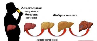
And among internal organs, the deserved leader in regeneration is the liver. The legend of the titan Prometheus, whose pecked liver grew back in just one night (Fig. 3), has a rational grain in it.
Figure 3. “Chained Prometheus.” Sculpture by Nicolas-Sébastien Adam, 1762 (Louvre).
"Wikipedia"
Indeed, the liver has the unique property of being restored to its original volume, even if more than 70% of the liver tissue is destroyed. A similar process occurs due to the work of liver cells - hepatocytes. These cells play a key role in modifying and eliminating toxic substances from the body. In a healthy organ, untouched by pathological processes, these cells are usually at rest. But if it is necessary to restore the integrity of the organ, for example, after partial resection (removal of part of the organ), almost all hepatocytes are activated and begin to divide. Moreover, they divide 1–2 times, and then return to a resting state again. This property underlies the treatment of some diseases, for example, cirrhosis of the liver or hepatitis, when the patient is transplanted a part of a healthy liver from a donor. However, such manipulations can lead to a number of health problems, including dilation of the veins of the esophagus and stomach, kidney failure and jaundice. Moreover, the appearance of rapidly dividing donor cells in the patient's liver can lead to cancer. Hepatocytes can no longer cope with a progressive disease on their own, because only healthy cells are capable of dividing, of which there are fewer and fewer in the patient’s body.
It turns out that, despite the powerful regenerative potential, the regenerative abilities of the liver have a limit. In cases where the pathological process goes too far, the effect of the work of hepatocytes is insufficient. For example, when a healthy liver is damaged as a result of toxic or viral influences, which provokes the proliferation of connective tissue (fibrosis). Are there other ways to restore the structure of this vital organ without the participation of hepatocytes? This question can be answered by a joint study by a team of scientists from the University of Edinburgh, the Massachusetts Institute of Technology and the Skolkovo Institute of Science and Technology [6].
Non-drug liver restoration
It is important to understand that the liver’s ability to recover after alcohol is fully activated only if a healthy lifestyle is followed. The same goes for nutrition and taking medications. The liver needs help, so to normalize all functions of the organ, it is extremely important to adhere to a diet. In particular, the diet should contain:
- vegetables,
- greenery,
- fruits,
- fish,
- lean meat,
- honey,
- nuts,
- dairy products,
- eggs.
Food can be consumed raw or fermented, boiled, steamed and baked. The preferred drinks are pure water, compotes of berries and dried fruits, and natural juices. It is necessary to completely exclude from the menu fatty and unhealthy foods, smoked foods, seasonings, canned food, butter, confectionery, bread, and sweets. Alcoholic and carbonated drinks, strong black tea and coffee are also prohibited. It is important to eat often and in small portions for better absorption of food. Vitamins (B, C, D, E) and minerals (zinc and selenium) play a huge role in normalizing liver function, therefore, as part of non-drug therapy, experts recommend taking complexes with an optimal ratio of nutrients.
Promising Reality: Accelerated Liver Regeneration
To study the processes of liver regeneration, transgenic mice of the tdTomato (tdTom) line were used. This line is modified with red fluorescent proteins, which allows visualization of liver cells [7]. However, the search for other “rescue” cells is complicated by the fact that hepatocytes in the affected body continue to work. To identify “non-hepatocytes,” the researchers used gene knockdown technology in mice. The knockdown effect is that it allows you to temporarily reduce the activity of specific genes without making changes to the chromosome structure and DNA sequence. To “turn off” the genes responsible for the division and migration of hepatocytes, special lipid nanoparticles with short interfering RNA (siRNA) were created [8], [9]. With their help, it was possible to block the expression of necessary genes.
Reducing the proliferation of hepatocytes by “turning off” the genes of interest to scientists was carried out using two models. In the first case, the ITGB1 , which encodes β1-integrin, was temporarily blocked. The second model is the simultaneous blocking of β1-integrin and stimulation of overexpression of the p21 protein. Both models had a similar effect, but their mechanisms differed: β1-integrin causes necrosis of hepatocytes, and overexpression of p21 suppresses their proliferation.
Figure 4. Liver regeneration using bile duct cells (highlighted in white).
[6]
Such a “blockade” of the main functional cells of the liver led to an unusual effect: when the main regeneration mechanism was turned off, a backup method was launched with the participation of bile duct cells. Thus, loss of β1-integrin and increased levels of p21 protein led to a significant increase in the number of cholangiocyte-derived hepatocytes. These epithelial cells of the intrahepatic bile ducts make up only 2-3% of the total population, but further observations showed that they are the ones that are able to “reprogram” and turn into hepatocytes, thereby restoring the liver (Fig. 4). Moreover, cholangiocytes showed a better ability to divide than hepatocytes. Previously, it was discovered that they are close to the oval cells of the liver—a kind of “stem” agent of this organ [10].
In order to track the regenerative abilities of cholangiocytes, three independent pathways were used:
- Modeling of cholestatic liver disease.
- Modeling of metabolic non-alcoholic steatohepatitis (an inflammatory process associated with excessive accumulation of triglycerides in the liver).
- Modeling of liver fibrosis (proliferation of connective tissue in the liver due to damage to hepatocytes).
To study liver regeneration due to non-hepatocytes along all three pathways, separate sections of the liver were visualized. It turned out that approximately 20–30% of hepatocytes are restored due to cholangiocytes, and small proliferative hepatocytes were identified already from the 7th day, and by the 14th day of regeneration their number increased significantly (Fig. 4–6).
Figure 5. Dynamics of restoration of liver tissue with the help of cholangiocytes (using the example of a model of cholestatic liver disease). Asterisks indicate necrotic areas; arrows indicate areas with cellular infiltrate.
[6]
Figure 6. Mouse liver cells transformed with the ITGB1 gene (day 14 of regeneration). Imaging with red fluorescent proteins. 1 — arrows indicate invasive cells; 2 - the area of the regenerative node is outlined with a dotted line.
[6]
It turns out that there is a reserve regeneration mechanism in the liver: the transformation of bile duct cells into functional hepatocytes when β1-integrin is blocked. It is possible that there are other target genes in the body, the “switching off” of which stabilizes the processes associated with the development of fibrosis and further cirrhosis of the liver. Scientists have yet to uncover the mechanisms of the signals that prompt bile cells to initiate “reprogramming” processes. However, the results of the study are already opening up a new direction in cellular regenerative medicine: the development of drugs that stimulate cholangiocytes. Perhaps in the future, medicine will eliminate the need to perform complex and expensive liver transplant operations. Instead, a more accessible technology will spread: “switching on” signals about the need for regeneration, when the processes of active division of liver cells will be launched with the help of the most active cholangiocytes.
Symptoms of liver pathology
If your liver hurts after drinking, this indicates the development of an inflammatory reaction, alcoholic hepatitis. According to various studies, this pathology develops in 60-90% of people who abuse alcohol, and in chronic alcoholism - in 100% of patients.
After a long binge, along with withdrawal symptoms, signs of liver damage may bother you:
- feeling of heaviness in the right hypochondrium
- bitter taste in the mouth, expressed in the morning
- belching, nausea
- feeling of a full stomach
- stool disorders
- yellowing of the skin and sclera of the eyes
- dark urine, discoloration of stool
- loss of appetite
Some of these symptoms may be present at the same time, but in the case of established alcoholic liver disease, binge drinking leads to the manifestation of all signs.