Anatomy of the larynx
In an adult, the larynx is located at the level of the IV-VI cervical vertebrae along the midline of the neck. At the top it comes into contact with the hyoid bone, at the bottom it passes into the trachea, at the back it is covered with fiber and communicates with the pharynx. The anterior surface of the larynx is covered with muscles, fascia and skin.
The organ has a complex anatomical structure - it contains cartilage, ligaments, many muscles and joints. The large thyroid cartilage, also called the Adam's apple, is palpated on the neck and protrudes significantly forward in men.
Functions of the larynx:
- respiratory – regulation of external respiration, its depth and rhythm;
- insulating (protective) - protection of the respiratory tract from food entering during swallowing, harmful impurities from the air (for this, a spasm of the larynx occurs), evacuation of foreign particles trapped in the respiratory tract by coughing;
- vocal (phonatory) - the formation of vowels and parts of consonant sounds when air passes through the glottis.
Laryngeal cancer is a malignant neoplasm, most often developing from squamous epithelium. Localized in all parts of the organ.
Anatomy of cartilage
When studying the structure of the larynx, special attention should be paid to the cartilage present.
They are presented as:
- Cricoid cartilage. This is a wide plate in the form of a ring, covering the back, front and sides. On the sides and edges, the cartilage has articular areas for connection with the thyroid and arytenoid cartilages.
- Thyroid cartilage, consisting of 2 plates that fuse in front at an angle. When studying the structure of a child’s larynx, these plates can be seen to converge in a rounded manner. This happens in women too, but in men it usually develops an angular protrusion.
- Arytenoid cartilages. They have the shape of pyramids, at the base of which there are 2 processes. The first, the anterior one, is the place for fastening the vocal cord, and the second, the lateral cartilage, is where the muscles are attached.
- Horn-shaped cartilages, which are located on the tops of the arytenoids.
- Epiglottic cartilage. It has a leaf-shaped form. The convex - concave surface is lined with mucous membrane, and it faces the larynx. The lower part of the cartilage extends into the laryngeal cavity. The front side faces the tongue.
Morbidity statistics
Laryngeal cancer accounts for 2.6% of all cancers. It is in first place in terms of incidence among head and neck tumors. In 95% of cases, malignant lesions of the larynx are squamous cell carcinoma, 2% each are glandular cancer and basal cell carcinoma, and 1% are rare types of cancer.
Men are more susceptible to the disease - they are diagnosed 9-10 times more often than women. 80-95% of patients are men from 40 to 60 years old. Most of them are heavy smokers.
The survival prognosis directly depends on the stage at which the cancer is detected and its location. If the tumor is detected at stage I, the five-year survival rate is 85%, at stage II – 70%, at stage III – 60%, at stage IV it decreases to 20%.
When chemoradiotherapy is started in the early stages, stable remission is achieved in 85-95% of cases, in late stages - in 30-40%.
Neoplasms of the upper part of the larynx give metastases to regional lymph nodes in 35-45% of cases, of the lower part - in 15-20%. In the area of the vocal cords, the lymphatic network is less developed, so the tumor in this area metastasizes rarely and late.
Causes and risk factors
Laryngeal cancer, like other cancers, develops from mutated cells of normal tissues or benign tumors. Cell malignancy, or malignancy, occurs under the influence of external factors; there are also diseases that have a high risk of degeneration.
External factors that provoke the occurrence of laryngeal cancer:
- smoking and chewing tobacco;
- drinking alcohol;
- occupational hazards - dust, high and low temperatures, benzene vapors, petroleum products, phenol resins.
Diseases prone to malignancy:
- long-standing papillomatosis;
- fibroma with a wide base;
- leukoplakia;
- pachydermia;
- dyskeratosis;
- ventricular cysts;
- chronic inflammatory processes.
Ultrasonography
Ultrasound of the esophagus is performed even in a regular clinic.
Ultrasound examination is an absolutely harmless and quite informative method, the duration of which is no more than 30 minutes.
The high resolution of the ultrasonic sensor allows you to examine areas less than a millimeter, and equipment with 3D or 4D visualization provides an accurate color image. This significantly speeds up and simplifies the research process.
During an ultrasound of the esophagus and stomach, information is obtained about the condition of:
- the walls of the stomach, intestines and the structure of their tissues;
- motility (passage of food along the tract)
- lymph nodes and blood vessels.
In a small child, an ultrasound of the gastrointestinal tract can reveal developmental abnormalities. The primary diagnosis of any disease in children through ultrasound is related to the safety of the ultrasound wave for the child’s body.
Symptoms of laryngeal cancer
The first signs of a tumor are nonspecific, they are similar to the symptoms of many inflammatory diseases, and it is difficult to suspect an oncological process from them and, even more so, to determine its location.
Early symptoms:
- low-grade fever;
- weakness, fatigue, general malaise;
- drowsiness.
Late signs vary depending on where the neoplasm develops.
Supraglottic cancer is characterized by:
- dryness and sore throat;
- discomfort and pain when swallowing, radiating to the ear on the side of the tumor, choking;
- sensation of a foreign body in the larynx;
- dull voice.
Symptoms of a neoplasm on the vocal cords:
- change in voice, loss of sonority and melody;
- hoarseness and hoarseness.
- When a tumor develops in the subglottic region, patients complain of:
- paroxysmal dry cough;
- voice disorders.
In the late period, when cancer of any localization grows into the lumen of the larynx, difficulty breathing, attacks of suffocation, putrid breath, and cough with blood clots appear. Due to discomfort when swallowing, the patient limits food intake, and exhaustion develops.
The sooner a person seeks help, the more effective the treatment will be. Even early signs (weakness, fatigue) should be a reason to visit a doctor. In this case, it is possible to diagnose the tumor at an early stage. If you experience coughing or difficulty swallowing, you should consult a doctor immediately.
Symptoms
Signs of the disease appear even with large benign tumors, when they directly begin to interfere with digestion or put pressure on neighboring organs. The most common symptoms of the pathology are:
- Difficulty swallowing (dysphagia). The tumor mechanically prevents food from passing through the esophagus. The patient must wash down food with water; it is difficult for him to eat meat, bread and solid foods.
- Mild or moderate chest pain in the projection of the esophagus. The discomfort is caused by compression of the nerve endings by the tumor.
- Belching, nausea, vomiting. These signs are a consequence of digestive disorders.
- Weight loss, general weakness. Caused by metabolic disorders due to the long-term existence of the tumor.
Classification
Classification of laryngeal cancer is carried out according to different criteria.
Localization of education
There are three anatomical sections of the organ:
- supraglottic (vestibular);
- middle (vocal cords);
- subglottic
Cancer of the supraglottic region develops most often - from 65% to 70% of all laryngeal tumors. It appears on one side and quickly spreads to the other. Neoplasms in this area are characterized by aggressive growth and rapid appearance of metastases.
A tumor of the middle section is diagnosed in 25-30% of cases. Usually develops on one vocal cord. Less aggressive than in the supraglottic. Voice disorders force patients to see a doctor quickly, which is why ligament tumors are often detected in the early stages. Localization of the formation facilitates surgical access to it.
Neoplasms of the subglottic region are the rarest - approximately 2% of cases. At the same time, they are characterized by fairly rapid infiltrative growth, and their location complicates surgical access and increases the risk of injury to the vocal cords during surgery.
Stages of laryngeal cancer, Russian classification
According to the prevalence of the process, malignant lesions of the larynx are divided into four stages - I, II, III and IV, stage III has substages a, b, IV - a, b, c, d.
| Stage | Characteristic |
| I | The formation is limited in size and does not extend beyond the mucous membrane of one anatomical part of the larynx. |
| II | The process completely covers one anatomical part of the larynx (all layers can be involved), does not spread beyond its limits, and does not metastasize. |
| III | a – the tumor extends beyond one anatomical part of the larynx, spreads to adjacent tissues, and causes immobility of half of the larynx. b – in addition to the spread of cancer to neighboring anatomical areas, regional lymph nodes are affected: one fixed or several mobile enlarged nodes are detected. |
| IV | a – spread of the tumor to neighboring organs. b – the formation occupies a significant part of the larynx and penetrates into the underlying tissue. c – fixed metastases are detected in the lymph nodes of the neck. d – tumor of any size, metastasizes to regional lymph nodes and distant organs. |
Growth pattern
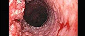
Exophytic cancer - grows into the lumen of the organ or outward. The formation usually occurs on the wall of the larynx and grows outward, blocking the lumen of the upper respiratory tract. It has no clear boundaries, the surface of the tumor is lumpy, with papillary growths.
Endophytic (infiltrative) cancer - grows inward, into the tissue of the organ. It looks like an infiltrate with ulcerations, without clear contours. Penetrates into the thickness of adjacent tissues.
Mixed - combines the features of exo- and endophytic growth.
Histological structure
Most often, laryngeal cancer arises from squamous epithelial cells. Glandular cancer, basal cell carcinoma and other rare types of tumor are diagnosed much less frequently. Some types are further subdivided:
- Squamous cell carcinoma : non-keratinizing – arises from non-keratinizing epithelium, grows quickly, has a high risk of metastases;
- keratinizing – develops slowly, metastases appear after a long period of time.
- poorly differentiated - it is difficult to determine the type of cells and tissues that make up the neoplasm, the tumor is characterized by a high degree of malignancy, grows quickly and metastasizes;
Malignant tumors
Malignant neoplasms develop in all anatomical parts of the pharynx. Tumors of the oropharynx and nasopharynx are approximately equally common and account for about 80% of all pharyngeal tumors. Malignant tumors of the pharynx are observed at different ages, and tumors of the nasopharynx and oropharynx are found even in children. Cancer of the hypopharynx occurs predominantly in men over 40 years of age. Tumors of the pharynx metastasize very early to the lymph nodes of the neck, and 30–70% of patients seek help with large and multiple regional metastases.
of squamous cell carcinoma occur more often .
Lymphomas often develop in places where lymphadenoid tissue accumulates (palatine tonsils, nasopharynx, root of the tongue, etc.) .
Malignant formations of the esophagus
Malignant tumors make up 95-99.5% of all esophageal tumors, these include:
- esophageal cancer (carcinoma), which has many types,
- sarcoma, which often develops from muscle tissue - leiomyosarcoma,
- lymphoma growing from lymphatic tissue, or formed as a result of a blood disease - lymphogranulomatosis or non-Hodgkin's lymphomas (NHL),
- metastatic tumors - carcinoma, sarcoma, which are metastases of tumors coming from other organs (stomach, liver, mediastinum).
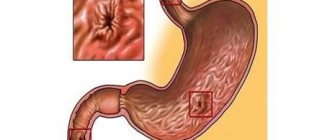
Esophageal cancer is a malignant neoplasm of the esophagus. The main morphological form of esophageal cancer is squamous cell carcinoma (keratinizing or non-keratinizing) (95%), in 5% of cases adenocarcinoma is observed. Most often, distant metastases are detected in the liver, lungs, bones, brain and adrenal glands. This is one of the most aggressive malignant diseases. Esophageal cancer is the 8th leading cause of death worldwide.
Treatment for cancer
Radical surgical intervention for common tumors consists of subtotal or total resection of the organ , tissues of the floor of the mouth, and if the tumor spreads to the larynx, resection or extirpation of the larynx. The operation is completed by applying a temporary tracheostomy.
Patients with advanced cancer are treated with a combination of chemoradiotherapy and chemotherapy. Surgical interventions are performed when it is possible to perform a radical operation with removal of regional lymph nodes or for residual tumor after chemoradiotherapy.
Diagnosis of laryngeal cancer
During the initial visit, the doctor collects an anamnesis of the patient’s life and illness, asks him about the presence of provoking factors, conducts a visual examination, palpation of the neck, indirect laryngoscopy - examination of the larynx with a mirror on a long curved handle.
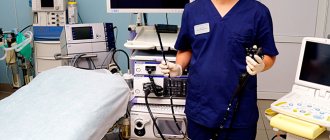
If there is still suspicion of a tumor formation, the patient is prescribed direct laryngoscopy . This is an invasive diagnostic procedure during which the larynx, trachea, and bronchi are examined using a laryngoscope (rigid method) or a flexible fiberscope. As a rule, during direct laryngoscopy, a biopsy of the neoplasm is performed - biomaterial is taken for cytological and histological analysis.
of tumor markers SCC and CYFRA 21-1 is considered an effective diagnostic method . To analyze tumor markers, venous blood is taken from the patient.
To assess the degree of tumor invasion, damage to the lymph nodes, and the presence of metastases in distant organs and tissues, additional procedures are used: CT or MRI , PET scan , biopsy of sentinel lymph nodes , scintigraphy , radiography .
Reconstruction of the pharynx
Despite the constant improvement of methods of combined treatment of advanced cancer of the upper digestive tract, extensive gaping defects of the hypopharynx and cervical esophagus are by no means a rare complication. In some cases, organ-removing operations on the larynx and hypopharynx result in the formation of a planned esophagostomy without subsequent reconstruction.
The purpose of pharynx reconstruction is to restore the natural passage of the bolus of food from the oral cavity to the stomach, as well as to compensate for aesthetic disorders. The operating surgeon is faced with the task of simultaneously replacing defects of the larynx and pharynx while maintaining adequate lumen of the formed section of the digestive tube.
The neck area is a functionally active anatomical zone that does not have a sufficient supply of plastic material. Tissue deficiency seems especially obvious after radiation therapy or radical surgery on the lymphatic tract of the neck and other surgical interventions that cause a fibrotic process. The aggressive environment of the oral cavity and pharynx against the background of suppression of reparative processes in weakened cancer patients significantly aggravates the course of the postoperative period.
Reconstruction methods
The issues of reconstruction of the pharynx and esophagus have been discussed by plastic surgeons for a long time. The earliest reports of pharyngeal reconstruction methods date back to the end of the 19th century. Yu.K. made a great contribution to the development of domestic plastic surgery. Szymanowski, who in 1865 became the author of the first fundamental manual, “Operations on the surface of the human body,” containing an analysis and generalization of the experience of Russian surgeons.
Attempts at total pharyngoplasty and reconstruction of the cervical esophagus with muscle flaps were painful for the patient and ineffective. Severe scarring and rigidity of the walls of the gastrointestinal tube led to difficulty swallowing up to grade III dysphagia and the need for bougienage or installation of a nasogastric tube. At the same time, the incidence of complications with total pharyngoplasty, according to summary data, is up to 71%, mortality is 6%, and the completion of plastic surgery is no higher than 40%.
The gastroepiploic flap is a good plastic material for reconstruction of the upper parts of the digestive tract, allowing the restoration of both the mucous membrane of the organ using a fragment of the stomach, and the volume of soft tissue with covering of vital structures due to the omentum. However, practice shows that in life there are often situations when the stomach is unsuitable for autotransplantation due to its previous resection, roughly applied gastrostomy, or in the presence of a peptic ulcer.
There is no doubt that the use of visceral grafts is a promising direction in the reconstruction of the pharynx and cervical esophagus due to the morphological and functional proximity of the plastic material to the reconstructed organs. However, microsurgical reconstruction of organs and tissues requires the use of expensive equipment, special microsurgical instruments, and is possible only in specialized and few centers with specially trained personnel who have the knowledge and practical skills of these operations, and therefore, the performance of such operations everywhere by a wide range of specialists is sharply limited.
At the Russian Scientific Center for Surgery named after. acad. B.V. Petrovsky performs surgical treatment of patients with cancer of the upper gastrointestinal tract with a simultaneous reconstructive stage.
You might be interested in
Esophageal diverticulum ⇒
Esophageal strictures ⇒
Cardiospasm/achalasia of the cardia ⇒
Treatment methods
For laryngeal cancer , radiation chemotherapy (rarely), targeted therapy , and surgery . A single method or an integrated approach can be used, depending on the stage of the tumor, its location, degree of aggressiveness, growth pattern, and extent of the process.
Conservative therapy
Almost always, the first stage of treatment is radiation therapy . It is used to treat cancer of the middle section of the larynx, which is highly radiosensitive, as well as for tumors of the upper and lower regions of the larynx of stages I-II. Radiation is sometimes combined with hyperbaric oxygenation - saturating the blood with oxygen in a special chamber. This procedure enhances the effect of rays on degenerated cells and reduces damage to healthy tissue.
Treatment of stage III-IV laryngeal cancer, localized in the upper region of the organ, begins with chemotherapy . Chemotherapy is ineffective for the lower and middle parts of the larynx.
Radiation and chemotherapy can be used in combination.
Targeted therapy is the directed effect of a drug on the epidermal growth factor receptor. In laryngeal cancer, a large amount of the EGFR receptor protein is often found on the surface of tumor cells, which stimulates cell division. The drug Cetuximab, used for targeted therapy of the disease, suppresses the activity of this receptor. The drug is administered intravenously, usually used in combination with radiation, and in later stages - together with chemotherapy.
Surgical treatment
Sometimes, for stages I-II of laryngeal cancer, conservative therapy is sufficient. If it turns out to be ineffective, as well as for tumors detected at stages III-IV, surgical intervention is recommended. Before surgery, radiation therapy is always indicated to reduce the size of the tumor.
For stage I-II tumors, doctors try to perform organ-preserving resection: hemilaryngectomy - removal of one vocal cord, supraglottic laryngectomy - removal of part of the larynx above the ligamentous apparatus.
In the early stages, laser removal of the tumor using an endoscope can be used. The advantage of this method is that it is less traumatic; the disadvantage is that it is not possible to take a tissue sample for histological examination.
In later stages of the disease, it is necessary to resort to radical operations: chordectomy - complete removal of the vocal cords, total laryngectomy. In this case, the patient completely loses his voice.
Auxiliary Operations
In addition to direct removal of the malignant tumor, other surgical operations are performed. When laryngeal cancer metastasizes to regional lymph nodes or there is a high risk of metastases, these nodes are excised along with the surrounding tissue. The operation is called a cervical dissection .
When the larynx is completely removed, the patient needs a tracheostomy , a surgically created hole in the trachea. When creating a tracheostomy, the upper end of the trachea is sutured to the skin of the neck.
If laryngeal cancer makes it difficult to eat, the patient will have gastrostomy tube placed directly into the stomach.
If necessary, after extensive surgery, reconstructive plastic - operations that allow at least partially restoring the functions of the removed organs.
Sore throat: possible causes, diagnosis and treatment
Sore throat is a common complaint of patients when visiting an otolaryngologist, therapist, pediatrician and general practitioner. The causes of sore throat can be infectious and inflammatory diseases of bacterial and viral etiology, inflammatory processes in the pharynx, larynx and surrounding organs caused by non-infectious factors, and non-infectious pathology (
). Before proceeding with pharyngoscopy, it is necessary to carefully detail the patient’s complaints and collect an anamnesis. The patient can call a sore throat the unpleasant sensations of scratching, burning, soreness, rawness, the feeling of a “foreign body,” the intensity of which is conveniently assessed on a 10-point scale, where the maximum manifestation of pain is rated at 10 points, the minimum at 1 point.
It is important to determine what, in the patient’s opinion, preceded the onset of pain and what other symptoms it is accompanied by. If there is hyperthermia, then the inflammatory nature of the disease is most likely: pharyngitis, tonsillitis (tonsillitis) [16]. Refusal of food and water can provoke severe pain in aphthous stomatitis (Fig. 2), tonsillitis of the lingual tonsil, paratonsillar and retropharyngeal abscess, Ludwig's angina (phlegmon of the floor of the mouth). With these diseases, drooling is possible, and inflammation of the paramygdaloid tissue and tissues of the floor of the mouth leads to trismus of the masticatory muscles (inability to open the mouth), a forced position of the head tilted to the painful side.
The occurrence of such a condition requires immediate contact with a medical specialist to sanitize the area of inflammation. Epiglottitis, an inflammation of the epiglottis caused by Haemophilus influenzae (more common in young children), gives similar symptoms. Along with refusal to eat, salivation, pain and hyperthermic symptoms, epiglottitis is accompanied by voice disturbance (it becomes dull, hoarse) and difficulty breathing. There is a forced position of the patient in a sniffing position, as if holding porridge in the mouth, the mouth opens freely, but a rough examination of the pharynx with a spatula can lead to laryngospasm and death. Therefore, with the above symptoms, pharyngoscopy is performed carefully; during it, you can see a hyperemic, edematous epiglottis behind the root of the tongue.
Other causes of sore throat accompanied by sore throat and cough can be in children and young people: drainage of discharge from the nasopharynx due to adenoiditis or sinusitis; irritation of the respiratory tract by dry air, smoke, including active and passive smoking; childhood infectious diseases [9]. In adults, a common cause of such complaints, often with a feeling of a lump in the throat, a “foreign body,” is an exacerbation of chronic pharyngitis associated with pathology of the gastrointestinal tract: gastritis, esophagitis, gastroesophageal reflux, cholecystitis, gastric ulcer. Severe dysphagia, regurgitation and pain when swallowing can be caused by esophageal varices [14].
A carefully collected anamnesis allows us to find out the dynamics of complaints, the time of their appearance, the connection with a previous injury or medical examination (gastroscopy), foreign body entry, contact with an infectious patient, hypothermia (drinking cold beer, ice cream), occupational or household hazards (irritants, dust , hot air, taking concentrated solutions of vinegar, spices, medications: corticosteroids, antibiotics, diuretics, local decongestants and others). Sore throat can occur as a manifestation of sexually transmitted diseases: gonococcal pharyngitis, syphilis, chlamydia of the respiratory tract. Infection with the human immunodeficiency virus contributes to the formation of inflammatory viral, tumor and mycotic lesions of the mucous membranes [3].
Chronic pathology of the kidneys, endocrine system, blood, previous radiation and chemotherapy can lead to the formation of a chronic inflammatory and atrophic process in the pharynx. The first manifestation of hyperglycemia [5] may be thirst and dry mouth, accompanied by catarrhal changes in the pharynx. Similar complaints occur with Itsenko–Cushing syndrome [5]. In patients with hypothyroidism, swallowing is often impaired, speech becomes slurred due to swelling and dryness of the tongue and lips, and it is difficult to perform pharyngoscopy.
Non-infectious pathology of the organs of the neck and chest cavity - angina pectoris, myocardial infarction - can manifest itself as intense pain in the pharynx and behind the sternum. Long-lasting dysphagia and unpleasant sensations in the form of a lump or foreign body in the throat, which are not amenable to anti-inflammatory therapy, can cause [14] tumors of the larynx, laryngopharynx, thyroid gland, and pharyngoesophageal (Zencker) diverticula. Many days of fasting, dieting, dyspepsia and heavy menstruation lead to a deficiency of vitamins and minerals. Vitamin A deficiency [5] causes dryness and erosion of the mucous membranes.
Vitamin B2 deficiency produces a triad of symptoms: dermatitis, cheilitis and glossitis (bright red, smooth and shiny dry tongue), accompanied by burning and pain in the mouth when talking and eating. Vitamin C hypovitaminosis occurs with dietary deficiency of ascorbic acid, inflammatory processes in the intestines and is manifested by pain, hemorrhagic and ulcerative-necrotic manifestations in the oral cavity and in the area of the palatine tonsils, mobility and tooth loss. Similar changes in the oral cavity and pharynx give rise to blood diseases (leukemia). Against the background of iron loss (with hyperpolymenorrhea), Plammer-Vinson syndrome is formed, characterized by superficial glossitis, dysphagia, cracks in the corners of the mouth, nail dystrophy, seborrheic dermatitis of the face, blepharitis, conjunctivitis, and decreased vision at dusk. B12 deficiency anemia associated with impaired absorption of this vitamin in the stomach due to anacid gastritis, tapeworm infestation, or increased consumption in pregnant women, manifested by Möller-Gunter glossitis (bright red tongue with smoothed papillae) and atrophy of the pharyngeal mucosa, burning pain in the tongue, weakness, crawling sensation in the limbs. A blood test reveals macrocytes, megalocytes, hyperchromic anemia, leukopenia.
Diseases of the spine [2] (cervical osteochondrosis, tuberculous spondylitis, radiculitis) can cause pain in the pharynx. Neuralgia of the glossopharyngeal nerve manifests itself as intense pain in the pharynx, especially against the background of chronic stress in anxious and suspicious patients. Metabolic disorders, intoxication, and trauma contribute to its occurrence. Characterized by unilateral pain in the root of the tongue, tonsil, lasting several minutes, accompanied by dry throat and subsequent hypersalivation. The therapeutic effect is achieved by lubricating the root of the tongue and pharynx with local anesthetics. Neuralgia of the superior laryngeal nerve [1, 6] gives similar symptoms, but also includes a painful dry cough and spasm of the vocal folds during inspiration [6].
Pain in the pharynx can be caused by an odontogenic process: periodontitis, teething pathology, galvanism [4, 17]. Rarely, unilateral pain in the pharynx occurs, the cause of which is the long styloid process (Eagle syndrome), accessible to palpation in the area of the palatine tonsil [15].
Rare causes of pain in the pharynx are ulcerations on the mucous membrane of tuberculous etiology [14]. In this case, there is a prolonged cough, weight loss, and swollen lymph nodes.
A final diagnosis is possible based on oropharyngoscopy. The main differential diagnosis for pain in the pharynx is between its most common causes - acute (or exacerbation of chronic) pharyngitis and sore throats caused by streptococcal infection (group A beta-hemolytic streptococcus - GABHS).
Acute pharyngitis is a viral infection of the pharynx in 90% of cases. Its main symptoms are: increased body temperature, sore throat when the throat is empty, when eating, soreness and a dry cough that does not bring relief. Patients indicate the localization of unpleasant sensations on the back wall of the pharynx. In the pharynx (Fig. 3), hyperemia of all parts is detected: the posterior wall, arches, tonsils, and there may be vesicular rashes (herpes, enterovirus). Plaques are not typical, there is often a runny nose and other catarrhal phenomena - nasal congestion, sneezing. If a rash is detected on the skin and mucous membranes, it is necessary to exclude an infectious disease - measles, scarlet fever, rubella [8]. Adenoviral infection manifests itself in the form of conjunctivitis, enlarged lymph nodes, fever, runny nose, and there may be plaque in the throat.
The disease progresses in waves: on the 7th–10th day of illness, a repeated increase in body temperature and a return of symptoms are possible. Enterovirus infection (“summer flu”) manifests itself in the form of dyspeptic, myalgic, and meningeal syndromes. Pharyngoscopy reveals bubbles on the mucous membrane of the oropharynx. In making the diagnosis of viral pharyngitis, the following help: knowledge of the epidemiological situation, dynamic observation of the patient, a variety of clinical manifestations (abdominal pain, vomiting, eye damage, meningeal symptoms, myalgia), lymphocytosis in the blood with normal ESR, lack of effect from antibacterial therapy, serological data research, polymerase chain reaction and others.
Sore throat is a general infectious disease with local manifestations in the form of acute inflammation of one or more components of the lymphadenoid pharyngeal ring, most often the palatine tonsils (tonsillitis), pharyngeal tonsil (adenoiditis), lingual tonsil, lateral ridges of the pharynx and larynx.
The classification of acute tonsillitis (according to I.B. Soldatov, 1975) involves the division into primary tonsillitis: catarrhal, lacunar, follicular, ulcerative-membranous; and secondary: for acute infectious diseases (diphtheria, scarlet fever, measles, tularemia, typhoid fever, infectious mononucleosis) and diseases of the blood system (agranulocytosis, alimentary-toxic aleukia, leukemia). There are special forms of tonsillitis [10]: viral, fungal, syphilitic.
With catarrhal tonsillitis, there is hyperemia and enlargement of the palatine tonsils, regional lymphadenitis, no plaque, a blood test shows slight leukocytosis, increased ESR. Catarrhal sore throat often has to be differentiated from viral pharyngitis, in which there is a cough, there may be a runny nose, and there is no enlargement or tenderness of the lymph nodes.
Follicular tonsillitis is manifested by bright hyperemia and swelling of the tonsils, subepithelial rounded yellowish elevations (follicles).
With lacunar tonsillitis, white-yellow plaques appear at the mouths of the lacunae of the tonsils (Fig. 4), which can merge with each other and cover the entire surface without going beyond the tonsils, being removed without leaving a bleeding surface, rubbing between two spatulas, dissolving in the vessel with water.
It is important to determine the streptococcal etiology of acute tonsillitis. This can be done using the McIsaac screening scale [12], which includes symptoms and their assessment in points (
).
A patient’s symptoms with a score of 3 means the probability of streptococcal etiology is 30%, and a score of 4 means about 70%. If the clinical picture is determined to be 0–1 points, then systemic antibiotic therapy is not indicated. At 2–3 points, antibiotics are necessary only if the infection is bacteriologically confirmed. Antibiotics should be prescribed when symptoms of 4–5 points are detected.
Diphtheria of the pharynx occurs in the form of localized, widespread, toxic I, II, III degrees and hypertoxic forms. With it, gray plaques are detected on the surface of the tonsils, tightly fused with the underlying tissues, they may spread to the arches, the mucous membrane of the posterior pharyngeal wall, the uvula (Fig. 5), and when you try to remove them, the mucous membrane bleeds. Plaques do not rub down and do not dissolve in water. In the toxic form, the neck is noticeably thickened due to swelling of the subcutaneous fat, pressure is painless and does not leave pits.
Infectious mononucleosis is an acute infectious disease caused by the Epstein-Barr virus, characterized by fever, sore throat, enlarged lymph nodes, pharyngeal tonsil, liver, and spleen. Plaques on the tonsils, as in lacunar tonsillitis (Fig. 6), but can spread beyond them. A general blood test (the appearance of atypical mononuclear cells), serological research methods (enzyme-linked immunosorbent assay (ELISA), Paul-Bunnel-Davidson reaction) help in diagnosing the disease.
Fungal infections of the pharynx in the form of curdled films, easily removed when scraped with a spatula, are caused mainly by yeast-like fungi of the genus Candida (about 90% of cases); less commonly, there are mold fungi of the genus Aspergillus, Penicillium [7]. C. albicans permanently or temporarily lives on human mucous membranes, skin and intestines. Factors contributing to the development of mycoses are: treatment with broad-spectrum antibacterial drugs, cytostatics and corticosteroids, diabetes mellitus, blood diseases, tumors, gastrointestinal diseases, vitamin imbalance. Candidiasis of the oral cavity, pharynx and esophagus occurs in more than 90% of AIDS patients (Fig. 7) [3].
Diagnosis of sore throats should include throat and nasal swabs for diphtheria (BL). Express determination of streptococcal antigen using test strips from the surface of the tonsils makes it possible to justify antibacterial therapy. A general blood test facilitates the differential diagnosis of primary and secondary acute tonsillitis. Opinions on the question “Should I do a smear?” controversial. It is needed to confirm the GABHS etiology of the disease in doubtful cases [16].
Therapeutic tactics for pain in the pharynx can be presented in the form of a diagram ().
For the treatment of acute pharyngitis, a gentle diet, hot foot baths, warm compresses on the front surface of the neck, warm alkaline drink (mineral water, milk with honey), steam inhalations, and smoking cessation are recommended. The attitude towards gargling is ambiguous [16]. Fresh infusions of mint, chamomile, calendula, eucalyptus, sage, and caragana are effective for relieving sore throat. Antiseptics of artificial origin (dichlorobenzene, metacresol, hexethidine, benzalkonium, thymol, ambazone, chlorhexidine) are bactericidal in their mechanism of action, which can lead to suppression of the normal microflora of the oral cavity, so they should be used with caution in children under 6 years of age [9]. For the treatment of sore throat, the most important painkillers are those containing menthol, tetracaine, lidocaine or flurbiprofen. Children from 6 months of age can use the herbal preparation Tonsilgon, which has an antiseptic and analgesic effect, but does not contain either menthol or lidocaine.
Menthol preparations and any sprays cannot be used before 3 years of age due to the possible development of laryngospasm.
Local combination drugs (antiseptics and painkillers) in the form of finished dosage forms are the most popular for the treatment of sore throat. One of them is TheraFlu LAR, a universal and highly effective local drug with an antiseptic and analgesic effect. TheraFlu LAR contains benzoxonium chloride and lidocaine. Available in the form of a spray and tablets, the content of lidocaine in which is 0–75 mg and 1 mg, respectively.
Recent in vitro and in vivo studies confirm the broad antiseptic spectrum of benzoxonium chloride and its activity against the main pathogens of the most common diseases of the oral cavity and pharynx. At the same time, the balance of the bacterial flora in the mouth is not disturbed, even with prolonged use of benzoxonium chloride.
Benzoxonium chloride has:
- bactericidal effect against aerobic and anaerobic gram-positive and gram-negative bacteria;
- fungicidal effect against Candida albicans, Aspergillus spp. and yeast fungi;
- antiviral activity against membrane viruses, including herpes virus, influenza virus, parainfluenza viruses, the causative agent of vesicular stomatitis.
Lidocaine is a local anesthetic that reduces pain in the throat when swallowing.
Adults should take the drug 1 lozenge every 2–3 hours (no more than 10 tablets per day) or as a spray, 4 sprays 3–6 times a day. Children aged 4 years and older are prescribed 1 lozenge every 2-3 hours (no more than 6 tablets per day) or as a spray, 2-3 sprays 3-6 times a day. The required duration of treatment, as a rule, does not exceed five days. The drug is well tolerated by patients, side effects are recorded extremely rarely. Possible short-term local irritation of the mucous membrane, allergic reactions.
Indications for its use are: pharyngitis, laryngitis, catarrhal tonsillitis, stomatitis, ulcerative gingivitis. Contraindications to the use of TheraFlu LAR: pregnancy (first trimester), breastfeeding, hypersensitivity to lidocaine, children under 4 years of age. TheraFlu LAR can be used by people with diabetes, as it does not contain sugar. Thanks to the combined effect - antibacterial and analgesic, TeraFlu LAR can relieve all symptoms of viral pharyngitis and be used in the complex therapy of bacterial sore throats.
Systemic antibiotic therapy in the treatment of patients with “sore throat” is indicated mainly for acute tonsillitis (angina) of suspected or established streptococcal etiology (GABHS) and is carried out with penicillins, and if they are intolerant, with macrolides; cefuroxime (Axetin) is effective. Clindamycin and lincomycin are reserve drugs [11]. Epiglottitis caused by Haemophilus influenzae is effectively treated with protected penicillins.
Literature
- Alimetov Kh. A. Secondary neuropathy of the upper laryngeal nerve / M materials of the anniversary All-Russian. scientific-practical conf. with international participation “Modern aspects and prospects for the development of otorhinolaryngology. M., September 29–30, 2005. P. 46.
- Alimetov Kh. A. Spondylogenic pharyngeal dyskinesia / Materials of the anniversary All-Russian. scientific-practical conf. with international participation “Modern aspects and prospects for the development of otorhinolaryngology. M., September 29–30, 2005. P. 22.
- Bessarab T.P., Yushchuk N.D., Anyutin R.G., Potekaev S.N. HIV infection in otorhinolaryngological practice // Attending Doctor, 2000. No. 1. P. 26–30.
- Inflammatory diseases of the mucous membrane of the pharynx, oral cavity and periodontium. Scientific review. Solvay pharma, 2002. P. 2.
- Diseases of the mucous membrane of the oral cavity and lips / Ed. E. V. Borovsky, A. L. Mashkilleyson. 1984. 400 p.
- Karpova O. Yu. Clinic, diagnosis and treatment of laryngoneurosis / Materials of the anniversary All-Russian. scientific-practical conf. with international participation “Modern aspects and prospects for the development of otorhinolaryngology. M., September 29–30, 2005. P. 53.
- Kunelskaya V. Ya., Kasimov K. On the issue of the clinic, diagnosis and treatment of candidal tonsillitis in children // Vestn. otorhinolaryngology. 1980. No. 4. P. 50–52.
- Nisevich N. I., Uchaikin V. F. Infectious diseases in children. M.: Medicine, 1985. 298 p.
- Acute respiratory diseases in children: treatment and prevention. Scientific and practical program of the Union of Pediatricians of Russia / Ed. A. A. Baranova. M., 2008.
- Otorhinolaryngology: national guide / Ed. V. T. Palchuna. M.: GEOTAR-Media, 2008. 960 p.
- Rational antimicrobial pharmacotherapy: Hand. for practicing doctors. Under general ed. V. P. Yakovleva, S. V. Yakovleva. M.: Litterra, 2003. 108 p.
- Sidorenko S.V., Guchev I.A. Tonsillopharyngitis: issues of diagnosis and antibacterial therapy // Consilium medicum. Infections and antimicrobial therapy. 2004. 4: 36–38.
- Folomeeva O. M., Amirdzhanova V. N., Yakusheva E. O. et al. Incidence of rheumatic diseases in the Russian population (analysis over 10 years) // Ter. Archive. 2002. No. 5. P. 5–11.
- Shevrygin B.V., Mchedlidze T.P. Handbook of otorhinolaryngology. M.: Triada-X, 1998. 448 p.
- Shulga I. A., Zaitsev N. V., Zaitseva V. S. Variants of the structure of the stylohyoid complex / Materials of the Anniversary All-Russian. scientific-practical conf. with international participation “Modern aspects and prospects for the development of otorhinolaryngology. M., September 29–30, 2005. P. 75.
- Etiopathogenetic therapy of diseases of the upper respiratory tract and ear: Methodological recommendations. Compiled by S.V. Ryazantsev, Kotserovets V.I. St. Petersburg: National Register, 2008. 100 p.
- Yakovleva V.I. Diagnosis and treatment of neurogenic diseases of the maxillofacial region: Textbook. manual for institutes and faculties. improved doctors. Minsk: Vysh. school, 1989. 102 p.
M. V. Subbotina , Candidate of Medical Sciences Irkutsk State Medical University , Irkutsk
Contact information about the author for correspondence
Forecast
The prognosis of the disease depends on how early the tumor is detected. Unfortunately, laryngeal tumors are often diagnosed late due to the nonspecificity of early symptoms.
Newly diagnosed stage III laryngeal cancer is 46.8%, stage IV – 17.0%. The mortality rate in the first year from the moment of diagnosis for lesions of the larynx is 24.2%.
A large number of patients develop resistance to radiation and chemotherapy. When conservative therapy is used, recurrent tumors occur in 20-40% of cases, the treatment of which is only possible through surgery.
Without treatment, laryngeal cancer lasts from one to three years. The prognosis of 85-90% of cases of complete recovery is given only if the tumor is detected early, treatment is started in a timely manner and completely completed.
Palliative treatment of advanced esophageal cancer
In this situation, the complete impossibility of nutrition through the esophagus, which is practically closed by the tumor, comes to the fore. Often they resort to expansion - recanalization or installation of a special stent in the esophagus. Stents are required when connecting the esophagus and trachea or bronchi with an anastomosis - a tumor fistula, which prevents the reflux of food into the respiratory tree and protects against pneumonia. In some cases, during endoscopy, the tumor is partially destroyed with a laser using photodynamic therapy. There are enough treatment options for esophageal cancer; you need to use them promptly and skillfully.
| More information about treatment at Euroonco: | |
| Oncologist-gastroenterologist | RUB 5,100 |
| Chemotherapy appointment | RUB 6,900 |
| Emergency oncology care | from 12,100 rub. |
| Palliative care in Moscow | from 44,300 rubles per day |
| Radiologist consultation | RUB 11,500 |
Book a consultation 24 hours a day
+7+7+78
Prevention of laryngeal cancer
Quitting smoking cigarettes, pipes, hookahs, and chewing tobacco is the basis for preventing the disease. Eliminating alcoholic beverages or reducing their consumption will help prevent not only laryngeal cancer, but also other pathologies.
There is an opinion that red meat and smoked meats increase the risk of cancer. You should reduce their number in the menu, eat fresh vegetables and fruits more often.
It is important to undergo medical examinations on time - medical examinations, medical examinations at enterprises. If you suspect a disease of the larynx, even if general symptoms appear, you should consult a doctor.
Information verified by an expert
Mikhailov Alexey Gennadievich operating oncologist, doctor of the highest qualification category, Ph.D. experience: 21 years
The information in this article is provided for reference purposes and does not replace advice from a qualified professional. Don't self-medicate! At the first signs of illness, you should consult a doctor.