The article was prepared by a specialist for informational purposes only. We urge you not to self-medicate. When the first symptoms appear, consult a doctor.
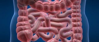
Antibiotics for diarrhea do not always need to be taken. You should not start taking antibacterial drugs without first finding out the cause of the problem. However, there are situations in which taking antibiotics is the only treatment option.
Indications for taking antibiotics for diarrhea
There are specific indications for prescribing antibacterial therapy against the background of diarrhea.
This applies to cases where stool liquefaction was caused by a bacterial disease:
- Salmonellosis.
- Cholera.
- Dysentery.
If diarrhea is a consequence of a viral infection, then you should stop taking antibacterial drugs. Failure to comply with this recommendation may result in the development of complications.
Taking antibiotics allows you to destroy pathogenic bacterial flora, but at the same time they kill beneficial bacteria that are always present in the intestines. That is why using them without good reason is prohibited. As a result of improper therapy, it will only be possible to worsen the course of the disease.
Symptoms that may indicate the need for antibiotic therapy:
- Copious loose stools.
- The presence of green impurities in the stool.
- The presence of mucus in large quantities in the stool.
- Presence of blood in stool.
Why does common diarrhea not require antibiotics?
Diarrhea is not always caused by bacteria.
The cause of thinning and frequent stools may be:
- Viral infection (enteroviruses, rotoviruses, caliciviruses).
- Parasitic infestation (infection with helminths).
- Toxic infection.
- Infection of the intestines with protozoan microorganisms.
Moreover, it is not always the infection that causes diarrhea.
The reason for its occurrence can be reduced to one of the following factors:
- Taking medications.
- Inflammation of the small or large intestine against the background of chronic damage to the gastrointestinal tract.
- Ischemic intestinal disease.
- Diseases of other organs included in the digestive system.
- Failure in food digestion processes.
- Impaired absorption of nutrients.
- Lactase deficiency.
- Enzyme deficiency, gallbladder dysfunction.
Sometimes diarrhea develops against the background of neurological disorders, for example, with severe emotional excitement or stress.
As a rule, intestinal infections that were not provoked by bacterial flora go away on their own. Taking antibiotics in this case is not required. Moreover, they can cause serious harm to health. For example, if diarrhea was caused by enterotoxins entering the body, then taking antibiotics can cause toxic and infectious shock.
It is dangerous to take antibacterial drugs if the intestines are damaged by the E.Coli bacterium. Under the influence of these drugs, bacteria begin to produce Shiga toxins. They have a hemolytic-uremic effect. This can lead to the infection spreading throughout the body and making it very difficult to get rid of.
Diarrhea that develops while taking antibacterial drugs or after their discontinuation also cannot be treated with these medications. This will lead to the fact that the entire intestinal microflora will be completely destroyed, and a metabolic failure will occur. Against this background, osmotic diarrhea often develops, which is fraught with serious complications for human health.
Causes of abdominal pain and diarrhea
These symptoms most often accompany diseases of the abdominal organs: stomach, intestines, liver, gallbladder and bile ducts, pancreas, internal genital organs and bladder. The appearance of pain is provoked by spasms or stretching of the walls of hollow organs (intestines, stomach, gall bladder), stretching of the outer membrane of the liver and pancreas, poor circulation and inflammation of the organs themselves, the membrane of the abdominal cavity covering them.
The causes of loose stools from a physiological point of view can be:
- increased secretion of fluid into the intestinal cavity due to its irritation, for example, by bacterial toxins during foodborne toxic infection,
- accumulation in the small and large intestines of substances that attract water, as happens, in particular, with lactose intolerance;
- inflammation of the intestine, accompanied by the release of various fluids into it, for example, mucus, pus in nonspecific ulcerative colitis;
- increased intestinal motility, for example, with irritable bowel syndrome,
- slowing of motor skills, for example, with scleroderma.
Due to the common causes of diarrhea and abdominal pain, they are often combined with each other. In particular, pain is caused by an increase in the volume of intestinal contents and the accumulation of gases that stretch the intestinal walls. Inflammation and spasms, in turn, increase the symptoms of the disease.
Up to contents
How to choose a drug to treat diarrhea?
To get rid of diarrhea and not harm your own health, you first need to get medical advice. Only after passing all the necessary tests will the doctor be able to select adequate therapy. It will be etiological, that is, based on the causes of the disease.
The goal of antibacterial therapy is to destroy pathogenic flora, eliminate symptoms of the disease, and restore normal intestinal function.
The antibiotic is selected depending on the sensitivity of the pathogenic flora to it. In addition, the doctor may prescribe intestinal antiseptics, which can help get rid of the infection faster.
If the patient has a history of an allergic reaction to a particular drug, then before taking an antibiotic, he is prescribed special allergen tests.
Antibacterial therapy is possible for the following indications:
- The patient has an infection caused by invasive bacterial flora. Diarrhea is characterized by large, loose stools containing mucus and blood.
- Against the background of an intestinal infection, the patient’s condition is complicated by other somatic diseases. The kidneys, heart, and lungs may be damaged. It is possible that a serious complication such as blood poisoning may develop.
It is mandatory to take antibiotics when treating dysentery and escherichiosis, when the infection spreads through the bloodstream throughout the body. Antibiotics are sometimes recommended for people suffering from traveler's diarrhea.
Effective drugs for treating diarrhea:
- Levomycetin is a drug that has been used to treat bacterial diarrhea for many years. Levomycetin has a wide spectrum of action, due to which it fights most pathogenic microorganisms.
- Metronidazole is an antimicrobial and antiprotozoal drug.
- Ciprofloxacin is a modern antibacterial drug that allows you to get rid of most diarrhea pathogens.
- Amoxicillin is an antibiotic that is effective against a number of intestinal and extraintestinal pathogens.
In any case, before taking any medication, you must obtain medical advice.
Infectious diarrhea in the practice of a general practitioner
Academician of the Russian Academy of Medical Sciences, Professor V.T. Ivashkin, professor A.A. Sheptulin
MMA im. THEM. Sechenov
Diarrhea of an infectious nature is currently one of the most common diseases and ranks second in frequency after acute inflammatory diseases of the upper respiratory tract. For example, in the countries of Africa, Asia (excluding China) and Latin America, more than 750 million cases of acute infectious diarrhea are registered annually in children under 5 years of age, which leads to death in more than 4.5 million children [1].
The causative agents of infectious diarrhea can be various agents that can determine the uniqueness of the clinical picture of the disease, features of diagnosis and treatment. The seriousness of the problem is also due to the fact that the diagnosis and treatment of infectious diarrhea is often carried out not by infectious disease specialists, but by general practitioners [2].
Bacterial diarrhea
Etiology and pathogenesis
Significant changes have currently occurred in the structure of etiological factors of bacterial diarrhea. The frequency of infectious diarrhea caused by common pathogens (Shigella, Salmonella) has decreased, and the number of cases of diseases caused by enteropathogenic strains of Escherichia coli and Campylobacter infection has increased [3].
Pathophysiological mechanisms of bacterial diarrhea include the production of an enterotoxin, which increases the activity of adenylate cyclase and thus stimulates the secretion of water and electrolytes by enterocytes (for example, in infections caused by Vibrio cholerae, clostridia, enterotoxin-producing strains of Escherichia coli), or direct invasion of bacteria into the epithelial cells of the mucous membrane intestines with their subsequent damage and the development of an inflammatory reaction (with shigellosis infection, infection caused by enteroinvasive strains of Escherichia coli, yersiniosis, salmonellosis).
Clinical picture
The incubation period for bacterial diarrhea can last from several hours (with salmonellosis or staphylococcal infection) to 10 days (with yersiniosis). The leading clinical symptoms of bacterial diarrhea, in addition to loose stools, are fever and cramping abdominal pain. In this case, some features of clinical manifestations may be noted, due to the etiological factor. For example, the clinical picture of Campylobacter ileitis may resemble acute appendicitis, and the surgeon who decides to operate in this situation finds an unchanged appendix and signs of mesenteric lymphadenitis. Yersinia infection sometimes occurs with the development of erythema nodosum and joint damage. Salmonella infection can be complicated by bacteremia with the occurrence of pneumonia, meningitis, and abscesses of internal organs. Infectious diarrhea caused by enterohemolytic strains of Escherichia coli (0157:H7) can lead to the development of hemolytic-uremic syndrome, manifested by acute renal failure, hemolytic anemia and thrombocytopenic purpura.
Diagnostics
Diagnosis of bacterial diarrhea involves a clinical blood test (leukocytosis with a shift of the formula to the left is detected) and sigmoidoscopy (a picture of acute proctosigmoiditis with shigellosis infection), as well as a search for the etiological factor that caused its development. Stool culture followed by microbiological examination gives a positive result in approximately 40-60% of patients with acute diarrhea, accompanied by fever and the appearance of leukocytes in the stool [4]. If culture results are negative, immunological diagnostic methods are used. Thus, the use of enzyme immunoassay methods makes it possible to detect antibodies to Campylobacter and Salmonella. Enterotoxins from pathogenic strains of Escherichia coli can be detected using polymerase chain reaction and latex agglutination. With shigellosis, antibodies to antigen 0 can be determined already in the first days of the disease using the hemagglutination method.
Treatment
Fighting dehydration
Treatment of acute infectious diarrhea should include, first of all (especially in children), the fight against dehydration. In mild cases of the disease, rehydration therapy comes down to sufficient fluid intake (tea, mineral water, etc.) containing glucose and electrolytes. The simplest rehydration solution is prepared as follows: add 1/2 teaspoon of table salt (3.5 g of sodium chloride) and 1 teaspoon of soda (2.5 g of bicarbonate) to 1 glass of orange juice (it contains 1.5 g of potassium chloride). sodium), after which the total volume of the solution is brought to 1 liter with boiled water [5].
For more severe dehydration, special rehydration solutions are indicated that have an electrolyte composition recommended by WHO (Na+ 90 mmol/l, K+ 20 mmol/l, CI- 80 mmol/l, HCO-3 30 mmol/l, glucose 110 mmol/l) . In pediatric practice, in order to combat dehydration during diarrhea, the drug rehydron is used, containing 3.5 g of sodium chloride, 2.9 g of sodium citrate, 2.5 g of potassium chloride and 10 g of dextrose in 1 sachet. After dissolving the contents of the sachet in 1 liter of warm boiled water, the patient is given the resulting solution to drink, based on the expected loss of body weight (with a loss of 57.5% of body weight, the volume of injected liquid is 4050 ml/kg body weight for 4 hours or up to 150 ml/ kg body weight per day). The total volume of orally administered fluid for adult patients should be at least 23 liters per day. In case of severe dehydration (loss of more than 10% of body weight within 24 hours), they additionally resort to intravenous administration of water and electrolyte solutions. For patients with acute infectious diarrhea, a light diet is recommended for a period of 23 days, including foods such as slimy soups, rice, dried bread, salted crackers, baked potatoes, eggs, etc. Avoid foods rich in plant fiber, dairy products, coffee and alcohol.
Antibacterial therapy
Antibacterial drugs, previously widely used in the treatment of bacterial diarrhea, are now prescribed differentially, taking into account the type of pathogen and the severity of the disease. It should be borne in mind that many forms of infectious diarrhea end on their own within 5 days with rehydration therapy [5].
In uncomplicated cases of salmonellosis, antibiotics are not indicated, since they do not reduce the duration of the disease and prolong the period of isolation of the pathogen. Antibiotic therapy is used in cases where the disease occurs with high fever, signs of severe intoxication, bacteremia and damage to other organs. The drugs of choice in this case are ampicillin (at a dose of 46 g per day) or chloramphenicol (1 g 3 times a day), which are used for 2 weeks. In case of severe intoxication, co-trimoxazole (160 mg trimethoprim and 800 mg sulfamethoxazole 2 times a day for 12 weeks) can be considered an alternative.
Antibiotic therapy administered to patients with shigellosis helps reduce the duration of fever and shorten the period of carriage of microorganisms. The drug of choice is co-trimoxazole, prescribed at a dose of 960 mg 2 times a day for 5 days. Taking into account possible resistance to this drug, nalidixic acid (1 g 4 times a day), norfloxacin (400 mg 2 times a day) or ciprofloxacin (500 mg 2 times a day) can also be used instead. Ampicillin and doxycycline are used only when the sensitivity of the cultured bacterial strains to them is confirmed. The use of ceftriaxone (1 g per day intravenously for 5 days) is considered as a backup treatment method.
In the treatment of uncomplicated campylobacteriosis, antibiotics usually do not play a significant role, since the clinical manifestations of this disease often completely subside in such cases even before the pathogen is identified. Antibacterial agents are usually used in cases of severe disease, severe intoxication, and the presence of blood in the stool. The main drug for the treatment of campylobacteriosis is erythromycin, which is prescribed at a dose of 1 g per day (in 2 or 4 doses) for 57 days. Tetracyclines (for example, doxycycline 200 mg on day 1 and then 100 mg per day) and fluoroquinolones are also effective. For campylobacter septicemia, gentamicin (at a dose of 45 mg per 1 kg of body weight), ceftriaxone (1 g per day) or chloramphenicol (3 g per day) is used.
For yersiniosis, antibacterial treatment is carried out only in severe cases of the disease. The drugs of choice in this case should be gentamicin (5 mg per 1 kg of body weight per day intravenously) or chloramphenicol (50 mg per 1 kg of body weight per day intravenously or orally). The duration of treatment should be at least 2 weeks.
The use of antibiotics for cholera contributes to the rapid disappearance of Vibrio cholerae from feces and a reduction in the duration of diarrhea. Tetracycline (250 mg every 6 hours for 4 days) remains the drug of choice. You can also use furazolidone (at a dose of 5 mg per 1 kg of body weight per day, divided into 4 doses, for 3 days), chloramphenicol (500 mg 4 times a day for 7 days) and co-trimoxazole (960 mg 2 times per day for 1 week).
The use of symptomatic antidiarrheals in the treatment of infectious diarrhea caused by enteroinvasive bacteria (Shigella and Salmonella) is not indicated because they slow the clearance of microorganisms and prolong the duration of the disease. Their use should also be avoided in case of high fever, severe intoxication, as well as in the presence of leukocytes and blood in the stool, due to the risk of developing toxic dilatation of the colon (toxic megacolon).
Viral diarrhea
Among the various types of viral diarrhea, diarrhea of rotavirus etiology (the most common form of infectious diarrhea in children), diarrhea caused by the Norfolk virus, as well as adeno- and astroviruses are of greatest clinical importance. In the clinical picture of viral diarrhea, attention is drawn to the frequent combination of diarrhea and fever with dyspeptic disorders, and often (with rotavirus diarrhea) with damage to the upper respiratory tract. The diagnosis is confirmed by detecting the virus in the stool using electron microscopy or by conducting special immunological studies (for example, with monoclonal antibodies). The course of diarrhea of viral origin is usually favorable. The duration of the disease usually does not exceed 35 days. Treatment is symptomatic and boils down to eliminating water and electrolyte disturbances.
Diarrhea of parasitic origin
In the group of diarrhea of parasitic origin, amebiasis and giardiasis are of greatest clinical importance.
The causative agent of amebiasis is dysenteric amoeba, which is characterized by almost universal prevalence. It is believed that about 10% of the entire world population is infected with these protozoa. Moreover, in 70% of infected people, colonization of the colon mucosa by parasites is asymptomatic, while in other cases the classic picture of amoebic colitis develops with abdominal pain, diarrhea and fever. The stool contains blood that is clearly visible to the eye. In severe cases of amebiasis, complications are possible in the form of perforation of the intestinal wall with the development of peritonitis, toxic megacolon, abscesses of the liver, lungs, and brain. The diagnosis of amebiasis is confirmed by detection of Entamoeba histolytica trophozoites or their cysts in the stool. Of additional diagnostic value is the determination of specific IgA, IgM and IgG by the enzyme immunoassay method. Treatment includes metronidazole (0.75 g 3 times a day for 5-10 days) or a combination of tetracycline (0.25 g 4 times) with chloroquine (0.25-0.5 g 34 times) for 15 days.
One of the most common infections that can cause acute or chronic diarrhea is giardiasis. When Giardia enters the intestines, in many cases asymptomatic carriage develops. The picture of giardiasis enteritis occurs only in 2550% of infected people, and the disease often takes a chronic course. The diagnosis of giardiasis is confirmed by detection of trophozoites or Giardia cysts in feces, bile or duodenal contents. It is also possible to determine antibodies of the IgA, IgM and IgG classes to G.lamblia. The drug of choice in the treatment of patients with giardiasis is metronidazole, used for 7 days at a dose of 0.25 g 3 times a day.
Special forms of diarrhea
Currently, special forms of infectious diarrhea have been identified:
- traveler's diarrhea;
- diarrhea in homosexual men;
- diarrhea in patients with AIDS;
- antibiotic-associated diarrhea;
- bacterial overgrowth syndrome.
The need for their separate consideration is dictated by various circumstances: wide prevalence (travelers' diarrhea), unique etiological factors (diarrhea in homosexual men, diarrhea in AIDS patients), and often uncontrolled use of antibiotics (antibiotic-associated diarrhea).
The risk of developing traveler's diarrhea is highest (30-70%) when traveling to countries in Asia, Africa and Latin America. In most cases, this form of the disease is caused by enterotoxigenic strains of Escherichia coli (less commonly Salmonella, Shigella and other microorganisms). Of additional importance in its development are changes in the nature of nutrition, climatic features, and nervous stress. Travelers' diarrhea usually begins suddenly, occurs with cramping abdominal pain and a slight increase in temperature. As a rule, there are no signs of dehydration and the symptoms of the disease disappear spontaneously within 34 days. For most patients, symptomatic treatment is sufficient. Loperamide (Imodium) has proven itself well. The antidiarrheal effect of the drug is associated with a decrease in peristalsis and an increase in the transit time of contents through the intestines. The drug increases the tone of the anal sphincter, promoting better fecal retention and reducing the imperative urge to defecate. In adults with acute diarrhea, the first dose of Imodium is 4 mg (in children 2 mg), then after each episode of loose stool - 2 mg. Antibacterial drugs (furazolidone, fluoroquinolones, co-trimoxazole) are prescribed only in case of high fever and symptoms of intoxication.
Infectious diarrhea in homosexual men is caused by specific pathogens (gonococci, chlamydia, herpes simplex virus). Thus, entry of gonococci into the rectum can cause sluggish gonorrheal proctitis with mucopurulent discharge. The diagnosis is confirmed by the detection of gram-negative diplococci in leukocytes obtained using a swab during sigmoidoscopy. Treatment in such cases is carried out with penicillins.
The combination of diarrhea syndrome with severe proctalgia, difficulty urinating, enlarged inguinal lymph nodes and characteristic vesicular rashes on the rectal mucosa in the anal area may indicate that the patient has an infection caused by the herpes simplex virus. The diagnosis is confirmed by examining the contents of the vesicles (using tissue culture), as well as determining the titer of the corresponding antibodies. Acyclovir is used for treatment.
Diarrhea in AIDS patients is one of the leading syndromes in the clinical picture of the disease, occurring in 30-40% of patients. The etiological factors of infectious diarrhea in patients most often are protozoa (cryptosporidium and isospores), opportunistic viruses (cytomegalovirus, herpes simplex virus), bacterial agents (most often Mycobacterium avium intracellulare). Infectious diarrhea in patients with AIDS often takes a life-threatening course (primarily due to significant loss of body weight) and is difficult to treat.
In addition, diarrhea in patients with AIDS can be caused by the direct effect of the human immunodeficiency virus itself on the intestinal mucosa with the development of HIV-associated enteropathy and malabsorption syndrome. The ability of the human immunodeficiency virus to enhance secretion processes in the intestines sometimes leads to the occurrence of secretory diarrhea with an increase in stool volume to 1214 liters per day. The only drug effective in such cases is octreotide. Finally, when looking for the causes of diarrhea in patients with AIDS, it is necessary to keep in mind that it may be associated with malignant lesions of the gastrointestinal tract (Kaposi's sarcoma, malignant lymphoma).
Antibiotic-associated diarrhea becomes important clinically when it is caused by Clostridium difficile. The frequency of carriage of these microorganisms, which is 315% among the adult population, increases significantly (up to 1540%) when taking antibiotics (primarily clindamycin, ampicillin, cephalosporins), which inhibit the growth of strains of intestinal flora that normally suppress the vital activity of Clostridium difficile.
The spectrum of clinical manifestations of this infection ranges from mild loose stools to severe pseudomembranous colitis associated with the damaging effect on colonocytes of enterotoxins A and B produced by these microorganisms. Pseudomembranous colitis usually occurs with high fever, cramping abdominal pain, loose stools (often mixed with blood), and high leukocytosis. The disease can be complicated by electrolyte disturbances, the development of arterial hypotension and toxic megacolon. The diagnosis is confirmed by endoscopic examination (characteristic plaques on the mucous membrane of the colon in the form of pseudomembranes) and detection of Clostridium difficile (using tissue culture) or its toxins (enzyme immunoassay). Treatment of patients with pseudomembranous colitis is carried out with vancomycin (0.1250.5 g 4 times a day) or metronidazole (0.25 g 4 times a day) for 714 days.
Bacterial overgrowth syndrome is a specific variant of infectious diarrhea, which is based on an increase in the content of bacteria in the small intestine (from 104107/ml to 1011/ml). Bacterial overgrowth syndrome occurs in cases where the passage of contents through the small intestine slows down (for example, after operations on the stomach and intestines, during adhesions, intestinal strictures) or the function of the ileocecal valve is disrupted (resection of the cecum and ileum), resulting in the contents the colon enters the lumen of the small intestine.
Bacterial overgrowth syndrome is clinically manifested by diarrhea followed by the development of malabsorption syndrome. Diagnosis is based on the results of a breath test performed with labeled lactulose, as well as on the detection of elevated levels of microorganisms (>106) in the duodenal aspirate. Treatment of bacterial overgrowth syndrome should be aimed at eliminating the diseases that caused its development (tumor of the small intestine, adhesions, fistulas, etc.).
Thus, the presented data show that in clinical practice, a general practitioner may encounter various forms and variants of infectious diarrhea, often having clinical features caused by the causative agent of the disease. Knowledge of these features turns out to be extremely necessary for making a correct diagnosis and prescribing adequate treatment.
Literature
1. Speelman P. Acute gastrointestinal infections and their complications. Current topics in gastroenterology and hepatology (Ed. GNJTytgat, M. van Blankenstein). Stuttgart-New York, 1990; 817.
2. Ivashkin V.T. Infectious diarrhea in the practice of a gastroenterologist. Ross. magazine gastroenterology, hepatology, coloproctology. 1997; 5; 517.
3. Slutsker L., Ries AA, Greene KD et al. Escherichia coli 0157: H7 diarrhea in the United States: clinical and epidemiologic features. Ann. Intern Med. 1997; 126:50513.
4. Bogomolov B.P. Diarrhea in the differential diagnosis of infectious diseases. Wedge. honey. 1997; 7:812.
5. McQuaid KR Diarrhea. Current medical diagnosis & treatment (Ed.LMTierney, SJMcPhee, MAPapadakis). 38th Ed. Appleton & Lange. Stamford, 1999; 54652.
Published with permission from the administration of the Russian Medical Journal.
What antibiotics are prescribed for diarrhea in children?
Antibiotics for the treatment of diarrhea in children are prescribed only when the disease is caused by a bacterial infection. Viral diarrhea is treated by taking other medications. Before starting treatment, the exact cause of diarrhea should be established, which will allow you to choose a targeted drug.
The following diseases accompanied by diarrhea require the prescription of antibacterial drugs in childhood:
- Botulism;
- Salmonellosis;
- Dysentery;
- Giardiasis;
- Typhoid fever and paratyphoid fever;
- Cholera, etc.
For botulism
The causative agent of botulism is Clostridium Botulinum. Infection occurs when eating foods containing this bacterium. Homemade canned food is especially dangerous in this regard.
Symptoms of botulism: diarrhea, vomiting, abdominal pain. Severe disease requires the following medications:
- Levomycetin.
Children 3-8 years old are given 125 mg of the drug, 3-4 times a day. Children 8-16 years old are prescribed 250 mg of the drug 3-4 times a day. The course of treatment lasts 7-10 days.
- Ampicillin.
The dose of the drug is calculated depending on the patient’s body weight (12-15 mg/kg). Take the drug 4 times a day. The course of treatment lasts 5-10 days.
For salmonellosis
The causative agent of salmonellosis is Salmonella. Infection occurs through consumption of food contaminated with this parasite.
Symptoms of salmonellosis: profuse and prolonged diarrhea, vomiting, nausea, high body temperature.
Salmonellosis requires the following medications:
- Furazolidone.
The dosage of the drug is calculated by the doctor. It depends not only on the age, but also on the weight of the child. The course of treatment lasts at least 10 days.
- Levomycetin.
Children 3-8 years old are given 125 mg of the drug, 3-4 times a day. Children 8-16 years old are prescribed 250 mg of the drug 3-4 times a day. The course of treatment lasts 7-10 days.
- Doxycycline.
The drug is prescribed for the treatment of children over 8 years of age. The dose calculation depends on the child’s weight (2-4 mg/kg). Frequency of administration: 1-2 times a day.
For cholera
The causative agent of cholera is Vibrio cholerae. Infection occurs by consuming food or water containing Vibrio cholerae.
The causative agent of cholera settles in the small intestine and provokes the following symptoms: diarrhea, vomiting, dehydration, and limb cramps.
The following medications are prescribed to treat cholera:
- Levomycetin.
Children 3-8 years old are given 125 mg of the drug, 3-4 times a day. Children 8-16 years old are prescribed 250 mg of the drug 3-4 times a day. The course of treatment lasts 7-10 days.
- Tetracycline.
The drug is prescribed for the treatment of children over 8 years of age. The dose is calculated based on the child’s body weight (25-50 mg/kg); Tetracycline is taken 4 times a day. The duration of treatment is 5 days.
For giardiasis
The causative agent of the infection is Giardia (Lamblia) intestinalis. Infection occurs through ingestion of food or water that contains the parasite. Giardiasis can be transmitted through unwashed hands.
Symptoms of infection: severe diarrhea, lack of appetite, nausea, bloating.
Treatment of giardiasis requires the following antibiotics:
- Metronidazole.
The drug is prescribed to children over 12 years of age. The dose calculation depends on the child’s body weight (7.5 mg/kg). The frequency of administration is 3 times a day.
For typhoid fever and paratyphoid fever
The causative agent of the infection is Salmonella. Infection occurs when food or water containing pathogenic microorganisms is ingested. They settle in the small intestine.
Symptoms of typhoid fever: diarrhea, which can be replaced by constipation, flatulence, lack of desire to eat food, headaches.
Typhoid and paratyphoid are treated by taking the following medications:
- Chloramphenicol.
Children under 3 years old: 15 mg/kg. Children 3-8 years old: 150-200 mg. Children over 8 years old: 200-400 mg. The drug is taken 3-4 times a day for a week (maximum 10 days).
- Amoxicillin.
Children 2-5 years old are prescribed 125 mg 3 times a day. Children aged 5 years and older are prescribed 250-500 mg 3 times a day. The course of treatment lasts for 5-12 days.
For dysentery
The causative agent of the infection is Shigella. The bacterium enters the body through unwashed hands or by drinking dirty water.
Dysentery is characterized by the following symptoms: profuse diarrhea with mucus, blood and pus, vomiting and nausea, headache, weakness.
To treat dysentery, the following medications are required:
- Furadonin.
The dose calculation depends on the child’s body weight: 5-8 mg/kg. The received daily dose will need to be divided into 4 doses.
- Ersefuril.
The drug is prescribed for the treatment of children aged 6 years and older. The patient should drink 3-4 capsules per day. The course of treatment lasts for a week, but no more.
- Intetrix.
Children are prescribed 1-2 tablets 2 times a day. The course of treatment is 10 days.
One of the complications that patients encounter after a course of antibacterial therapy is antibiotic-associated diarrhea and colitis. During the COVID-19 pandemic and the outbreak of pneumonia of various origins, the problem has become even more urgent. The head of the Department of Infectious Diseases and Children's Infections of BelMAPO, Candidate of Medical Sciences, spoke about this in an interview with an MV correspondent. Sciences Yuri Gorbich.
Yuri Gorbich, Head of the Department of Infectious Diseases and Children's Infections of BelMAPO, Candidate of Medical Sciences. Science Yuri Leonidovich, why should patients with viral pneumonia be prescribed antibiotics? What is the purpose of taking them for COVID-19?
“In most cases, with viral infections, including COVID-19, prescribing antibacterial drugs really does not make sense. However, when a patient with pneumonia is admitted to a hospital, it is not always possible to immediately exclude the bacterial component of lung damage; signs of mixed origin, i.e., viral-bacterial pneumonia, are often encountered. And if a doctor sees a seriously ill patient in front of him, he is afraid to leave him without antibacterial therapy, since the condition of such a patient can sharply worsen within a few hours.
In principle, when prescribing any medicine, one should be guided by a simple rule: the potential benefit must exceed the potential risk. As world practice has shown, prescribing antibiotics for COVID-19 in most cases does much more harm than good. Therefore, the latest WHO recommendations clearly state: for mild and moderate forms of coronavirus infection, antibiotics are not prescribed, but the patient’s condition is assessed over time. And only in severe forms, in cases where a bacterial component cannot be excluded and there is a risk of developing life-threatening conditions, is the prescription of antibacterial drugs indicated.”
What indicators should be monitored over time to make a decision on prescribing antibiotics to a patient with a coronavirus infection or suspected of it?
“If the leukocyte level is normal, there is no shift of the formula to the left, and the procalcitonin level is less than 0.5 ng/ml, then the likelihood of bacterial origin is minimal and antibacterial therapy is not needed.”
And if during treatment according to approved regimens the patient’s condition worsens and respiratory failure increases?
“The decision-making algorithm for prescribing AB therapy is the same. First of all, this is a general clinical and laboratory assessment of the patient’s condition, risk factors and the possibility of developing bacterial complications, and additionally determining the level of procalcitonin. If it is below 0.5 ng/ml, then you should not think about a secondary bacterial infection, but urgently look for other reasons for the deterioration of the patient’s condition. The most common of them is thromboembolism of small branches of the pulmonary artery.”
What is the algorithm of action if pneumonia is mixed and a decision is made to prescribe AB therapy, but it is necessary to act quickly and there is no way to determine the sensitivity of the pathogen to a group of antibiotics?
“If we are talking about a primary patient admitted to a hospital, then standard treatment regimens for community-acquired lower respiratory tract infections are used, since “surprises” in terms of resistance in community-acquired microorganisms are extremely rare. There are only two of these schemes (see figure).
The tactics are extremely simple, and they were brought to medical institutions in three successive orders of the Ministry of Health under numbers 488, 557 and 615.”
Even if everything was done correctly and the right antibiotics were prescribed, is there still a risk of complications from taking them?
“There is a principle of clinical pharmacology: if a drug has no side effects, most likely it does not have the main one. This applies to almost all medications. A side effect of antibiotics is the suppression of normal intestinal microflora and the development of various complications against this background. Infection caused by the bacterium Clostridioides difficile (CDI) is particularly dangerous. This microorganism accounts for up to 1/4 of antibiotic-associated diarrhea, 3/4 of antibiotic-associated colitis and almost all pseudomembranous colitis.”
How does this bacterium enter the body?
“Its spores are present in the human environment, mainly in the soil. According to many foreign studies, they can be found in tap water. You can bring them into your apartment on the sole of your shoe. American researchers have discovered this microorganism in large quantities in the kitchens of ordinary families. The explanation is simple: people come with bags from the store, open the door, put the bags on the floor, and then take them to the kitchen.
The spores are quite stable in the environment. In hospital settings, they can be present on various surfaces, door handles, clothing of patients or staff, and examination objects such as a stethoscope. And, of course, in the hands of patients or staff. Through the hands they enter the digestive tract. But the point is that in a person with a normal intestinal microbiota, Clostridioides difficile causes absolutely no symptoms. Even if he is a carrier, this does not manifest itself clinically. The danger arises only under certain conditions, one of which is excessive prescription of antibiotics and suppression of normal intestinal microflora.”
How long after the start of antibacterial therapy can an intestinal infection caused by Clostridioides difficile occur, and how does it manifest itself?
“Intestinal symptoms in most patients debut during antibiotic therapy or within the next three weeks after its discontinuation. For some, manifestations of CDI may occur even on the first day of taking antibiotics; sometimes the disease develops 1–3 months after completion of antibiotic therapy. This depends on a number of factors, primarily on the state of the intestinal microbiota. For example, if a patient admitted with COVID-19 has had a sore throat in the previous 3 months, has taken antibiotics, and already has certain microbiota disorders, then the risk of CDI increases. Risk factors for the rapid development of antibiotic-associated diarrhea also include: taking drugs that reduce the acidity of gastric juice, antitumor and immunosuppressive therapy, previous surgery or frequent hospitalizations in the immediate history, errors in diet, chronic gastrointestinal diseases. In a patient with a serious concomitant pathology, but with a normal microbiota, antibiotic-associated diarrhea will either not develop at all (the body will cope with it) or will arise only by the end of the second - beginning of the third week of AB therapy.
In the vast majority of cases, CDI is manifested by loose or watery stools. The severity of diarrhea varies depending on the size of the affected area of the intestine. Some patients may experience constipation, even intestinal obstruction. Most often the large intestine is affected and in very rare cases the small intestine. Unlike rotavirus intestinal disorder, antibiotic-associated diarrhea is a serious disease that, if severe, can lead to infectious-toxic shock and even death.”
It is known that the onset of coronavirus infection can also be manifested by intestinal upset. How not to make a mistake here?
“With antibiotic-associated diarrhea, the large intestine is predominantly affected, causing colitis; with coronavirus infection, enteritis is most often observed. In addition, when examining and examining a patient, the doctor evaluates the symptoms and clinical picture as a whole, and not any individual symptom.”
What laboratory tests are needed if antibiotic-associated diarrhea is suspected? How is this diagnosis correctly formulated?
“The diagnosis is formulated as follows: infection caused by Clostridioides difficile. Next, its form is indicated through a colon (antibiotic-associated diarrhea, antibiotic-associated colitis, pseudomembranous colitis) and the severity of the disease.
The laboratory confirmation algorithm for CDI involves the simultaneous determination of the enzyme glutamate dehydrogenase (GDH) and toxins A/B in unformed stool samples. In case of intestinal obstruction, a smear from the rectum can be examined.
The simultaneous analysis of the GDH enzyme and toxins is of fundamental importance, because the presence of GDH in the stool implies the presence of Clostridioides difficile in the intestines. However, there are strains that produce toxins and cause symptoms, and strains that do not produce toxins.
- If neither GDH nor toxins are detected, the diagnosis is excluded.
- If GDH is positive and at least one of the toxins is detected, the diagnosis is confirmed.
- Detecting toxins in the absence of GDH is a challenge with the test because toxins cannot be produced without the presence of the bacterium.
- The presence of GDH and the absence of toxins requires clarification using PCR detection of toxin-forming genes.
When genes responsible for the synthesis of toxins are identified, the diagnosis is confirmed. If they are not present, then the strain of Clostridioides difficile present in the body does not produce toxins, the diagnosis is excluded and it is necessary to look for another cause of loose stools and intestinal upset in the patient.
What should you do if tests are not available?
“In the practice of medical institutions in Belarus, the most common method for determining toxins is simple, quick and inexpensive tests. Determination of GDH is also carried out, but mainly at the level of regional and large city medical institutions. The recommendations of the European and American infectious disease associations indicate that if tests are not available, but you see a clinical picture of an intestinal disorder and there is a recent history of AB therapy, then you should prescribe active therapy for CDI and monitor the patient. If within 48–72 hours his symptoms of intoxication decrease and stool becomes less frequent, then your tactics are correct. If, despite adequate therapy, there is no improvement, then there is a different diagnosis.
What is adequate treatment for infection caused by Clostridioides difficile?
“If this happens in a hospital and the patient is taking antibiotics, the first thing to do is stop them. If the symptoms are mild and you have a young or middle-aged person with no concomitant pathology, then after stopping the antibiotics the body will be able to return to normal on its own. This process is facilitated by following a diet that does not irritate the intestines, as well as probiotics. Saccharomycetes or lactobacilli are most preferable (complexes containing lactobacilli and bifidobacteria are also possible).
If all this does not help or the symptoms were initially more severe, then specific antimicrobial therapy for CDI is prescribed. In Belarus, these are two drugs: vancomycin - for the first episode of infection (non-severe) 250 mg orally 4 times a day or teicoplanin (orally 200 mg 2 times a day, 10 days). Metronidazole (500 mg orally 3 times a day, 10 days) can be used as an alternative therapy in case of unavailability or presence of allergic reactions. If the patient has a severe infection, vancomycin is prescribed orally or by nasogastric tube (500 mg 4 times a day) or teicoplanin (orally or by nasogastric tube 400 mg 2 times a day) and metronidazole (intravenous 500 mg 3 times a day) for 10 -14 days.
For severe symptoms, it is recommended to replace fluid and electrolytes lost by the patient through oral rehydration or saline infusion therapy. Fluid loss should be assessed over time (diarrhea, vomiting, perspiration) to determine the amount of additional rehydration.
It is strictly forbidden to prescribe drugs that inhibit gastrointestinal motility (loperamide and its analogues) in case of CDI. Against their background, the risk of developing toxic megacolon and infectious-toxic shock sharply increases.
The infection is cured by the absence of diarrhea for two consecutive days after the end of the standard treatment regimen.”
What mistakes do outpatient and hospital doctors most often make when identifying and treating antibiotic-associated diarrhea? Are there any methods to prevent this infection?
“The main reason for the development of Clostridioides difficile infections is the overuse of antibiotics. This is especially abused by doctors in clinics where patients come with a respiratory infection. There is a lot of talk about this at all levels, but the problem remains. Plus, the population often self-medicates. When you have a cold, you buy antibiotics at the pharmacy, available without a prescription, or take last year’s medicines out of the medicine cabinet. During the pandemic, this situation has acquired even greater proportions.
Another point is the lack of alertness among doctors regarding the fact that a patient may develop an antibiotic-associated complication in the form of diarrhea during antibacterial therapy. Sometimes this is attributed to rotavirus or errors in diet. It happens that CDI diarrhea is regarded as an intestinal infection and antibiotics or drugs that inhibit intestinal motility are prescribed, which is completely unacceptable.
Patients with bacterial pneumonia and other pathologies who are prescribed AB therapy according to indications and who have risk factors for developing CDI can be prescribed probiotics containing Saccharomycetes or a combination of lacto- and bifidobacteria for preventive purposes.”
Restoring bowel function after taking antibiotics
Diarrhea after taking antibacterial drugs is very common. This can happen when high doses of the drug are prescribed, or when it was chosen incorrectly.
Diarrhea can begin after long-term treatment with antibacterial drugs, even if the therapy was completely justified and prescribed by a doctor. The fact is that such drugs are destructive not only for pathogenic, but also for the intestinal microflora. To prevent such a situation, it is necessary, even at the stage of antibiotic treatment, to take drugs to restore and maintain the intestinal biocenosis. These are the so-called probiotics and prebiotics.
Many patients wonder: how can we ensure that the treatment of diarrhea does not lead to disruption of the intestinal microflora? To prevent antibiotic-associated diarrhea, or to quickly get rid of it, you need to know and follow certain recommendations.
The intestinal microflora is sensitive to the components included in antibacterial agents.
To restore the natural biocenosis of the small and large intestines, you should take into account the following tips:
- Follow a diet.
- Take medications aimed at correcting intestinal microflora.
- Avoid dehydration.
- Avoid severe intoxication of the body.
Drugs that help restore intestinal microflora:
- Probiotics, which contain live bacteria.
- Prebiotics, which contain components that help normalize intestinal microflora.
- Synbiotics, which contain substances that are part of probiotics and prebiotics.
In addition to the fact that these drugs allow you to restore your own intestinal microflora, they also help to inhibit the growth of pathogenic microorganisms, thereby speeding up recovery. Taking probiotics and prebiotics allows for better absorption of vitamins from food, and also creates favorable conditions for the body to produce the vitamins necessary for its functioning. At the same time, digestive functions are normalized and the intestines are cleansed of toxic substances.
Probiotics
After entering the intestines, the beneficial bacteria contained in the drug begin to actively grow and multiply.
All probiotics are presented in 4 groups:
- First generation (monobiotics). These drugs contain only 1 type of bacteria: bifidobacteria, colibacteria or lactobacilli.
- Second generation (antagonists). These drugs contain yeast and bacteria that help inhibit the growth of pathogenic flora. They themselves do not settle in the intestines and are excreted naturally.
- Third generation (polycomponent drugs). These probiotics contain several types of bacteria, which, after entering the intestines, begin to grow and develop.
- Fourth generation (probiotic sorbents). In addition to beneficial bacteria, these drugs contain sorbents, which increases the effectiveness of the treatment.
Prebiotics
Prebiotics contain components that are the basis for nutrition of beneficial flora, which promotes its growth and reproduction.
This can be pectin, fiber, sorbitol, xylitol and other carbohydrates. Prebiotics should be taken in combination with probiotics.
Synbiotics
These drugs combine complexes of beneficial bacteria and the basis for their growth and development. That is, symbiotics simultaneously have the effects of probiotics and prebiotics. It is this group of medications that doctors most actively prescribe for the treatment of diarrhea caused by taking antibacterial drugs.
Diet
Diet is a must when treating diarrhea. A light menu allows you to reduce the load on the digestive organs, provides the basis for quick recovery, and saturates the body with nutrients.
When a person suffers from profuse diarrhea, he needs to focus on the following foods:
- Boiled eggs.
- Steamed omelette.
- Mucous porridges: semolina, rice, buckwheat.
- Rice broth.
- Kissel with fruits and berries.
- Baked apples.
- Crackers.
In order for porridges to have the necessary astringent properties, they should be boiled in more water than usual. Cereals should be boiled thoroughly. This will avoid straining the intestines.
Kissels should be cooked using sweet berries and fruits. You can dilute the juice with water.
When the acute stage of diarrhea has passed, you are allowed to add fish and meat to your menu. Low-fat varieties should be given preference. Meat dishes should be prepared in the form of soufflés, cutlets, casseroles, and meatballs. The heat treatment method should be chosen as gentle as possible - steaming or boiling.
Soups and purees are made from vegetables such as carrots, potatoes, broccoli. Avoid eating sweet peppers, white cabbage, legumes and mushrooms.
In order not to increase diarrhea and gas formation, the menu should not include products such as coffee, carbonated drinks, black bread, spices. You can start eating them no earlier than 10 days after recovery. The same rule applies to fatty, fried, salty and pickled foods. Dairy products should be treated with caution.
Lactic acid drinks perfectly restore the intestinal microflora. Therefore, it is useful to drink yoghurt, kefir, and sourdough. You should purchase those dairy products that have a short shelf life. On the packaging you can read which microorganisms a particular drink is enriched with. Kefir and yogurt can be administered after diarrhea has completely stopped.
Prevention of dehydration and intoxication
During each bowel movement, the patient loses a large amount of fluid, so it must be replenished in a timely manner. Otherwise, the person faces dehydration. In addition to water, you can drink lightly brewed green and black tea, herbal decoctions, and juices diluted with water.
Lemon juice has an excellent antiseptic effect, so it can be added to drinks, but in limited quantities.
Compotes based on quince, bird cherry and blueberry have an astringent effect.
Antibiotic-associated diarrhea: myths and reality
At the same time, there are no two identical opinions regarding the attitude towards antibiotic-associated diarrhea (AAD), from the definition to the tactics of patient management. Definition Today, we can talk about AAD if, during or within 4 weeks after taking antibiotics, there were more than three episodes of loose stool for more than 2 days in a row [1]. In foreign literature one can find such synonyms for AAD as “antibiotic-associated colitis”, “nosocomial colitis”. Prevalence of AAD The incidence of AAD depends on the type of antibiotic prescribed and ranges from 2–30% [2]. It has been proven that almost all antibiotics can cause diarrhea, but there are the most “evil” culprits of this syndrome. So, according to V.T. Ivashkin, diarrhea most often occurs while taking clindamycin and amoxicillin/clavulanate [3] (Table 1). Classification of AAD It is fundamentally important to distinguish two forms of AAD, which differ significantly not only in the mechanism of development, but also in clinical manifestations and prognosis: 1. Idiopathic AAD (IAAD). 2. Diarrhea caused by the microorganism C. difficile – pseudomembranous colitis (10–20% of all AAD). So, let’s take a closer look at each of the ADD options. Idiopathic AAD IAAD is a condition in which the specific pathogen causing diarrhea cannot be identified. As a rule, these are mild forms of the disease that do not require specific therapy. Pathogenesis The following mechanisms for the development of idiopathic diarrhea can be distinguished, different depending on the “culprit” antibiotic: 1. Increased intestinal motility (hyperkinetic diarrhea). Thus, clavulanic acid and its metabolites stimulate small intestinal motility. Macrolides are stimulants of motilin receptors, which leads to contraction of the antrum of the stomach and duodenum, causing diarrhea. 2. Increase in osmotic pressure (hyperosmolar diarrhea): a) due to disruption of carbohydrate metabolism (a decrease in the number of anaerobes during antibacterial therapy leads to disruption of the metabolism of carbohydrates and fiber; accumulating in the lumen of the large intestine, carbohydrates and fiber promote the secretion of water and electrolytes; as a result osmotic diarrhea develops); b) due to incomplete absorption of the antibiotic (cefoperazone, cefixime). 3. Increased secretion in the colon due to impaired deconjugation of bile acids (secretory diarrhea). Bile acids, under the influence of altered microflora, undergo excessive deconjugation and stimulate the secretion of chlorides and water, resulting in the development of secretory diarrhea. 4. Toxic diarrhea. Thus, penicillins can cause segmental colitis, tetracyclines can have a toxic effect on the gastrointestinal mucosa. 5. Changes in the composition and quantity of normal intestinal microflora. During antibacterial therapy, along with pathogenic microorganisms, normal intestinal flora also dies. This leads to a decrease in colonization resistance and active reproduction of microorganisms that are insensitive to the antibiotic used. The risk of developing dysbiosis increases with long-term antibacterial therapy (more than 10 days), frequent use of antibiotics, as well as when using low-quality drugs or an incorrect administration regimen. A fairly common mistake is the combined use of fungicidal agents (nystatin, levorin) with antibacterial drugs, which leads to the proliferation of lactose-negative Escherichia, Proteus. It is important to understand that if diarrhea develops in the first few days of taking an antibiotic, it is unlikely to be caused by changes in the microflora, since they will not have time to develop. Risk factors for idiopathic AAD 1. Age under 5 years and over 65 years 2. History of severe somatic diseases 3. Chronic gastrointestinal diseases 4. Non-compliance with the dosage regimen (too short or long course, frequent changes of antibiotics) 5. High doses of antibiotics Clinical picture Symptoms in most patients they appear during treatment (hyperkinetic or hyperosmolar diarrhea), in 30% - 1–10 days after its cessation (microflora disturbances are added). The main and often only symptom of idiopathic AAD is watery diarrhea, usually without fever or leukocytosis. Sometimes bowel dysfunction is accompanied by abdominal pain syndrome associated with increased intestinal motility. Treatment In most cases, diarrhea stops on its own after taking the drug. Therefore, the main medical measures are aimed not so much at treatment, but at the prevention of diarrhea in patients at risk. However, in some cases additional drug therapy is required. According to indications, antidiarrheal drugs, adsorbents, and agents aimed at correcting dehydration are prescribed. The choice of drug will largely depend on the expected mechanism of diarrhea. 1. For the hyperkinetic and hyperosmolar type, the use of antidiarrheal drugs such as loperapide is permissible. However, it should be prescribed with great caution and with absolute confidence that there is no infectious mechanism for diarrhea. Otherwise, there is a risk of developing such a formidable complication as toxic megacolon. 2. If the leading mechanism of diarrhea is changes in microflora, it is advisable to use such groups of drugs as pre- and probiotics. There is information about the effective treatment of AAD with large doses of probiotics, which include representatives of normal intestinal microflora [4,5]. Due to the fact that the number of microbes providing a therapeutic effect is several orders of magnitude higher than the doses of conventional bacterial preparations, the issue of delivering probiotics using enemas, through a nasoduodenal tube or colonoscope is being discussed. One of the promising directions in the treatment of AAD is the use of the drug Enterol®. Enterol® is fundamentally different from other probiotics in that the medicinal yeast Saccharomyces boulardii it contains is not destroyed by the acidic environment of the stomach and has genetically determined resistance to antibiotics. This allows Enterol® to be prescribed simultaneously with antibacterial therapy not only for treatment, but also, what is especially important, for the prevention of dysbiosis and diarrhea. Basic measures to prevent AAD Given the fairly high prevalence of AAD, an urgent task is to develop effective measures for its prevention. 1. Rational use of antibiotics 2. Use of probiotics with proven effectiveness in adequate doses from the first day of antibiotic therapy, especially in patients at risk. Preference should be given to drugs containing S. boulardi (Enterol®), as they can be prescribed simultaneously with antibiotics. The effectiveness of using medicinal yeast S. boulardi has been proven in numerous studies. It has been shown that the administration of Enterol from the first day of antibiotic therapy reduces the likelihood of developing AAD by 2 times [6]. Pseudomembranous colitis (PMC) PMC is a variant of AAD associated with the anaerobic bacterium Clostridium difficile. A similar clinical case was first described in 1893 by the American surgeon J. Finney, but the etiological role of C. difficile in the development of MVP was established only in 1997 by JG Bartlett. C. difficile is part of the normal intestinal microflora in 2–3% of the adult population and usually does not exhibit pathogenic properties. However, antibiotic therapy creates favorable conditions for their transition to toxin-forming forms. Since C. difficile is resistant to most antibiotics, they actively multiply in the intestines, producing cytotoxins and enterotoxins that affect the intestinal mucosa and cause the development of secretory diarrhea. Prevalence of MVP More than 30% of cases of AAD are due to infection with C. difficile. The frequency of carriage of this microorganism increases sharply in hospitalized patients. Thus, 1–2 weeks after hospitalization, C. difficile inoculation is recorded in 13% of patients, after 4 weeks or more – in 50%. On average, 20–30% of patients become carriers of C. difficile during hospitalization, and 1/3 of them develop AAD. ABs are a cofactor for the development of MVP in 60–85% of cases, and immunosuppressive and chemotherapy in 36%. Risk factors for developing MVP Risk factors are somewhat different from those for idiopathic AAD. The role of the following has been proven [1]: • use of antibiotics for more than 3 days. • hospitalization • staying in the same room with a patient with MVP • history of MVP Despite the fact that MVP can be provoked by almost any antibiotics, most often it develops while taking third generation cephalosporins, clindamycin, ampicillin, amoxicillin with clavulanic acid, fluoroquinolones. Macrolides and rifampicin rarely act as co-factors in the development of MVP [7]. It should be remembered that, unlike idiopathic AAD, the risk of developing MVP does not depend on the dose of the antibiotic, the frequency of administration, or the method of administration of the drug. Cases of the development of MVP after a single administration of an antibiotic have been described. Pathogenesis of MVP Despite the fact that MVP most often develops after treatment with antibiotics, nevertheless, the interaction between the body's protective factors, the external environment and C. difficile remains poorly studied, due to which many aspects of the pathogenesis are still not clear. The pathogenesis of MVP can be imagined as follows: as a result of antibacterial therapy, the composition of the intestinal microflora changes, which leads to a decrease in colonization resistance and active reproduction of resistant toxigenic strains of C. difficile. The leading factors of the pathogenicity of C. difficile are two toxins: toxin A and toxin B, which damage epithelial cells, which leads to pronounced inflammatory changes in the intestinal mucosa, a decrease in barrier function and activation of fluid secretion into the intestinal lumen. It is interesting that some antibiotics, especially clindamycin, lincomycin, ampicillin, can themselves stimulate the production of toxins A and B without an increase in the overall population of the microorganism. The morphological changes in the mucosa detected in the colon are caused by the action of toxins only, since C. difficile itself does not have invasive properties and, as a rule, does not penetrate the submucosal layer. The extent and depth of morphological changes detected in the colon determine the severity of the infectious process. Pathomorphological changes in MVP are detected mainly in the colon and are characterized by diffuse hyperemia, as well as swelling of the intestinal mucosa with thickening of the intestinal wall and the formation of characteristic fibrinous deposits in the form of yellowish-white plaques (pseudomembranes). Clinic of MVP The spectrum of clinical manifestations varies from asymptomatic carriage to mild diarrhea and severe MVP [8]. 1. Profuse watery diarrhea. The frequency of bowel movements per day reaches five or more times, sometimes reaching 20–30. Diarrhea is persistent and can persist for up to 8–10 weeks. Often the stool contains mucus and sometimes blood. 2. Cramping pain in the abdomen, subsiding after defecation. Most often, the pain does not have a clear localization and is determined along the intestine. 3. Febrile or even hectic fever, 38–40°C. 4. Severe leukocytosis, reaching 15x109/l, and in some cases even leukemoid reactions are detected, in which the number of leukocytes can reach 40x109/l. Rarely, patients develop leukopenia, which is a predictor of a fulminant course. Complications of MVP Given the prolonged and persistent nature of diarrhea, patients with MVP are likely to develop complications such as: • electrolyte disturbances • dehydration • arterial hypotension • toxic megacolon • colon perforation • intestinal bleeding • peritonitis • sepsis • reactive polyarthritis • exudative enteropathy with loss protein, leading to the development of hypoalbuminemia, edema, even anasarca. Without treatment, mortality reaches 15–30%. Cases of a fulminant course of MVP, with the development of uncorrectable dehydration and death within a few hours, have been described. Diagnosis To make a diagnosis of MVP, a combination of 3 factors is necessary: 1) the connection of diarrhea with taking antibiotics; 2) the corresponding clinical picture with severe diarrhea syndrome, fever; 3) evidence of the etiological role of C. difficile. • Isolation of the pathogen, although it seems important, is of secondary importance due to the slow growth of bacteria. • The most important method for verifying MVP is the detection of toxins A and B in feces [9]. The “gold standard” is considered to be an enzyme-linked immunosorbent assay, which is characterized by its simplicity, speed of obtaining results (2–3 hours), low cost, good sensitivity (63–99%) and specificity (75–100%) [10]. Endoscopic methods. Pathological changes are localized predominantly in the distal part of the colon, and to identify them it is sufficient to perform sigmoidoscopy, however, in one third of patients, pathological changes are localized only in the proximal part of the colon; in this case, a colonoscopy is required. In this case, characteristic yellowish-white fibrinous plaques with a diameter of 2 mm to 2 cm or more are found, covering ulcers of the intestinal mucosa. These plaques (pseudomembranes) are a specific sign of PMC and serve as a differential diagnostic distinction from Crohn's disease and UC. However, given the severity of MVP, colonoscopy in the first days of the disease is not always justified, and currently endoscopy is recommended only if specific laboratory tests for C. difficile cannot be used, as well as in unclear cases for differential diagnosis [11]. Microscopic examination of pseudomembranes reveals necrotic epithelium, cellular infiltrate and mucus. In milder forms of the disease, changes in the mucosa may be limited only to the development of catarrhal changes in the mucosa. Methods such as computed tomography and ultrasound are of secondary importance and can only detect thickening of the colon wall and effusion in the abdominal cavity. A clinical blood test can detect: • neutrophilic leukocytosis, • acceleration of ESR, • sometimes – hypochromic anemia. Treatment of MVP When a diagnosis of MVP is made, treatment begins immediately. The therapy carried out by MVP patients has three main goals: 1. Relief of the acute inflammatory process in the intestines. 2. Sanitation of the intestines from C. difficile spores. 3. Prevention and treatment of complications of MVP. The main principles of management of patients with MVP are: • isolation of the patient • antibiotic • antibacterial therapy • enterosorption of toxins and bacterial bodies • restoration of water and electrolyte balance • restoration of normal intestinal biocenosis • prevention of relapse of the disease 1. Considering the high resistance of C. difficile to antibacterial drugs, the choice of antibiotics is not great. The main drugs for the treatment of MVP are metronidazole and vancomycin, which are comparable in clinical effectiveness. • Metronidazole is prescribed 500 mg 3 times a day for 10–14 days. • Vancomycin is practically not absorbed in the intestine, which makes it possible to create its high concentration when taken orally. Vancomycin is prescribed at a dose of 125–500 mg 4 times a day for 7–10 days. 2. In order to remove clostridial toxins and microbial bodies from the intestinal lumen, it is recommended to prescribe enterosorbents and drugs that reduce the adhesion of microorganisms to colonocytes. It is important to remember that the use of antidiarrheals and antispasmodics is strictly contraindicated due to the risk of developing a serious complication - toxic megacolon. 3. In order to restore normal biocenosis, the following are used: • preparations of microorganisms or non-pathogenic fungi “eubiotics” that transit through the gastrointestinal tract and at the same time eliminate metabolic niches for enteropathogenic microorganisms (S. boulardii). • preparations of microorganisms related to “probiotics”, normal inhabitants of the intestine, creating favorable conditions for the restoration of resident microflora (some strains of Lactobacillus, Bifidobacteria). These drugs are prescribed in maximum dosages during the first 3–5 days. Then long-term, in some cases up to 3 months, maintenance therapy continues. However, none of the etiotropic therapy regimens guarantee complete sanitation of the intestines from C. difficile spores. 20% of patients with MVP have a relapsing course, 2–5% have multiple relapses. Risk factors for the development of recurrent MVP V.A. Malov includes [7]: • a history of previous episodes of diarrhea caused by C. difficile; • diseases caused by type 1 or 2 strain of C. difficile; • history of long courses of antibiotic therapy; • female. 4. The search for drugs that prevent relapses of the disease is extremely important. The complete reorganization of the intestines from the S. Difficile dispute can only be achieved by the prescription of biological drugs, among which only therapeutic yeast Saccharomyces Boulardii (Enterol®) has proven an undoubted effect in the treatment of AADs. Fundamentally, S. Boulardii differ in genetically determined resistance to antibiotics. At the same time, therapeutic yeast does not colonize the intestine and are completely excreted from the body 2-5 days after the cessation of its administration. The mechanism of action of Enterol® is realized through the effects on several links of the pathogenesis of C. difficile - an accelerated colitis: - direct antimicrobial effects in relation to a wide spectrum of pathogens and conditionally pathogenic microorganisms (C. Difficili, E. Coli, S. Typhi, etc.). - The antitoxic effect of Enterol® is due to the ability of S. Boulardi neutralized terotoxins A and B C. Difficile by developing serin proteases that destroy them and at the same time blocks the receptors on the ciliated intestinal epithelium, to which they are fixed. - The antisecretory effect of Enterol® is associated with protein products that can inhibit the synthesis of cyclic AMF, which leads to a decrease in the secretion of water and electrolytes into the intestinal lumen and a decrease in diarrhea [12]. - Improving local immune protection is carried out by increasing IGA and mucosal mucus [13]. - Enterol® has a trophic effect on enterocytes by releasing polyamins of sperm and spermin [14]. - Enterol® increases the activity of intestinal proteases, increasing the products of short -stinging fatty acids and improving parietal digestion [15]. - Enterol® reduces the activity of the inflammatory process in the intestinal wall by suppressing the products of anti -inflammatory agents [16]. Each dose (250 mg) of Enterol® contains approximately 5 billion Saccharmyces Boulardii viable cells. The drug is prescribed in a daily dose of 1 to. 2 p./Day. For 7-10 days. Laboratory and clinical studies show high biological efficiency of Enterol®. So, it was shown that the intake of Enterol® reduces the risk of developing AAD by 2 times, and the probability of the occurrence of AAD associated with C. Difficile is 3 times [6]. Thus, Enterol® is an important component of etiotropic therapy of PMK necessary to prevent relapse of the disease. It can also be successfully used for the prevention of idiopathic AAD and as monotherapy for its correction.
References 1. Wistrom J, Norrby SR, Myhre EB et al. Frequency of antibiotic–associated diarrhea in 2462 antibiotic–treated hospitalized patients: a prospective study. J Antimicrob Chemother 2001; 47:43–50. 2. Gilbert DN Aspects of thensikium profile of oral antimicrobial agents. Infect Dis Clin Pract 1995; 4 (Suppl. 2): 103–9. 3. Ivashkin V.T., A.A. Sheptulin. Diarrhea syndrome. M.: GEOTAR-medicine, 2000; 135. 4. Persky SE, Brandt LJ Treatment of recurrent Clostridium difficile–associated diarrhea by administration of donated stool directly through a colonoscope. Am J Gastroenterol 2000; 95:3283–5. 5. Gustafsson A., Lund–Tonnesen S., Berstad A. et al. Faecal short-chain fatty acids in patients with antibiotic-associated diarrhea, before and after faecal enema treatment. Scand J Gastroenterol 1998; 33: 721–7. 6. Surawicz CM, McFarland LV, Elmer G. et al. Treatment of recurrent Clostridium difficile colitis with vancomycin and Saccharomyces boulardii. Am J Gastroenterol. 1989;84:1285–1287. 7. V.A. Malov, V.M. Bondarenko, S.G. Pack. The role of Clostridium difficile in human pathology.//Journal of microbiol.–1996.–No. 1.– P.91–96. 8. Grigorieva G.A. Diarrhea and pseudomembranous colitis associated with antibiotic treatment (diagnosis and treatment). Ter. arch. 2001; 8:75–8. 9. Borriello SP Virulence factors of Clostridium difficile. In “Recent advances in Clostridium difficile and its toxins” Societe Fransaise de Microbiologie, Tours, May 4 1995. 10. Vanpoucke H., De Baere T., Claeys G. et al. Evaluation of six commercial assays for the rapid detection of Clostridium difficile toxin and/or antigen in stool specimens. Clin Microbiol Infect 2001; 7:55–64. 11. Bergstein JM, Kramer A, Wittman DH et al. Pseudomembranous colitis: How useful is endoscopy? Surg Endosc 1990; 4: 217–9. 12. Czerucka D., Roux I., Rampal P. Saccharomyces boulardi inhibits secretagogue–mediated adenosine 3'5' cyclic monophosphate induction in intestinal cells. Gastroenterol 1994; 106:65–72. 13. JP Buts et al. (Digestive Diseases and Sciences, 1990). 14. JP Buts & al. – Pediatric Research, 1994. 15. F. Girard-Pipau & al. – 2002 Microbial Ecology in Health and Disease. 16. Dahan S. & al. Saccharomyces boulardii interferes with enterohemorrhagic Escherichia coliinduced signaking pathways in T84 cells. Infect Immun. 2003 Feb;71(2):766–73.
Contraindications to antibiotic treatment
In 80% of cases, intestinal infections go away on their own, so taking antibacterial drugs is justified in no more than 20% of cases. Antibiotics should be prescribed by a doctor, and only on the basis of laboratory tests. It is important to evaluate the benefits of taking the drug and the possible harm it can cause.
Not every diarrhea of infectious origin requires antibacterial therapy. Sometimes it is enough to provide the patient with high-quality rehydration so that the intestines cope with the pathology on their own.
In addition, taking antibiotics can harm the body. So, there is a group of bacteria that produce enterotoxins. When these bacteria quickly die, which is facilitated by antibacterial therapy, a person may develop infectious-toxic shock. This condition poses a threat not only to the health, but also to the life of the patient.
A similar condition can develop with the massive death of E. Coli, which produces Shiga toxin, which has a hemolytic-uremic effect.
For salmonellosis, taking antibacterial drugs is associated with an increase in the time period during which the bacteria will be eliminated from the body.
Taking antibiotics may not prevent, but may provoke increased bowel movements. This condition is called antibiotic-associated diarrhea. Thus, antibiotics have a detrimental effect on the intestinal microflora, which disrupts the absorption of carbohydrates. As a result, the patient develops osmotic diarrhea. This state of the body is a favorable environment for the growth and development of pathogenic flora.
Clavulanic acid and Erythromycin affect intestinal motility. Drugs from the tetracycline and penicillin groups can provoke an inflammatory process on the thick intestinal wall.
Therefore, diarrhea develops either while taking antibacterial drugs or after their withdrawal. To prevent the development of more severe complications, it is necessary to carry out rehydration therapy and follow the instructions given by the doctor.
Antibiotic-associated diarrhea and Helicobacter pylori eradication: ways of prevention
Antibiotic-associated diarrhea (AAD) is one of the common complications of antibacterial therapy. In 10–30% of cases, AAD is caused by Clostridium difficile
, in 80–90% – by other microorganisms.
C. difficile
causes pseudomebranous colitis.
In the absence of timely diagnosis and pathogenetic therapy, the disease leads to the development of life-threatening complications. To prevent AAD, it is necessary to optimize the use of antibacterial drugs and develop prevention regimens. The article describes the role of probiotic preparations both for prevention and for increasing the effectiveness of therapy using the example of treatment of Helicobacter pylori
.
The widespread use of antibacterial agents has led to an increase in the number of side effects. The most common adverse events include gastrointestinal disorders, such as nausea and diarrhea, due to the negative effects of antibiotics on the intestinal mucosa and intestinal microbiota. Antibiotic-associated diarrhea (AAD) occurs when an antibiotic disrupts the ecology of the gut microbiota, altering the diversity and abundance of bacteria in the gut. Such changes may affect the ability of the resident microbiota to resist invasion by pathogenic microorganisms or the proliferation of opportunistic microbial species that are endogenously present in the microbiota [1, 2].
AAD is diagnosed when three or more episodes of loose stool occur within two or more consecutive days while taking antibacterial drugs or for eight weeks after taking antibacterial drugs [3].
The incidence of AAD among children is 5–30% [4], among adults – 5–70% [5, 6] and depends on the specific type of antibiotic, the health status of patients and the impact of pathogenic factors.
The antibiotics most commonly associated with AAD include cephalosporins, clindamycin, broad-spectrum penicillins, and fluoroquinolones such as ciprofloxacin and levofloxacin. In 8–26% of cases, AAD develops when using clindamycin or lincomycin, up to 9% when treating with penicillins, mainly amoxicillin and ampicillin [7].
AAD can occur several hours after taking an antibiotic or several months after stopping it [8].
AAD can be of an infectious nature (pathogen-specific) or non-infectious (idiopathic).
10–33% of all cases of AAD are associated with Clostridium difficile
[9, 10].
80–90% of AAD are caused by other bacterial pathogens, including C. perfringens, Staphylococcus aureus
, and
Klebsiella oxytoca
[11].
For example, the Gram-positive spore-forming anaerobe C. perfringens
can cause numerous diseases, including food poisoning, gas gangrene, necrotic enteritis, sudden death syndrome, and enterotoxemia [12].
Most patients have C. difficile
causes mild to moderate disease, but in some cases severe forms can develop, such as pseudomembranous colitis, or even complicated by toxic megacolon [13]. This condition is quite rare (
Over the past decade, the incidence rate of C. difficile
has increased dramatically worldwide in both frequency and severity.
In addition to broad-spectrum antibiotic therapy, many other potential risk factors for C. difficile
(older age, female gender, comorbidities, hospitalization, prolonged hospital stay, immunosuppressive therapy, and use of proton pump inhibitors) [11].
The pathogenesis of AAD is caused by a disruption of the normal microflora, which leads to excessive growth of pathogenic microorganisms that produce cytotoxins and enterotoxins [15]. Bacterial toxins reduce the activity of membrane digestive enzymes in the small intestine, leading to impaired digestion. Colonization of the small intestine with opportunistic pathogenic flora promotes the deconjugation of bile acids and disrupts carbohydrate metabolism. Macrolide antibiotics, primarily erythromycin, stimulate the synthesis of motilin, which enhances peristaltic activity. C. difficile toxins
cause an inflammatory response by recruiting neutrophils and mast cells that secrete cytokines. As a consequence, the formation of pseudomembranes. These mechanisms lead to the development of diarrhea syndrome, disruption of the integrity of the intestinal epithelium and inflammation [16, 17].
Currently, the following classifications are accepted: AAD without signs of colitis, antibiotic-associated colitis and pseudomembranous colitis caused by C. difficile
.
In turn, colitis caused by C. difficile
is divided into diarrhea without colitis, colitis without pseudomembranes, pseudomembranous and fulminant colitis [18].
Antibiotic-associated conditions should be suspected in any patient with diarrhea who has received antibiotics in the previous two months. An important component of diagnosis and differential diagnosis is a thorough assessment of the characteristics of the clinical picture of the disease.
For the purpose of diagnosing colitis associated with C. difficile
, use laboratory research methods aimed at identifying bacterial waste products in stool samples.
According to international recommendations, two- and three-stage algorithms are preferable. An enzyme-linked immunosorbent assay is recommended for screening to detect C. difficile
A and B (method sensitivity is 75–95%), as well as glutamate dehydrogenase. This method is used less frequently, but its sensitivity exceeds 90%. In addition, a highly sensitive polymerase chain reaction method is used [3].
Endoscopic examination with assessment of histological material is considered a reliable method for diagnosing pseudomembranous colitis in the presence of typical changes in the colon.
The main principle of treatment of idiopathic AAD is the abolition of antibacterial drugs. If necessary, antidiarrheal drugs are prescribed, as well as drugs to correct dehydration, probiotics that help restore normal intestinal microflora. The main drugs for treating C. difficile
are metronidazole and vancomycin with different modes of administration. Oral administration allows maintaining maximum concentration in the intestinal lumen.
Given the urgency of the problem, the search for new effective drugs continues. The literature describes the use of fidaxomicin and tigecycline, biological drugs, etc. [19].
Sometimes there are relapses of the disease associated with the germination of C. difficile
into vegetative forms after cessation of antibacterial therapy. Antibacterial agents used for treatment are effective only against vegetative forms. According to the literature, relapse occurs in 15–25% of cases within two months after the initial episode [20].
To reduce the likelihood of a relapse, it is proposed to use the method of gradual withdrawal of vancomycin with a decrease in its daily dose depending on the starting dose or pulse therapy with vancomycin at a dose of 125–500 mg every other day or every two days (every second or third day) for three to four weeks
As shown by a retrospective analysis of different regimens, the best results in preventing repeated relapses are associated with the vancomycin pulse therapy regimen (relapse rate - 14%). The results of treatment with a gradual reduction in the dose of vancomycin are somewhat lower (31%). With the standard treatment regimen, the relapse rate reaches 54% [8].
For the treatment of recurrent C. difficile
Fecal microbiota transplantation is proposed.
This was first described by B. Eisman in 1958 in the treatment of pseudomembranous enterocolitis. The first randomized trial, published in 2013, demonstrated the effectiveness of infusing donor stool into the duodenum via a nasoduodenal tube in patients with recurrent C. difficile
.
According to the results obtained, in relation to the treatment of recurrent C. difficile
, donor stool transplantation is significantly more effective than the use of vancomycin [21]. The mechanism explaining the high efficiency of fecal microbiota transplantation is multifactorial and not fully understood [22, 23].
Prognosis for antibiotic therapy depends on the severity of C. difficile
- associated diarrhea/colitis, the nature of the course, the presence of complications. For mild to moderate cases, the prognosis is favorable. In the absence of serious complications, such as intestinal perforation, intestinal obstruction, toxic megacolon, almost all patients respond to therapy.
The most common cause of AAD in the practice of a gastroenterologist is treatment of Helicobacter pylori
- associated diseases.
Standard triple therapy consisting of clarithromycin, amoxicillin or metronidazole, and a proton pump inhibitor for 14 days remains the gold standard for the treatment of H. pylori
[24].
H. pylori
in both adults and children
between 2009 and 2014 The main reasons for the decline in the effectiveness of therapy were increasing resistance to antibiotics and poor adherence to treatment due to the development of side effects (nausea, vomiting and diarrhea caused by antibiotics) [26]. The development of side effects forces patients to interrupt the course of antibacterial therapy, which leads to treatment failure and the development of H. pylori
to antibiotics.
Eradication therapy is usually well tolerated. However, all components of triple H. pylori
are potential risk factors for developing
C. difficile
. However, severe side effects such as pseudomembranous colitis have been reported very rarely after eradication therapy [13].
Thus, when prescribing treatment regimens for H. pylori
The doctor faces a number of problems. The key to solving them is the development of preventive measures. In this regard, it is proposed to use probiotic supplements both to prevent AAD and to increase the effectiveness of treatment. The use of probiotics can improve drug tolerance and patient adherence to treatment.
In recent years, issues of preventing AAD with probiotic preparations have been actively considered. Many authors consider the prescription of probiotics in the complex therapy of AAD to be completely justified, since these agents help restore normal intestinal flora and thereby prevent the progression of clostridial infection [27, 28]. Probiotics in the absence of antibiotics are recommended for use in mild cases of AAD that occurs without signs of colitis. For moderate to severe AAD, including those caused by clostridium, probiotics are considered as an addition to standard therapy for C. difficile-
associated diarrhea [29]. Maastricht V consensus experts noted the positive effect of individual probiotics in reducing the incidence and severity of side effects during eradication therapy. At the same time, a decrease in the frequency of AAD while taking probiotics helps to increase patient adherence to treatment [24].
Probiotics are live microorganisms that, when administered in adequate quantities, confer health benefits on the host [30].
The main benefit of probiotics is that they help maintain a balanced microbiota and therefore create a favorable environment for the gut. In addition, probiotics support the health of the digestive tract and immune system [31].
The positive effects of probiotics on gut health in various conditions (antibiotic-associated and infectious diarrhea, irritable bowel syndrome, necrotizing enterocolitis, etc.) have been evaluated in a number of randomized controlled clinical trials [32]. Probiotics counteract pathogens by competing with pathogens for nutrients and adhesion sites on the gastrointestinal (GI) mucosa through a process of competitive binding. Probiotics can also reduce pathogenicity by interfering with signaling between bacteria through molecular degradation. In addition, direct antagonism may result from the production of bacteriocins or metabolites that have antimicrobial activity against pathogenic microorganisms. Finally, probiotics are able to modulate and stimulate local and systemic immune responses [33].
The most studied are Lactobacillus, Bifidobacterium
and
Saccharomyces
.
Some Lactobacillus
to modify the immune response by reducing the levels of proinflammatory cytokines, stimulating mucin secretion, inhibiting the growth of pathogenic bacteria, and inhibiting the adhesion of
H. pylori
to the gastric epithelium.
L. salivarius
reduces interleukin (IL) 8 secretion by gastric epithelial cells,
L. acidophilus
inactivates the Smad7 and NFB inflammatory pathways, and
L. bulgaricus
inhibits TLR4 signaling pathway activation and IL-8 production.
L. acidophilus
contains autolysin, a protein compound and antibacterial substance that is released after cell lysis.
L. reuteri
produces reuterin, which inhibits bacterial growth and also inhibits bacterial adhesion and colonization by binding the coiled bacterium to glycolipid protein receptors [34, 35].
The composition of the microbiota differs between children and adults at different periods of life. Interindividual differences were found in microbial composition at the genus and species levels. For example, it has been established that the microbiota of children's feces is bacteriologically less complex. At the same time, a decrease in bifidobacteria and an increase in the diversity of bacteroid species are associated with older age. It has been suggested that changes in gut microbial composition with age may alter the metabolic capacity of the gut microbiota, which is important for disease [36].
There is growing evidence that lactobacilli and bifidobacteria inhabiting the gastrointestinal tract develop antimicrobial activity that is involved in host defense.
The effectiveness of probiotics may depend on the type, dose and disease, and the duration of therapy may depend on clinical indications. Dosages of 5 billion colony-forming units or more per day have been reported to be significantly more effective than lower dosages [37].
A systematic review and meta-analysis of 31 randomized controlled trials involving 8672 patients demonstrated that probiotics are effective in preventing AAD and reduce the risk of clostridial infection by 60% [38].
Each probiotic strain has a unique mechanism of action that may be more or less effective in preventing AAD. Probiotic L. rhamnosus
GG effectively prevents antibiotic-associated diarrhea in children and adults and reduces the risk of developing AAD in patients treated with antibiotics from 22.4 to 12.3% [5].
A Cochrane review of probiotics for the prevention of AAD in children (23 studies, 3938 participants) found that antibiotic-induced diarrhea was less common in the probiotic group compared with the control group. Review shows effectiveness of L. rhamnosus
or
S. boulardii
at 5 to 40 billion colony-forming units per day. Side effects were reported rarely [39].
Evidence for the effectiveness of probiotics as an adjunct to antibiotic therapy to improve H. pylori
contradictory.
A meta-analysis of nine RCTs involving 1163 children and adults found that the use of probiotics containing lactobacilli as an adjunct to antibiotics increased the eradication rate of H. pylori
compared with controls [40].
However, according to a meta-analysis of 21 randomized clinical trials involving 3452 adult patients, probiotics as an adjunct to antibiotics did not improve the eradication of H. pylori
(odds ratio 1.44; 95% confidence interval 0.87–2.39) compared with placebo [41]. However, when probiotics are added to eradication regimens, side effects, including nausea, vomiting, epigastric pain and diarrhea, are significantly reduced [27, 34].
Along with probiotics, prebiotics also have a beneficial effect on the microbial composition of the gastrointestinal tract, selectively stimulating the growth and activity of one or a limited number of bacteria in the colon [42].
Currently, multi-strain and multi-species probiotics are becoming increasingly popular. The prospects for their use for the prevention and treatment of AAD, as well as the possibility of use in treatment regimens for H. pylori
is being studied. The effectiveness of therapy may depend on the type of strain and number of strains in the probiotic, dose, duration of treatment, etc.
The multiprobiotic Bio-Kult (UK), developed by Protexin, registered in Russia under the name BAK-SET, was used in many studies of the company, mainly in children with gastrointestinal diseases, atopic dermatitis and food allergies. Taking into account the age-related characteristics of the composition of the intestinal microbiota, the probiotic complex is produced in two balanced forms - BAK-SET Baby and BAK-SET Forte. BAK-SET Baby contains seven strains of microorganisms - L. casei, L. rhamnosus, L. acidophilus, B. breve, B. infantis, B. longum, S. thermophilus
, as well as the most optimal prebiotic for the digestion of young children (fructooligosaccharides from inulin), which enhances the effect of probiotic bacteria.
Approved for use in children from birth. BAK-SET Forte contains 14 types of live probiotic bacteria in high concentrations - L. casei, L. plantarum, L. rhamnosus, B. bifidum, B. breve, B. longum, L. acidophilus, L. lactis, S. thermophilus, B. infantis, L. bulgaricus, L. helveticus, L. salivarius, L. fermentum
, which allows them to reach the colon without loss of activity and viability.
In gastroenterological patients, the multiprobiotic BAC-SET was studied by Protexin for irritable bowel syndrome with diarrhea, AAD, which arose against the background of H. pylori
, as well as metabolic syndrome and obesity.
In the BAK-SET study, led by Dr. E. Harper, 66 children aged three to 14 years were given H. pylori eradication therapy.
The addition of a multiprobiotic to triple therapy significantly increased the eradication efficiency - up to 90% (in the group without probiotic - 70%) (p = 0.04). In addition, in the group of patients who did not receive a multiprobiotic, adverse events occurred (nausea and vomiting - 27%, diarrhea - 24%). During therapy with the inclusion of a multi-strain probiotic, adverse events occurred in only 6% of cases [43].
A new concept of probiotics – multiprobiotics. Their multispecies composition ensures microbial diversity, increasing biological activity due to the synergy of different types of bacteria.
Thus, the development of AAD limits the possibilities of antibacterial therapy, including the eradication of H. pylori
, which uses high daily doses of two antibiotics for 14 days. This has prompted intensive research into the benefits of using different probiotics as adjuncts in eradication therapy regimens. Published data are heterogeneous and are largely determined by the properties of the probiotic strains. The results of most studies demonstrate the benefits of this strategy, primarily in reducing the frequency and severity of adverse events of antibiotic therapy. The multiprobiotic drug BAK-SET is considered one of the promising ones, the evidence base for its effectiveness will expand in the course of further research.
Precautionary measures
Diarrhea can pose a threat to human life. Therefore, if it does not go away within several days, you should consult a doctor. Acute intoxication, severe pain and disturbances in the functioning of other organs require immediate consultation with a specialist.
A serious symptom is the presence of pus and blood in the stool. Self-medication in this case is an unacceptable measure. This is especially true for self-prescription of antibacterial drugs.
Author of the article:
Alekseeva Maria Yurievna |
Therapist Education: From 2010 to 2016 practicing physician at the therapeutic hospital of the central medical unit No. 21, the city of Elektrostal. Since 2016 he has been working at diagnostic center No. 3. Our authors