The food consumed by a person is first crushed in the mouth, moistened with saliva and passing through the digestive system, converted into feces in the large intestine. Various sections of the gastrointestinal tract are responsible for the gradual digestion and absorption of nutrients.
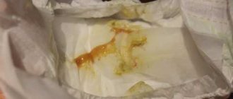
The composition of feces can indicate not only disturbances in the digestive process, but also indicate which part of the gastrointestinal tract has ceased to function normally. Therefore, in order to diagnose some diseases, the doctor resorts to prescribing a stool analysis - a coprogram. When do muscle fibers appear in feces?
What do muscle fibers in feces mean?
This means that protein foods are not digested properly and at the slowest possible speed. This may be a consequence of various pathologies, which will be discussed below.
Muscle fibers in the stool, in turn, can be distinguished by the absence or presence of stripes. It's important to know what each of these signs means and why you develop gonorrhea.
With striations
Each coroutine has a dedicated column for muscle fibers, which identifies them by how much they are separated or absent. In the first case, we mean the presence of pieces of meat food, partially processed in the stomach or intestines. The muscle fibers in the stool appear as elongated cylindrical structures with smoothed corners.
Without striations
Speaking about non-corrugated fibers, you should pay attention to the fact that
- these are small lumps in size;
- Normally, this indicator should disappear in the stomach area - under the influence of its juice;
- However, this does not happen, which may indicate a malfunction of the digestive system, especially the pancreas.
For this reason, you should never underestimate the fact that muscle fibers in stool appear unstreaked.
Norms
Macroscopic examination
| Parameter | Norm |
| Quantity | A healthy person produces an average of 100-200 g of feces per day. Normally, feces contain about 80% water and 20% dry matter. With a vegetarian diet, the amount of feces can reach 400-500 g per day; when using easily digestible food, the amount of feces decreases. |
| Consistency | Normally, formed feces have a dense consistency. Pasty feces can occur normally and are caused by the intake of predominantly plant foods. |
| Form | Normally cylindrical. |
| Smell | Normally, stool has a mild odor, which is called fecal (ordinary). It may intensify with the predominance of meat products in the diet, with putrefactive dyspepsia and weaken with a dairy-vegetable diet, constipation. |
| Color | Normally, stool is brown in color. When eating dairy foods, stool turns yellowish-brown, and meat stool turns dark brown. Ingestion of plant foods and some medications can change the color of stool (beets - reddish; blueberries, blackcurrants, blackberries, coffee, cocoa - dark brown; bismuth, iron color stool black). |
| Slime | Normally absent (or in scanty quantities). |
| Blood | Normally absent. |
| Pus | Normally absent. |
| Leftover undigested food (lientorrhea) | Normally none. |
Chemical research
| Parameter | Norm |
| Fecal reaction | Normally neutral, less often slightly alkaline or slightly acidic. Protein nutrition causes a shift in the reaction towards the alkaline side, while carbohydrate nutrition causes the reaction to shift towards the acidic side. |
| Reaction to blood (Gregersen reaction) | Normally negative |
| Reaction to stercobilin | Normally positive. |
| Reaction to bilirubin | Normally negative. |
| Vishnyakov-Triboulet reaction (for soluble protein) | Normally negative. |
Microscopic examination
| Parameter | Norm |
| Muscle fibers | Normally absent or single in the field of view. |
| Connective tissue (remnants of undigested vessels, ligaments, fascia, cartilage) | Normally absent. |
| Fat is neutral. Fatty acid. Salts of fatty acids (soaps). | Normally there are no or scanty amounts of fatty acid salts. |
| Plant fiber | Normally, there are single cells in the p/z. |
| Starch | Normally absent (or single starch cells). |
| Iodophilic microflora (clostridia) | Normally, single in rare areas (normally, iodophilic flora lives in the ileocecal region of the large intestine). |
| Epithelium | Normally, there are no or single columnar epithelial cells in the p/z. |
| Leukocytes | Normally, there are no or single neutrophils in the p/z. |
| Red blood cells | Normally none. |
| Worm eggs | Normally none. |
| Pathogenic protozoa | Normally none. |
| Yeast cells | Normally none. |
| Calcium oxalate (oxalic lime crystals) | Normally none. |
| Triple phosphate crystals (ammonium phosphate-magnesia) | Normally none. |
Causes of deviations and normal muscle fibers in feces
Factors causing disturbances in children and adults should be considered separately. This will allow us to fully understand the etiology of each of the processes.
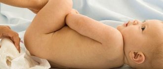
The child has
In children younger than 12 months who have already started eating meat, the stool may contain large amounts of muscle fibers. Such reactions are detected due to the immaturity of the digestive tract in general and digestive functions in particular.
As they mature, these processes will improve and the food will begin to be almost completely digested.
At the same time, if a large number of muscle fibers or numerous negative symptoms are found in the child’s stool, the reasons for these abnormalities are very serious. This list may include:
- Forced removal of chyme from the intestines. This anomaly is common in children and results in incomplete digestion of food.
- Diseases of the pancreas associated with a lack of external secretion. The disease can be congenital or acquired, but treatment is necessary regardless of the developmental conditions.
- Complex forms of gastritis, for example, hypoacid gastritis, in which there is excessive excretion of hydrochloric acid. As a result, the digestive capacity of the stomach decreases. In addition, there is acid gastritis, associated with inflammatory damage to the gastric mucosa, which also contributes to the appearance of undigested muscle fibers in the stool.
If there is a suspicion of any of the presented diagnoses, and the child is confirmed to have creative diarrhea, it is important not only to start treatment early enough, but also to undergo examinations every six months. This will allow you to monitor the healing process and prevent complications, which will be discussed below.
In an adult
The presence of muscle fibers in the stool in adults can be caused by bile deficiency, ulcerative colitis and a form of the disease accompanied by constipation. The list of triggers has been added:
- chronic form of pancreatitis, in which there is a decrease in the production of enzymes necessary for the breakdown of proteins;
- chronic atrophic gastritis;
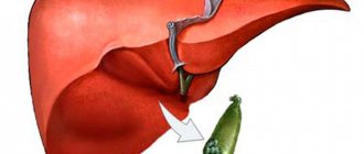
- hepatitis or cholelithiasis;
- dysbacteriosis, i.e. putrefactive or enzymatic dyspepsia.
Another predictable factor is what experts call gastrectomy. He may also have pathologies that are accompanied by a lack of gastric secretion (achylia) or a lack of components that make up hydrochloric acid. These are chlorine and hydrogen ions, and their absence is called achlorhydria. After determining the specific cause of the appearance of muscle fibers in the stool, treatment should begin.
Diseases for which a doctor may prescribe a general stool test (coprogram)
Anal fissure
With an anal fissure, feces may take the form of small lumps (“sheep feces” indicates a spastic condition of the intestines). Due to bleeding, scarlet blood and slightly changed red blood cells may be present in the stool.
Crohn's disease
With Crohn's disease, you may find blood in your stool. The Vishnyakov-Triboulet reaction reveals soluble protein in it. Crohn's disease affecting the colon is characterized by the presence in the feces of a large number of red blood cells in combination with leukocytes and columnar epithelium.
Colon diverticulosis
With diverticular disease, due to the long stay of stool in the colon, it takes the form of “large lumps”.
Duodenal ulcer
With a duodenal ulcer, the feces have the form of small lumps (“sheep feces” indicates a spastic condition of the intestines).
Stomach ulcer
With a stomach ulcer, the feces have the form of small lumps (“sheep feces” indicates a spastic condition of the intestines).
Chronic pancreatitis
In chronic pancreatitis with exocrine insufficiency, stool may have a pasty consistency.
Hemolytic anemia
With hemolytic jaundice (anemia), due to massive hemolysis of red blood cells, the amount of stercobilin in the feces increases.
Benign neoplasms of the colon
With a tumor accompanied by bleeding from the distal colon, the stool may have a pronounced red color. With disintegrating colon tumors, blood may be found in the stool. Pus on the surface of the stool occurs when there is severe inflammation and ulceration of the colon mucosa (disintegration of an intestinal tumor), often along with blood and mucus. When a colon tumor is in the stage of decay due to bleeding, the reaction to blood (Gregersen reaction) is positive.
Intestinal helminthiases
With helminthic infestation, the feces contain eggs of roundworms, tapeworms, etc.
Cirrhosis of the liver
In liver failure, including liver cirrhosis, the stool is grayish-white, clayey (acholic).
Ulcerative colitis
With colitis, there is mucus covering the outside of the stool in the form of thin lumps. With ulcerative colitis, blood may be found in the stool; pus on the surface of the stool, often along with blood and mucus; soluble protein in the Vishnyakov-Triboulet reaction; a large number of leukocytes (usually neutrophils); a large number of red blood cells in combination with leukocytes and columnar epithelium.
Constipation
With prolonged constipation caused by chronic colitis, peptic ulcers and other conditions associated with increased absorption of fluid in the intestine, the amount of feces decreases. With constant constipation due to excessive absorption of water, the consistency of stool is dense. With constipation, there may be mucus covering the outside of the stool in the form of thin lumps.
Malignant neoplasm of the colon
The form of feces in the form of “large lumps” - when feces remain in the colon for a long time - is noted in colon cancer. Pronounced red color of stool - with a tumor accompanied by bleeding from the distal parts of the colon and rectum. Blood in the stool can be found in disintegrating colon tumors. Pus on the surface of the stool occurs when there is severe inflammation and ulceration of the colon mucosa (disintegration of an intestinal tumor), often along with blood and mucus. A positive reaction to blood (Gregersen reaction) indicates bleeding with a tumor of the colon in the stage of decay. A large number of red blood cells in combination with leukocytes and columnar epithelium is characteristic of malignant neoplasms of the colon.
Irritable bowel syndrome, chronic colitis
With colitis with diarrhea, the amount of feces increases. The amount of feces decreases with prolonged constipation caused by chronic colitis. Mucus covering the outside of formed feces in the form of thin lumps is found in colitis. An alkaline reaction (pH 8.0-10.0) occurs in colitis with constipation. A large number of leukocytes (usually neutrophils) are observed in colitis of various etiologies.
Cholera
In cholera, stool looks like a gray inflammatory exudate with fibrin flakes and pieces of the colon mucosa (“rice water”).
Amoebiasis
With amebiasis, the stool is jelly-like, deep pink or red.
Typhoid fever
In typhoid fever, the stool looks like “pea soup.”
What to do if there is a deviation from the norm in the analysis
Treatment must be comprehensive. Outpatient treatment is usually recommended, while hospitalization and other more serious interventions are appropriate only in the most complex cases. The main treatments are the use of digestive enzymes, diet and antibiotics. Surgery may also be required.
When talking about digestive enzymes that may be needed for muscle fibrosis, keep in mind that their use is important for pancreatitis and some forms of gastritis. Please note that:
- names such as Pancreatin, Festal, Mezim and some others were taken;
- Each of these drugs helps relieve the pancreas, improves digestion and neutralizes the feeling of heaviness in the stomach;
- It can be used after consultation with a specialist in long courses during or after meals.
The next step is to follow a diet. It is known that any disease of the digestive system can be cured only by changing the diet. For example, it is very important for the patient to avoid heavy foods, spicy foods and bread.
Fast food, alcoholic drinks, strong tea and coffee are prohibited. If the general condition worsens, fasting can be considered complete.
As mentioned earlier, if the stool sample does not match the muscle fibers, antibiotics may help you deal with this along with the rest of your treatment. They are prescribed mainly for ulcerative lesions, gastritis and in the most severe cases of pancreatitis. Probiotics can also be used along with antibiotics to support and improve gut microflora.
In some cases, surgery is necessary. Experts note that in the case of mole rats:
- Surgery is performed only in extremely severe cases, when the pathology progresses rapidly or when basic treatment is ineffective for a long period of time.
- The procedure is performed for perforation of a stomach ulcer or bile duct obstruction. It may also be necessary for severe complications of cholecystitis and inflammatory processes in the pancreas.
- Whenever possible, specialists try to perform laparoscopy. This will prevent significant blood loss, as well as complications after surgery.
What will the coprogram tell you?
2014.05.27 10:16
Using a coprogram, you can evaluate how the digestive system and the body as a whole work, identify diseases of the pancreas, stomach, liver and gallbladder, small and large intestines. And also, using stool analysis, you can detect parasitic intestinal diseases (helminthiasis, giardiasis, etc.).
Feces are the end product of food digestion and are formed during the passage of food through the entire digestive tract and therefore contain information about the functioning of all digestive organs.
Correct preparation of the patient, correct collection, storage and delivery of research material are very important for the results of the study.
Feces for research are collected after spontaneous defecation in a dry, clean container; it is advisable to use plastic jars with a wide neck. Do not collect feces in containers with a narrow neck, cardboard or matchboxes. It is advisable to conduct the study immediately after defecation, but no later than 8-10 hours. It is not recommended to take the test after taking oil enemas, barium, taking medications that affect intestinal motility, using Vaseline or castor oil, after inserting suppositories into the anus. Also, urine or menstrual blood should not be present in the stool. To obtain reliable analysis results, preliminary preparation is necessary . To do this, the patient must be on a certain diet for 2-3 days. It is recommended to consume vegetable puree (preferably potato), dairy products, cereals, and a small amount of fruit.
Feces are collected 3-4 days from the start of the diet.
To conduct a study for hidden bleeding , the patient must exclude meat, fish, all types of green vegetables, as well as tomatoes and eggs for 3-4 days. Since these nutrients can be catalysts in those reactions that are used to detect blood.
Let's consider the indicators of scatological research.
Color. Normally, stool is brown in color. Depending on the diet, the color may change: a dairy diet gives a lighter color, a meat diet gives a darker color. The greenish color is given by plant pigments contained in sorrel, spinach, etc. Beets, blueberries, and black currants are colored black or reddish. Some medications taken orally, such as barium, give a light yellow or whitish color, bismuth - black, purgen - reddish.
In pathological processes, stool color: gray or white “clayey” - with obstruction (blockage) of the bile ducts; bright yellow – for acute enteritis and when taking antibiotics orally; red color - the presence of unchanged blood occurs with bleeding from the lower intestines (tumor, hemorrhoids, ulcers); black color “tarry” - bleeding from the stomach or small intestine. In some infectious diseases, for example, in typhoid fever, stool looks like pea soup; in cholera, it looks like rice water.
Consistency. Normally, an adult's stool is quite dense and well-formed. Pasty stools can be observed when the secretion of the pancreas is impaired and there is a lack of bile flow. Pasty stools occur when there is a violation of the evacuation of the colon, colitis, or fermentative dyspepsia. “Sheep feces”, small hard lumps, are a sign of colitis with constipation. Foamy stools are characteristic of intestinal infections and fermentative dyspepsia. Liquid is observed with insufficient digestion in the intestines and diarrhea.
Smell. Normally, stool does not have a strong, characteristic fecal odor. A foul odor is possible if there is a violation of the secretion of the pancreas, the flow of bile, or increased secretion of the glands of the large intestine. A putrid odor characterizes insufficiency of gastric digestion and weak intestinal motility, putrefactive dyspepsia, colitis with constipation. A sour smell appears with fermentative dyspepsia.
Reaction. Normally, stool has a neutral or slightly alkaline reaction. An acid reaction occurs when bile secretion is impaired and when absorption in the small intestine changes. A sharply acidic reaction is observed with fermentative dyspepsia. An alkaline reaction is characteristic of indigestion in the stomach, constipation, colitis, and increased secretory function of the colon. Strongly alkaline is a sign of putrefactive dyspepsia.
Muscle fibers. Normally they are absent or present in small quantities. An increase in level indicates pancreatic insufficiency, decreased secretory function of the stomach, and accelerated peristalsis.
Plant fiber is digestible. Not found normally. It is detected in cases of accelerated evacuation, diseases of the pancreas, impaired absorption of carbohydrates in the intestine, and increased fermentation processes in the large intestine.
Starch. It shouldn't be normal. A large amount can occur when consuming a diet rich in carbohydrates, with damage to the pancreas, impaired absorption in the small intestine, or accelerated evacuation of starch through the intestines.
Fat is neutral. It shouldn't be normal. A large number is noted with damage to the pancreas and with accelerated evacuation of fat through the small intestine.
Fatty acid. Normally they should not be present. They are detected when absorption processes through the wall of the small intestine are disrupted, in diseases of the biliary system, in the absence or deficiency of bile acids entering the intestinal cavity.
Soap. Normally available in small quantities. They are found in large quantities during intestinal congestion, malabsorption, and constipation.
Crystals. Not found normally. Tripelphosphates are found in feces during putrefactive processes in the large intestine. Oxalates appear during anacid conditions in the stomach and with insufficient gastric secretion. Bilirubin is detected during accelerated evacuation from the intestines, with diarrhea, and dysbacteriosis. Normally, it can be found in the feces of newborns. Hematoidin crystals appear in the stool during various bleeding in the digestive canal, and can occur when consuming foods rich in blood. Charcot-Leyden crystals are detected when the body is sensitized, in the presence of parasites and protozoa in the intestines.
Leukocytes. Normally, the presence of single leukocytes is allowed. A number over 10 indicates an inflammatory process in the intestines, such as ulcerative colitis or dysentery.
Red blood cells. It shouldn't be normal. Their appearance indicates bleeding. They are found in polyps and fissures of the rectum, hemorrhoids and colitis with ulcers.
Iodophilic flora. Not found normally. Its appearance indicates a violation of the absorption of starch and digestible fiber in the small intestine, the entry of a large amount of plant substances into the large intestine, an increase in fermentation processes in the intestine, and dysbiosis.
Laboratory assistant of the clinical department Bondareva Liana Aleksandrovna
Make an appointment
Add a comment Cancel reply
Can there be complications?
In some cases, when the patient has a large number of muscle fibers in the stool, the presented pathology is not without complications. If gastritis and then stomach ulcers are not treated in time, bleeding and even neoplasms are possible. In addition, peritonitis and appendicitis are considered serious complications of the pathology.
Such complications of diarrhea in children and adults can be avoided with timely treatment of diseases of the digestive system and preventive examinations. A proper diet, a healthy lifestyle, as well as physical activity and giving up bad habits are ideal preventive measures.
How to treat?
The establishment of creatorrhea is not an independent disease, but merely a symptom that indicates dyspepsia (functional digestive disorders) or serious gastrointestinal pathologies. Therefore, there is no need to treat creatorrhea if the analysis showed the presence of undigested protein fibers; it is necessary to look for the primary cause through a thorough examination.
In severe cases, antibiotic therapy or even surgery may be necessary. However, the first thing to start with when dealing with creatorrhoea and indigestion is diet.
You will have to forget about buns, cakes, fast food and other foods that are difficult to digest. A gastroenterologist can prescribe enzymes, the intake of which will reduce the load on the pancreas and help digest food. Enzymatic preparations for creatorrhoea can be taken for a long time without harm to the body, but even such simple treatment should be agreed with a doctor.
When creative rhea is detected in young children, sometimes it is enough just to adjust the diet and reduce the protein (specifically meat) load on the gastrointestinal tract. Until the age of three, it is better to give preference to dairy and fermented milk products, as well as low-fat fish, from the protein diet.