Stool with mucus looks like gray-white jelly-like inclusions enveloping the stool. A variant of this pathology may be small flakes of mucus mixed with feces. Less common is mucus with a yellowish tint.
Its amount may be small, or it may have such an impressive volume that it alarms a person with such a symptom and forces him to see a doctor. Consultation with a specialist - gastroenterologist is necessary for any amount of mucus in the stool, because the reasons for this can be quite dangerous conditions of the human body.
Causes of colitis
The development of the disease is influenced by a combination of negative, both exogenous (external) and endogenous (internal) factors.
These include:
- congenital predisposition;
- fetal pathologies;
- improper diet;
- poor quality diet;
- infection with intestinal parasites;
- predisposition to allergies;
- intestinal infections;
- infection of the digestive tract with the bacterium Helicobacter pylori;
- uncontrolled use of antibacterial and other drugs;
- intestinal dysbiosis.
Colitis in children under one year of age most often develops against the background of congenital malformations of the gastrointestinal tract with the addition of frequent viral infections, a tendency to allergies and lactose intolerance. In the case of formula-fed children, risk factors also include incorrect selection of formula.
Causes
Diarrhea with mucus is a symptom whose appearance indicates damage to the digestive system. Excessive mucus secretion is characteristic of diseases of the large intestine - colitis. This is due to the fact that in the wall of the colon there are many more goblet cells, which are responsible for the production of mucous secretion1.
The causes of diarrhea with mucus in a child can be divided into infectious and non-infectious2.
Up to contents
Infectious causes
Intestinal infections last 1-3 weeks2. Infectious factors include viruses, bacteria and pathological fungi that affect the intestinal mucosa and cause diarrhea2. Also, often mucous diarrhea is a manifestation of the active activity of parasites in the intestines2. In addition to mechanical injury to the intestinal wall, worms can have a toxic effect on the body with the substances they secrete. Parasites can provoke allergies and maintain constant inflammation in the gastrointestinal tract9.
Depending on the mechanism of development, the following types of diarrhea are distinguished:
- Secretory . The basis of diarrhea of secretory origin is enteritis - damage to the wall of the small intestine, primarily of viral origin. With this pathology, stool becomes liquid and watery14.
- Invasive diarrhea. Invasive is associated with damage to the large intestine by pathogenic bacteria - for example, Salmonella, Shigella or Escherichia. They penetrate the intestinal wall and cause active inflammation - colitis, which is manifested by the appearance of diarrhea with mucus14
It is the large intestine that normally ensures the formation of stool, so disruption of its function leads to the appearance of loose stools with abundant mucus1.
Up to contents
Non-infectious causes
Non-infectious diarrhea lasts more than 3 months and becomes chronic2.
Mucous diarrhea in a child often occurs as a result of an allergic reaction2,11. In young children, the main food allergens are cow's and goat's milk proteins. The list of allergens is expanding as new dishes are introduced; schoolchildren and teenagers are often allergic to eggs and gluten-containing products10. Food allergies can be triggered by an unbalanced diet, frequent or, conversely, too infrequent meals11.
Liquid, light stool mixed with mucus and blood is observed in the following cases2:
- Ulcerative colitis is a disease in which active damage to the mucous and submucosal layer of the large intestine occurs with the formation of ulcers. It is based on a disruption of the body’s immune response, which leads to chronic inflammation16.
- Crohn's disease is a long-term pathology that occurs with exacerbations. Unlike ulcerative colitis, this disease can affect all parts of the digestive system. In children and adolescents, the disease begins in the large intestine. In this case, chronic inflammation affects all layers of the intestinal wall16,17.
- Juvenile polyposis is the formation of multiple polyps in the small and large intestines, in which epithelial glands that produce mucus are actively working5.
Up to contents
Irritable bowel syndrome
Another cause of diarrhea in children and adolescents is irritable bowel syndrome. It develops due to a functional disorder of the digestive system. In this case, the child usually experiences mucous diarrhea with particles of undigested food1.
In most cases, symptoms appear in the morning, immediately after breakfast - for example, on the way to school. This phenomenon is called “morning rush syndrome.” In this case, the teenager first passes formed stool, then the stool becomes mushy or liquid feces are released. After bowel movements, the child feels well and, as a rule, diarrhea does not recur during the day1.
Up to contents
Antibiotic-associated diarrhea in children
It is customary to separately distinguish antibiotic-associated diarrhea - a bowel disorder that occurs while taking antimicrobial drugs or several months after a course of treatment2. When using drugs, the composition of the intestinal microflora is disrupted - dysbacteriosis develops. Against this background, clostridia actively multiply, and pseudomembranous colitis develops. Because of this, the child develops painful, repeated diarrhea with the release of mucus or blood6,15.
Up to contents
Symptoms of colitis
Determining intestinal inflammation is more difficult the younger the child is. Often the manifestations of the disease are mistaken by parents for a temporary problem. This is especially true for infants - in their case, the symptoms are blurred and may resemble a common digestive disorder due to a mild intestinal infection or a violation of the diet by the nursing mother.
In children older than one year, it is easier to determine the disease, since the symptoms become more pronounced and it is easier to determine from the child’s behavior at this age what exactly is bothering him.
Symptoms common to children of all ages include:
- Intestinal disorder. Digestive disorders can manifest themselves in different ways and alternate with each other: from watery, frequent stools to constipation that lasts several days.
- Increased gas formation. Due to disruption of the mucosal structure, intestinal immunity suffers, which causes an imbalance of microflora. It notes the predominance of pathogenic microorganisms, the result of whose vital activity is an increase in the volume of gases in the intestines. The child’s belly becomes swollen, the skin on it becomes tense, belching appears, and gases are often passed out.
- Nausea and vomiting occur at stages when a child’s colitis is at the development stage - this is how the body signals the onset of a pathological process in the gastrointestinal tract (GIT). Vomiting can also be a companion to chronic colitis during periods of exacerbation.
- Impurities in feces - pus, blood, bile, mucus. Sometimes the number of inclusions is so small that their presence can only be determined using laboratory analysis (coprogram).
- Dehydration occurs with frequent loose stools. In this case, a large amount of water leaves the body along with feces. Dehydration can be determined by dry, flaky skin, the smell of acetone from the breath, pallor, and lethargy.
- Abdominal pain localized below the navel.
Mucus in the stool of an infant
The cause of mucus in the stool of infants may be ARVI.
In the first month of life, the newborn’s intestines are populated by microflora, and the child’s gastrointestinal tract adapts to the changed operating conditions. During this transitional period, stool can have a very different appearance, have a different color, consistency, and frequency. Foam and mucus appear in it.
These phenomena in the feces of a newborn are caused by the establishment of a balance between beneficial lacto- and bifidobacteria and pathogenic flora, which is always present in varying quantities in the intestines of any person.
Mucus in an infant's stool during this period is normal, and there is no need to worry about it. It’s another matter when this symptom appears in a child several months after birth. Causes of mucus in stool in infants:
Diagnosis of colitis
The diagnosis is based on anamnesis, clinical picture, physical, laboratory, instrumental (x-ray, endoscopic) examination.
Blood tests in children suffering from colitis reveal anemia, hypoalbuminemia, and decreased levels of electrolytes in the blood serum.
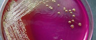
A scatological examination reveals the presence of leukocytes, mucus, steatorrhea, amilorrhea, and creatorrhoea in the stool. Bacteriological examination of stool allows us to exclude the infectious nature of acute and chronic colitis in children. Analysis of stool for dysbacteriosis, as a rule, demonstrates a change in the microbial landscape of the intestines due to an increase in opportunistic agents - staphylococci, Proteus, Candida.
An endoscopic examination of the intestines (colonoscopy, rectoscopy) in children often reveals a picture of catarrhal colitis: the mucous membrane of the colon is hyperemic and edematous; lymphoid follicles are enlarged; a large amount of mucus, pinpoint hemorrhages, and vulnerability of the mucous membrane upon contact are detected.
Stool with mucus in children
Constipation is the cause of mucus in the stool in children.
If mucus appears in the stool of children older than one year, then in addition to the addition of mucus to the intestinal contents during colds, allergic reactions and intestinal infections, this problem may have other causes. Reasons for the appearance of mucus in the stool in children:
- The child has worms. In this case, this symptom is accompanied by unexplained weight loss, tearfulness and frequent fatigue, and poor appetite. With helminthic infestation, mucus may appear as a reaction of the mucous membrane to irritation by the waste products of parasites.
- Intestinal flu or rotavirus infection. Additionally, hyperthermia, nausea, weakness, and vomiting are diagnosed. The stool is sparse, light and watery, containing mucus.
- Inflammatory process of the intestinal or stomach mucosa (catarrh).
- Constipation. Bloody discharge may accompany mucus during constipation.
- Diseases of the large intestine. Copious white mucus may be diluted with bloody inclusions.
The most important rule when detecting mucus in a child’s stool is that you should not self-medicate; a mandatory consultation with a specialist is required: a pediatrician, a pediatric gastroenterologist. It’s better to be safe than sorry about wasted time!
Treatment of colitis
Treatment of colitis in children under three years of age and older is carried out to a large extent by normalizing the diet and diet. To alleviate the child’s condition and reduce the intensity of symptoms, a dairy-free diet enriched with meat, fish, and eggs is prescribed. Artificial children up to one year old are transferred to lactose-free hypoallergenic milk formula.
In the case of breastfed children, identifying the etiology of the disease is of great importance, since some cases of allergic colitis require an urgent transfer of the child to artificial nutrition or a strict diet for the nursing mother.
From the diet of older children during periods of exacerbation, it is necessary to exclude all foods that complicate the digestion process, corrode the walls of the mucous membrane, and contribute to increased gas formation.
Drug treatment for colitis involves taking oral medications that improve digestion, protect and restore the intestinal mucosa.
Your child's health is in the capable hands of our doctors. Make an appointment with the medical pediatricians in Alexandrov by calling 8 (49244) 9-32-49.
Causes of mucus
Dysbacteriosis can cause the appearance of mucus in the stool.
The mucous membrane lining the walls of the stomach and intestines is constantly renewed. Cells that have fulfilled their purpose are rejected and move along with food through the intestines.
Normally, a certain amount of mucus, consisting of epithelial cells and leukocytes, is always present in the stool, but it is almost invisible.
Increased mucus secretion is associated with functional or organic damage to the distal intestine. Sometimes this symptom appears due to non-hazardous body conditions, for example, the inclusion of certain foods in the diet. Reasons for the appearance of mucus in stool:
- Mucous colitis. Impurities in feces look like films that can be confused with worms.
- Gluten intolerance (celiac disease) or lactose.
- Impaired absorption of fats in the intestines.
- Haemorrhoids. For this reason, mucus comes out after the bowel movement, remaining on the toilet paper.
- Dysbacteriosis. The inability to naturally remove waste and toxins from the body forces the body to intensively produce mucus.
- Tumors of the large intestine of various etiologies. This can be either benign intestinal polyps or malignant neoplasms.
- . When swallowed, the mucus released from the upper respiratory tract enters the stomach and intestines.
- Eating oatmeal, cottage cheese, watermelon, bananas.
- Severe stress or constant emotional overstrain. The human nervous system and the gastrointestinal tract are closely interconnected; a failure in the nervous system inevitably affects their condition.
- Allergies to food, medications and other factors. Goblet cells of the intestinal mucosa intensively produce mucus to protect it from undesirable factors.
- Chronic constipation. Toxic substances that are not eliminated in time with feces irritate the intestinal walls with the products of the activity of pathogenic bacteria; mucus is produced in response to irritation.
In addition to these reasons, increased mucus secretion may be an individual characteristic of the body, its reduced resistance to the effects of toxins and pathogenic microorganisms.
Clinical picture of ascariasis and enterobiasis in children at the present stage
What are the difficulties in diagnosing helminthiases? What is the treatment for helminthiasis?
Against the background of global environmental changes, the widespread use of immune, antibacterial and other drugs, frequent violations of adaptation processes (adaptation diseases), constant stress, increasing levels of education and culture of the population, many long-known human diseases have changed their clinical picture. Some symptoms practically ceased to occur, while others, on the contrary, began to come to the fore. This also applies to diseases caused by helminths, in particular parasitic roundworms (nematodes). In the temperate zone, the most widespread nematodes are roundworms and pinworms.
Helminths can cause dysfunction of the gastrointestinal tract (GIT), cause allergic (or pseudoallergic) reactions or aggravate them, cause intoxication, and also be a factor that weakens the immune system [2]. Nowadays, cases of massive infestation are rare, when diagnosis does not cause difficulties, and helminth balls cause intestinal obstruction - much more often helminths have become the cause of the development of allergic problems. At the same time, helminthiases are among those diseases that are difficult to diagnose due to objective and subjective difficulties (long periods of absence of oviposition, the possibility of the absence of females among the parasitic individuals, the likelihood of technical errors). Therefore, it is important to know the clinical picture of these diseases in order to be able to prescribe an in-depth examination or effective therapy based on a combination of indirect signs, even without direct evidence of the presence of helminthiasis.
In order to evaluate the clinical picture of nematodes, complaints, anamnesis and examination results of 150 children were analyzed in whom roundworms (116 children), pinworms (27 children), roundworms and pinworms (7 children) were found. Helminths were identified using standard methods: detection of roundworm eggs in feces using a smear method or pinworm eggs in a scraping for enterobiasis, as well as visual detection of roundworms or pinworms in feces, vomit, or during surgical or endoscopic intervention in the abdominal cavity or rectal area [3].
Among the children there were 67 boys and 83 girls. The age distribution is presented in table. 1. Children of primary preschool age (from one to three years old) predominated, their number was 63%. It is at this age that the greatest epidemiological preconditions for the appearance of nematodes occur.
In 150 children with proven infestations of roundworms and/or pinworms, the following clinical manifestations were noted.
107 children (71.3%) had allergic problems: skin rashes, diathesis, atopic dermatitis, neurodermatitis - 99 (66%), of which 18 had periodic problems, two children, in addition to skin rashes, had conjunctivitis ; ten (6.7%) had a proven food allergy to some foods with high levels of specific IgE in the blood serum; Six children (4%) had an asthmatic component or were diagnosed with bronchial asthma.
113 children (75.3%) had gastrointestinal dysfunction: unstable stool, undigested stool, the presence of “green” and mucus in the stool - in 66 (44%); constipation or a tendency to constipation - in 55 (36.7%), 17 children (11.3%) had unstable stool with a tendency to constipation, i.e. periods of normal or liquefied stool alternated with periods of constipation; flatulence, manifested by increased gas formation, bloating, rumbling, belching, occurred in 30 children (20%); nausea, vomiting, and regurgitation were observed in 29 children (19.3%).
In 60 (40%) children who, by age, could complain of abdominal pain, abdominal pain syndrome was noted: most often so-called “flying pains” were noted, which occurred regardless of food, periodically, usually without a specific localization or localized around navel (in the paraumbilical zone). Such pains went away on their own without medications or while taking sorbents and antispasmodics; some children needed to lie down for a while; Pain syndrome of this nature was observed in 54 children (36%). In eight children (5.3%), abdominal pain was constant, often associated with food (occurring after eating), intense, and often required drug treatment and hospitalization to exclude acute surgical pathology; one child underwent an appendectomy for this reason, and roundworms were found in the appendix with catarrhal changes; Two children had abdominal pain syndrome of both types.
66 children (44%) had appetite disturbances: in 54 (36%) appetite was often reduced or selective; in 12 (8%) - increased (the child is constantly hungry). Often, when asked about appetite, parents answered “when and how,” i.e., there were periods of normal appetite and decreased or increased appetite. In assessing this indicator, there is a large amount of subjectivity, since parents assess their children’s appetite differently.
Bruxism (teeth grinding) - a symptom that is often associated with helminthic infestations, but in fact is a nonspecific sign of intoxication of the central nervous system and can accompany any chronic intoxication - was noted in 25 children (16.7%).
Night sleep disturbances were observed in 81 children (54%): restlessness in the evening, difficulty falling asleep - in 15 children (10%); restless night's sleep (screaming, moaning, tossing and turning in sleep, waking up, crying, insomnia, nightmares) - in 76 (50.7%). Some parents of older children found it difficult to characterize the child's nighttime sleep, because the child sleeps in another room. Problems with falling asleep and sleeping at night are an important symptom of helminthic infestations, since it is known that intestinal nematodes (including roundworms and pinworms) are often activated at night.
54 children (36%) had itching and/or redness in the anus (anal escoriation) - in 43 children (28.7%); itching - in 38 (25.3%); both symptoms - in 27 (18%). Anal escoriation and itching are considered symptoms of enterobiasis (pinworms), but out of 54 children who had these symptoms, enterobiasis was proven in 17 children (31.5% of the number of children with these symptoms). In another 37 children (68.5% of the number of children with these symptoms), the presence of ascariasis was proven, but pinworms were not detected either visually or in tests. This may indicate either that in these cases there were pinworms that were not diagnosed, i.e., helminthiasis was mixed, or that anal escoriation and itching are symptoms characteristic not only of enterobiasis, but also of ascariasis.
29 children (19.3%) showed signs of general weakening of the immune system: frequently or long-term ill children (according to the generally accepted classification of Monto J. et al., 87) - 18 children (12%); recurrent stomatitis and gingivitis were observed in 13 children (8.7%); dental caries - in six (4%); recurrent purulent diseases of the skin or mucous membranes - in three (2%).
15 children (10%) had the results of a blood test of their immune status: 13 children had a reduced number of T cells; in all 15 children the number of T-helper cells was reduced, and in six of them it was significantly reduced; in 12 children the helper-suppressor ratio was reduced; in seven children there was a decrease in the level of IgA (secretory immunoglobulin), including in three a significant decrease, while in the remaining eight children the level of IgA in the blood serum was either normal or elevated; Six children had a decrease in the number of lymphocytes, including one child who had severe lympho- and neutropenia. These results confirm the known data that roundworms and pinworms have a depressing effect on immune function, and that people with weakened immune systems are more likely to develop helminthiasis [1].
49 children (32.7%) had various symptoms that can also be associated with the effects of helminths on the body: “geographical” tongue - in 12 children (8%); retardation in physical development, insufficient weight gain or weight loss over a period of time - in 21 children (14%); a painful appearance (blue under the eyes, pallor) was detected during examination in seven children (4.7%); Ten children (6.7%) had bad breath, especially in the morning; increased fatigue, asthenic syndrome - in eight children (5.3%); emotional lability, capriciousness, irritability of the child - in four (2.7%), and this indicator is subjective, as is the parents’ assessment of appetite; hypersalivation, another symptom usually associated with helminthiasis, was observed in three children (2%); various manifestations of hypovitaminosis, including rickets - in five children (3.3%). Often, during examination, changes in the skin in the form of “goose bumps” were revealed.
Most children had more than one symptom. Summarized data on the clinical picture are presented in table. 2.
Therapy in all cases consisted of two stages: first, anthelmintic therapy, and two different drugs were prescribed (decaris and vermox) with an interval of three to five days between them in order to maximally cover the different stages of the life of helminths; some time after anthelmintic therapy - drugs for microbiological and functional correction. Accordingly, the results of anthelmintic therapy, the results of the entire treatment, as well as follow-up within six months after therapy were assessed. In most cases, improvement occurred quickly during treatment. In 36 children, only after anthelmintic therapy, allergic manifestations significantly decreased or completely disappeared, in 39 children, after the first stage of therapy, the functioning of the gastrointestinal tract normalized, in 41 children, abdominal pain immediately stopped, and other symptoms also improved after anthelmintic drugs. This confirms that the symptoms were caused by the presence of helminths.
92 children had no complaints after the entire treatment; about 37 there is no data on changes in condition during and after treatment; in 16 children the effect was incomplete, i.e. some symptoms persisted after the end of treatment; in four children the effect of therapy was unstable, since relapses occurred after the end of treatment; Three children had no effect from therapy.
Literature
1. Blagova N. N. Some immunity factors in patients with ascariasis and enterobiasis during treatment with albendazole.
Author's abstract. dis. ...cand. honey. Sci. St. Petersburg, 1997. 2. Bogoyavlensky Yu. K., Rachkovskaya I. V., Chebyshev N. V. Nematodes and anthelmintics. M., 1994. 3. Laboratory diagnosis of parasitic diseases: https://www.infectology.spb.ru/. Bulletin of infectology and parasitology. Table 1. Age distribution
| Age | Up to 1 year | 1 – 3 years | 37 years | 7 – 12 years | 12 – 15 years |
| Number of children | 9 | 94 | 23 | 18 | 6 |
back
Table 2. Clinical picture of intestinal nematodes
| Complaints | Amount of children | % of total |
| Allergic problems | 107 | 71,3 |
| Gastrointestinal dysfunction | 113 | 75,3 |
| Abdominal pain syndrome | 60 | 40 |
| Appetite disorders | 66 | 44 |
| Bruxism (teeth grinding) | 25 | 16,7 |
| Night sleep disorders | 81 | 54 |
| Anal escoriation and/or itching | 54 | 36 |
| Signs of weakened immunity | 29 | 19,3 |
| Other symptoms | 49 | 32,7 |
back
Basic principles of treating diseases with blood in the stool
- stopping bleeding;
- correction of anemia (anemia);
- eliminating the causes of pathology;
- preventing relapses.
The impact on bleeding areas is carried out with drugs that increase coagulation (not suitable for people with thrombosis), coagulation with an endoscope, and gastric lavage with cold water. If non-invasive methods do not work, or the disease cannot be cured except by invasive means (for example, polyps, diverticula, intussusception), then surgery is involved.
Is occult blood in stool dangerous?
Hidden blood is difficult to detect visually, but it can signal bleeding from the gastrointestinal tract, duodenal ulcer, or stomach ulcer. If you suspect occult blood, you need to take an immunochemical stool test. With preliminary preparation: do not eat foods with barium, bromine, iodine, iron, or ascorbic acid. Avoid fibrous and hard foods that can injure the mucous membranes of the mouth and stomach. Change your toothbrush to a soft one three days in advance, and do not use rectal suppositories or do enemas the day before.
Prevention of diseases with blood in the stool
Change power mode:
in small portions on average 5 times a day. Have dinner 3 hours before bedtime. Exclude fast food, fatty foods, baked goods, sweets, canned foods, smoked foods, pickles, hot seasonings. Drink up to 2 liters of liquid per day: clean water, compotes, juices, fruit drinks, herbal or regular weak tea. Include foods containing fiber in your diet (fruits, peeled vegetables).
Create the right microclimate:
use air purifiers and humidifiers, regularly ventilate, wipe off dust and do wet cleaning.
Lead a healthy lifestyle:
give up smoking, alcohol, coffee and other bad habits. Maintain physical activity of the body, avoid stress. Empty the intestines in a timely manner to avoid chronic overstretching and unnecessary trauma to the wall. Protect the body from hypothermia. Be regularly examined by a proctologist for preventive purposes, especially in the presence of such hereditary diseases.
In the proctology department of the Altermed clinic network, diagnostics are carried out using the most modern equipment. The best doctors in St. Petersburg, both men and women, and a delicate approach are at your service.
Other articles by the author
- Blood in stool
- Hemorrhoids: symptoms and treatment methods
- Prolapse of the rectum (rectal prolapse)
- Pain in the anus
- Anal fringes
- Treatment of anal fissure
Who should I contact? How to prepare for examination and tests?
If blood is detected in the stool, you need to go to a coloproctologist. If this is the only symptom that bothers you, there is no need to do a thorough bowel preparation. It is advisable to administer a small cleansing enema (“pear”) only 1-2 hours before the initial examination. There are no restrictions on food intake. If you have an appointment after work in the evening, and there is no opportunity to prepare, then a cleansing enema can be given after the morning bowel movement.
If sigmoidoscopy is planned during the initial examination, you need to prepare more carefully. A couple of days before the procedure, clear the intestines of feces and gases and do not consume foods that provoke flatulence (peas, cabbage, soda, alcohol, rye bread, etc.). The day before the procedure, you can eat slag-free diet products: lean turkey and chicken meat, fermented milk products, eggs. You should not eat food 12 hours before the examination. Immediately before the procedure, it is necessary to give an enema (“pear”) or cleanse the intestines with a laxative.
Preparation for tests: 3 days before the test, do not eat foods containing iron - tomatoes, meat, buckwheat, pomegranate, fish, apples.
Preparing for an appointment with a proctologist at the Altermed clinic
Preparation for examination by a proctologist and sigmoidoscopy can be carried out directly in the proctology departments of the Altermed clinic network. If independent bowel preparation is difficult for you or you can only come to see a doctor in the evening after work, we have the opportunity to carry out the preparation in a specially equipped room. Such preparation will lengthen the reception by about 20 minutes, but this will save you from fasting and other problems associated with self-preparation.
IMPORTANT:
Normally, blood cannot be released from the rectum. Bloody feces are always a sign of disease. Therefore, your visit to the doctor should be urgent!