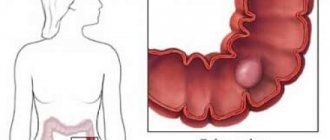
One of the most dangerous diseases of the digestive tract is gastric adenoma (adenomatous polyps). This is a benign neoplasm formed from epithelial glandular tissue. The tumor can appear in any part of the organ, most often affecting the antrum or the vestibule of the transition to the duodenum. The neoplasm is classified as a precancerous disease. According to research, the risk of its malignancy is 8-59%.
Types and their features
Depending on the location, adenoma is distinguished:
- thyroid gland – follicular, papillary nodes, formed from Hurthle cells and toxic with hyperfunction of the organ;
- prostate – benign prostatic hyperplasia BPH in men;
- mammary glands – phylloid, near-ductal, growing into the lumen of the flow and mixed type;
- salivary glands - rare encapsulated tumors near the ear glands with accumulations of lymphoid tissue;
- sebaceous glands;
- adrenal glands – adenocortical, pigment and oncocytic;
- pituitary gland;
- bronchial mucosa;
- gastrointestinal mucosa;
- liver - solitary, localized in the right lobe, found mainly in women.
Depending on the structure, neoplasms are distinguished:
- cystic – sac-like;
- papillary - with papillary growths into the lumen of the gland;
- polypoid - polyps of glandular tissue;
- solid - with a poorly developed supporting connective tissue structure and continuous glandular epithelium;
- tubular - from narrow epithelial canals and intercellular supporting stroma.
Depending on the number of nodes, they can be single or multiple (adenomatosis).
Diagnosis and treatment of esophageal tumors
99% of formations in the esophagus are malignant, and only 1% are benign, amenable to surgical removal. To diagnose pathological changes in the esophagus, contrast-enhanced radiography is used. To confirm the diagnosis, histology is required. Benign formations of the esophagus include polyps, adenomas, papillomas, lipomas, fibromas, hemangiomas, etc. Treatment is surgical, endoscopic or abdominal. Malignant tumors are treated with a combination of various methods: resection, extirpation of the esophagus, radiation and polychemical therapy.
After the operation, SM-Clinic patients are provided with a stay in a comfortable double room, which usually does not take more than a day.
Reasons for development
Most adenomas exhibit primary hormonal imbalances.
For example, hormone-dependent prostate hyperplasia occurs due to an imbalance of testosterone, and mammary glands due to an imbalance of estrogen. Sometimes the formation of adenomas of the thyroid gland is associated with mechanical lesions, and of the pituitary gland - with head injuries or previous infections. The risk of developing adrenal hyperplasia is higher in smokers.
Breast adenoma can be caused by a large number of abortions, taking oral contraceptives, unfavorable pregnancy, pathologies of the female reproductive system. Hepatic hyperplasia is associated with taking OCs or steroids.
Symptoms of adenoma
After a long asymptomatic course, the disease begins to manifest itself only if the tumor grows: it begins to compress surrounding organs, nerves, blood vessels, urinary tract, etc. Signs of adenoma appear depending on the location of the pathological process:
- Bronchial tumors are characterized by shortness of breath and wheezing cough.
- When the prostate gland is damaged, urination disorders, frequent urge to go to the toilet, intermittent urination, and constipation occur. According to the severity of manifestations, 3 stages of prostate adenoma are identified.
- An adenoma of the pituitary gland or thyroid gland is characterized by headache, irritability, tearfulness, fatigue, decreased libido, and excess weight.
- Cystic can be located in the pancreas, bile duct or appendix. When it opens spontaneously, a person feels severe attacks of pain.
- With adenoma of the lymphatic system, the lymph nodes increase in size and become denser.
- With adrenal adenoma, bone fragility and the risk of developing osteoporosis increase.
- With adenoma of the salivary glands, recurrent otitis is possible.
- Hepatic adenomas cause severe abdominal pain.
Symptoms of the development of adenomatous tumor
In the early stages of development, the pathology does not manifest itself in any way. It usually becomes an incidental finding during a preventive endoscopic examination of the stomach.
Later, symptoms of the disease similar to “gastritis” appear:
- periodic pain in the abdominal cavity of varying intensity;
- nausea;
- discomfort (sometimes vomiting) immediately after eating or when hungry;
- decreased appetite;
- increased pain due to consumption of fatty, spicy foods, pickles;
- foul belching;
- heartburn;
- feeling of heaviness in the epigastric area;
- weakness, general deterioration of health (not always observed);
- feeling of overeating, bloating after a small amount of food.
When an adonematous benign tumor grows to 2 cm or more, the patient is plagued by constant aching pain that is not associated with eating. Signs of internal bleeding appear:
- tar-colored feces;
- paleness of the skin, mucous membranes (anemia);
- tachycardia;
- fast fatiguability;
- dizziness;
- bloody vomiting.
Large adenomas cause a number of dangerous complications.
When the leg of an adenomatous polyp is twisted, vascular thrombosis and tissue necrosis develop. The patient's pain increases, body temperature rises, and signs of intoxication increase. Infringement of the tumor is indicated by cutting, cramping pain caused by stopping the movement of food through the digestive tract. Both of these complications are life-threatening conditions and require emergency medical care.
With malignancy of benign stomach tissue (degeneration of adenoma into carcinoma), all the above symptoms of the disease intensify, and regular jumps in body temperature to 38℃ and above are observed. The patient suddenly loses weight, stomach pain becomes constant and painful. To avoid the onset of dangerous consequences of the disease, it is important to identify adenomatous polyps in the early stages of their growth.
Diagnosis of adenomas
Diagnosis is made using:
- biopsy of a tissue sample – allows you to determine the type of neoplasm;
- CT and/or ultrasound, including transrectal TRUS, help determine the exact size and location of the tumor;
- Clinical blood and urine tests allow you to assess the overall picture and identify foci of inflammation.
Prostate adenoma is detected using digital examination, radiography and contrast urography, cystometry of the bladder and uroflowmetry of the urinary tract, determination of the level of the prostate-specific hormone PSA and a biochemical blood test. Palpation and mammography are also used to diagnose breast adenoma.
Diagnosis of benign stomach tumors
Gastric tumors can be diagnosed using medical history, X-ray and endoscopic examinations.
The presence of polyps on an X-ray of the stomach may be indicated by a filling defect that follows the contours of the tumor: clear, even contours, round or oval shape, its displacement in the presence of a pedicle or immobility in the case of a polyp with a wide base. In the case of gastric polyposis, a large number of filling defects of varying sizes are detected. The peristalsis of the stomach walls is preserved. Signs of a lack of peristalsis, an increase in size, a change in shape, and the appearance of blurred contours of a filling defect during dynamic observation may indicate malignancy of the polyp. The diagnosis is clarified by fibrogastroduodenoscopy (FGDS), which allows for a visual examination of the condition of the gastric mucosa, recognition and differentiation of polyps from other diseases. Visual differentiation of a benign polyp from a malignant one is difficult. Typically, malignancy can be indicated by the presence of a polyp larger than 2 cm, with a lumpy, lobulated surface and irregular, corroded contours. To accurately determine the nature of the polyp, during FGDS, a biopsy of suspicious areas is performed with a morphological examination of the biopsy samples. The diagnosis of non-epithelial benign tumor of the stomach in most cases can be established only after surgery and morphological examination of this neoplasm. The presence of clinical manifestations (for example, bleeding) indicates the possibility of a tumor process. FGDS is more informative for endogastric growth of non-epithelial gastric tumors. For intramural or exogastric tumors, endoscopic examination reveals compression of the stomach from the outside. X-ray of the stomach for non-epithelial benign tumors helps to detect rounded or irregular contours of the filling defect with preservation of peristalsis and folds in the submucosal layer; exogastric growth of the tumor with retraction of the stomach wall; ulceration with the formation of a niche at the apex of the tumor, etc. Ultrasound and CT of the abdominal cavity can be used to detect exogastric tumors of the stomach.
Treatment methods for adenomas
At the first stage, hormonal levels are adjusted using drug treatment. Then surgical intervention is performed - the main method of effective treatment of the pathology. During the operation, the tumor is completely removed. Preference is given to laparoscopic techniques with mini-access without incisions.
How are some types of adenomas treated?
In addition to surgery, alternative methods for removing prostate adenoma are often used:
- expansion of the narrowed area by inflating a special balloon;
- installation of expanding stents;
- thermo- or MV coagulation;
- UZT;
- cryodestruction with liquid nitrogen;
- transurethral needle ablation.
Drug treatment of the disease is additionally aimed at improving blood circulation in the pelvis, eliminating stagnation of urine and constipation, reducing inflammation, inhibiting hyperplasia and preventing infection.
With breast adenoma in young women, reverse development (resorption) of the tumor is possible. Therefore, if the tumor does not cause fear of malignancy and inconvenience for the patient, dynamic observation is recommended.
In the first weeks after discharge from the hospital, it is recommended to avoid any heavy physical activity and sudden movements, which may disrupt the healing of the postoperative wound.
Spread of the disease
This pathology is quite common, accounting for 25% to 40% of all polypoid-type neoplasms. Moreover, 15–58% are affected by multiple adenomas, the percentage of which increases with age.
The danger of such lesions is the high prevalence of parallel development of polyp and cancer. The provoking factors for this outcome are the following:
- villous structure of the adenoma;
- dimensions exceeding 1 cm;
- high grade dysplasia.
The percentage of damage to multiple adenomas of the colon is quite high. We can talk about the ratio of cases when the number of tumors reaches 100, from 1: 17,000 to 1: 5,000 patients.
Familial polyposis, when the number of tumors varies from 30 to 100, is being detected more and more often today.
A thematic video will tell you how to treat colon polyps:
Prevention
Prevention of pathology - undergoing regular preventive examinations and screenings - for example, monthly self-examinations and annual visits to a mammologist, as well as proper wearing of a bra to prevent breast adenoma. Prevention of prostate adenoma - timely treatment of prostatitis, monitoring PSA levels at the age of 50 years or more, control cholesterol and BMI, quitting smoking and alcohol abuse, balanced nutrition and physical activity.
After removal of the adenomasmal glands, medical supervision is necessary, since the disease is associated with Muir-Torre syndrome, which increases the risk of developing cancer. To consult with a specialized specialist in St. Petersburg, fill out the online appointment form.
Sources:
- Clinical guidelines of the European Association of Urology, 2017. https://urologi.kiev.ua/wp-content/uploads/2018/10/EAU_2017.pdf
- Urology. Russian clinical recommendations / ed. SOUTH. Alyaeva, P.V. Glybochko, D.Yu. Pushkar. — 2022.
- Colorectal adenomas - A.D. Borsuk, E.G. Malaeva: Problems of health and ecology, 2011.
- Thyroid adenoma. Pinsky S.B., Beloborodov V.A.: Siberian Medical Journal (Irkutsk), 2010.
- Aggressive pituitary adenomas (literature review and clinical observation. Kalinin P.L., Trunin Yu.Yu., Fomichev D.V., Chernov I.V., Ryzhova M.V.: Head and neck tumors, 2022.
The information in this article is provided for reference purposes and does not replace advice from a qualified professional. Don't self-medicate! At the first signs of illness, you should consult a doctor.
STOMACH CANCER
The incidence of stomach cancer varies from country to country. In recent decades, there has been a downward trend in the number of cases. Gastric cancer ranks fourth in the structure of mortality among all malignant neoplasms. Men and women are equally affected, most often at the age of 60 years and older. The leading cause has not been identified. It often develops against the background of chronic gastritis associated with HP, but the role of HP in the development of cancer has not been fully clarified. The resulting atrophy and dysplasia of the mucosa are considered precancerous conditions. A recurrent and, especially, long-lasting ulcer is the basis for repeat endoscopy and targeted biopsy. Precancerous diseases also include adenomatous polyps of the stomach, which are quite rare. There is a direct relationship between the number of sick people and their standard of living (5 to 1). Patients who have previously undergone gastric surgery have a 4-fold increased risk of cancer. A benign gastric ulcer is not a precancerous disease. Most often, gastric cancer is localized in the antrum (60-70%), less often on the lesser curvature (10-15%), in the cardiac region in 8-10%. Stomach cancer is divided according to its growth pattern:
- polypoid form with pronounced growth into the lumen of the stomach;
- in the form of ulceration with clear boundaries and a roll-like edge;
- ulcerative-infiltrative form;
- diffuse, total form of gastric damage.
Histological examination of the biopsy allows one to judge the degree of differentiation of the tumor (differentiated or undifferentiated cancer) and its belonging to a particular histological structure (glandular, fibrous cancer). In Russia, they use the TNM classification of tumors, which evaluates the prevalence of the tumor process according to three criteria: T - the size of the primary tumor, N - the presence of metastases in regional lymph nodes, M - distant metastases. The combination of these indicators makes it possible to classify and judge the prevalence and stage of the disease.
Manifestations of cancer largely depend on the local extent of the process, the transition of the tumor to neighboring organs (esophagus, duodenum), as well as on the nature of metastasis (local and distant lymph nodes, lungs, bones, brain, etc.):
- dyspepsia is often noted - loss of a sense of satisfaction from the food taken, a feeling of fullness in the stomach, bloating, a feeling of heaviness, pain in the upper abdomen, occasionally nausea, vomiting;
- anorexia, progressive loss of body weight, asthenia and other symptoms usually appear in the late stage of the disease;