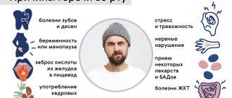
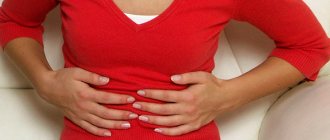
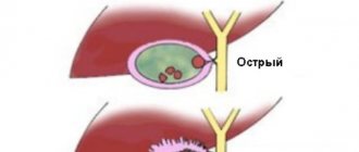
Compression of the biliary tract by malignant neoplasms leads to the development of obstructive jaundice. In patients, bilirubin levels increase and intoxication develops. Oncologists perform drainage of the bile ducts in case of obstructive jaundice. The Yusupov Hospital employs professors and doctors of the highest category. Drainage of the biliary tract is performed by leading oncologists. All complex cases of the disease are discussed at a meeting of the expert council.
Drainage of the bile ducts is performed to create an anastomosis between the bile ducts and the small intestine. Surgery can prolong the life of patients suffering from gallbladder or liver cancer. Oncologists perform drainage of the gallbladder in patients with neoplasms of the bladder and bile ducts, tumors in the area of the major duodenal papilla, and cancer of the head of the pancreas.
The following types of bile duct drainage are distinguished:
- external - the outflow of bile occurs through a specially installed external receiver;
- external-internal - most of the bile enters the intestine, flowing through the near channel, and the smaller part collects in the receiver located outside;
- internal drainage - requires endoprosthetics (biliary drainage is used as a palliative method for the treatment of inoperable cancer).
Oncologists at the Yusupov Hospital take an individual approach to choosing the method of drainage of the bile ducts for obstructive jaundice.
Methods of drainage of the biliary tract
Oncologists prefer to perform external internal or, if technically feasible, external drainage of the bile ducts in patients with obstructive jaundice of tumor origin. Both methods are quite effective in preoperative preparation for radical surgery or as a final treatment method. Their advantage is:
- constant monitoring of bile flow;
- the possibility of active removal of blood, pus, and clots from the bile ducts;
- washing the ducts with aseptic solutions;
- dynamic x-ray monitoring of the location of the drainage tube.
Unlike external-internal drainage, bile completely flows out through the external drainage of the gallbladder. The disadvantage of external drainage of the bile ducts compared to external internal drainage is the complete flow of bile through the drainage to the outside. To compensate for the vital substances contained in bile, patients are forced to drink their own bile or medical personnel administer it through a nasogastric drainage. With external internal drainage, the distal end of the tube is located further than the narrowing site and most of the bile flows directly into the intestine. It remains possible to control the patency and flush the drainage and replace it with an internal transpapillary endoprosthesis.
Internal endoprosthetics of the bile ducts is performed after the elimination of jaundice. This is the final stage of treatment for inoperable patients. To successfully perform external or external internal cholangiostomy, oncologists use a set of special instruments: wire guides, special puncture needles, bougies and catheters.
Under local anesthesia using a Shiba needle, the surgeon tightly fills the bile ducts with a contrast agent. A long needle with a diameter of 1.5-1.7 mm performs a puncture of one of the segmental ducts. A conductor wire is then passed through it. The end of the conductor is passed beyond the narrowing, the narrowed area is widened with bougies, and a drainage tube is installed. It is fixed to the skin and the bile ducts are washed with sterile solutions.
This drainage method has disadvantages: there is a risk of bile and blood leaking into the abdominal cavity when the needle is removed, when passing a guidewire or bougienage the canal. Additionally, this complication may be encountered if the outer diameter of the needle is larger than the outer diameter of the guidewire. In order to reduce the number of complications associated with liver puncture, oncologists use the technique of installing a cholangiostomy using a stylet catheter.
Introduction
Obstructive jaundice and acute cholangitis are among the most severe complications of biliary tract diseases. According to various authors, postoperative mortality in these diseases can reach 7.2-60% [3, 9, 14, 15].
It is well known that treatment of obstructive jaundice and cholangitis should primarily be aimed at eliminating bile stagnation and fighting infection [3, 10]. Most often, after eliminating the causes of jaundice, external drainage of the common bile duct (CBD) is used. Although technically this is the simplest way to complete a choledochotomy, along with its advantages, it also has disadvantages. Among the objections to external drainage, the most significant is the issue of bile loss, the danger of the formation of biliary fistulas after removal of the drainage tube, and the long stay of patients in the hospital. Some authors [10, 14] consider choledochotomy with external drainage of the CBD to be an outdated method that does not contribute to the final cure of obstructive jaundice and its consequences. They prefer one-stage internal drainage of the CBD with its radical sanitation. The main type of surgical treatment of benign strictures of the extrahepatic bile ducts, according to various authors [7, 13, 16], is also the creation of a biliodigestive anastomosis. However, long-term results of a large number of observations in which choledochoduodenoanastomoses were formed showed a number of unfavorable consequences: irregular flow of bile, residual pathological changes in the terminal part of the CBD, reflux cholangitis, cicatricial stenosis of the anastomosis. In addition, the formation of choledochoduodenoanastomosis in conditions of infiltration of the walls of the CBD and duodenum is fraught with the risk of developing insolvency and the formation of biliary and duodenal fistulas [5]. ON THE. Maystrenko et al. [14] believe that instrumental revision of the hepaticocholedochus with removal of stones and prolonged nasobiliary drainage should be performed in all patients with choledocholithiasis complicated by obstructive jaundice or cholangitis.
Treatment of acute cholangitis in patients with obstructive jaundice by systemic administration of antibacterial drugs is ineffective. Many researchers [1, 4, 11] note the effectiveness of using intraductal treatment methods in the complex treatment of cholangitis: cholesorption, ozone therapy, electrophoresis, laser irradiation. In recent years, methods of laser or quantum influence on the pathological focus, as well as ozone therapy, are increasingly used in practical surgery [1, 3, 4, 11]. According to a number of authors [11], for cholangitis, ozone therapy has antibacterial, antiviral, anti-inflammatory and immunomodulatory effects, enhances microhemodynamics, helps correct lipid peroxidation disorders and increase the activity of the antioxidant defense system, detoxification, and supports the body's energy homeostasis. Laser radiation with local exposure helps to reduce the duration of the inflammatory process due to increased tissue respiration, increased intensity of metabolic processes, normalization of the permeability of vascular tissue barriers, stimulation of phagocytosis, and increased protective and adaptive reactions of the body [6].
The purpose of our study was to develop an effective method for early elimination of the inflammatory process of the biliary tract in the postoperative period after the application of biliodigestive anastomoses in patients with obstructive jaundice and acute cholangitis.
Material and methods
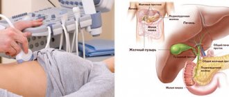
Under our supervision were 79 patients with obstructive jaundice (48 women and 31 men), operated on at the Scientific Center for Surgery named after. M.A. Topchibashev from 2006 to 2010. The age of the patients ranged from 25 to 77 years. Based on ultrasound, CT, MRI, endoscopic and radiopaque studies of the digestive tract, it was found that the cause of obstructive jaundice in most cases (47) was choledocholithiasis. Less commonly, obstructive jaundice occurred due to compression of the CBD by the enlarged head of the pancreas as a result of concomitant pancreatitis (12 patients), iatrogenic damage to the duct during cholecystectomy (6), cicatricial stricture of the papilla of Vater (3), narrowing of biliodigestive anastomoses (5), stenosis of the terminal part of the CBD (3 ), compression of the bile ducts by an hydatid cyst of the liver (2) and the presence of a microcystic adenoma of the head of the pancreas (1). In 56 patients, pathological changes in the bile ducts developed against the background of acute (31) and chronic (25) cholecystitis. The duration of jaundice before patients were admitted to the clinic ranged from 2 to 31 days, and in 57 (72.2%) patients the level of bilirubin in the blood serum upon admission exceeded 100 µmol/l. In 32 (40.5%) patients, obstructive jaundice was complicated by the development of cholangitis.
The patients were conditionally divided into 2 groups - control and main. The control group included 36 patients in whom pre- and postoperative conservative measures were carried out using traditional basic therapy, including antibiotic, detoxification and restorative therapy. 43 patients of the main group intraoperatively, as well as in the postoperative period, in addition to traditional therapy, received endocholedocheal laser irradiation of the biliary tract with a monofilament quartz light guide helium-neon laser with a wavelength of 0.83 μm, radiation power at the end of the light guide 8 mW, exposure time 10 min. Once a day using the Igla device. Endocholedocheal laser irradiation sessions were carried out for patients through bile duct drainages. In addition, patients in the main group underwent daily lavage of the bile ducts through drainage with an ozonated solution with an ozone concentration of 40 mg/l. To relieve the manifestations of severe endotoxicosis associated with obstructive jaundice, purulent cholangitis and liver failure, the main group also received pre-, intra- and postoperative daily intravenous infusions of ozonated saline solution at a concentration of 5 mg/l. Ozone was obtained in the medical ozone installation “Medozon-4MP-02”. The selected groups were comparable in the nature of pathological changes in the bile ducts and the surgical interventions performed.
All patients were operated on within a period of several hours to 18 days after admission to the hospital. All patients underwent choledochotomy, which was supplemented with external or internal drainage of the biliary tract. External drainage was performed using the Kerr method in 29 patients, of whom 15 were included in the control group, 14 in the main group, using the Halstead-Pikovsky method in 2 cases (1 in the control group, 1 in the main group), using the Vishnevsky method in 2 patients from the main group . Internal drainage was carried out using choledochojejunostomy in 31 patients, creation of choledochojejunostomy in 2 cases, hepaticojejunostomy in 9, and in 4 of them with drainage using the Saipol-Kurian method (2 from the main group and 2 from the control). A patient with narrowing of the choledochoduodenoanastomosis underwent duodenotomy followed by bougienage of the choledochoduodenoanastomosis. In one patient with obstructive jaundice, choledochotomy was completed by placing a blind suture on the wall of the CBD after removing stones from its terminal section and restoring the passage of bile into the duodenum. In one observation, in the presence of an hydatid cyst located in the central segments of the liver and causing compression of the main bile ducts, the operation was completed with echinococcectomy with external drainage of the cyst cavity. A patient with a microcystic adenoma of the head of the pancreas underwent pylorus-sparing pancreaticoduodenectomy - Traverso operation.
Since patients with obstructive jaundice and acute cholangitis with internal drainage in the postoperative period were not able to perform flushing of the CBD with an ozonized solution and intracorporeal laser irradiation of the bile ducts, we have developed a method for external drainage of the bile ducts, which consists of the following: intraoperatively after opening the CBD and creating the posterior wall of the biliodigestive anastomosis under With vision control, a tube consisting of a monofilament quartz light guide and nasobiliary drainage was inserted through the nose. Under visual control, the tube was passed into the bile ducts and installed in the area of the biliodigestive anastomosis. In those situations in which it was not possible to pass the tube into the duodenum, it was inserted through a nasogastric tube, which was subsequently removed. In patients in the control group, from the moment of installation, the drainage tube was used only for the purpose of collecting bile for bacteriological and biochemical studies. In patients of the main group, nasobiliary drainage served for drainage, taking bile samples and washing the bile ducts. A monofilament quartz light guide was used for endocholedocheal laser irradiation of bile ducts and biliodigestive anastomoses.
In all patients, bacteriological and biochemical studies of bile and biochemical blood tests were performed intraoperatively, as well as on days 2-4, 6-8, 9-11, and 13-15 after surgery. Material was collected under sterile conditions during surgery by puncture of the CBD before opening it or after choledochotomy. The antibacterial and anti-inflammatory effects of ozone therapy and endocholedocheal laser radiation were monitored using microbiological and biochemical tests, determining the levels of bile lipid peroxidation products: malondialdehyde [2] and diene conjugates [8], as well as the activity of the enzyme of the antioxidant defense system catalase [12].
The obtained digital data were subjected to statistical processing using medical statistics methods taking into account modern requirements. The average values of the obtained samples (M), standard errors (m), minimum (min) and maximum (max) values of the series were calculated. To assess the significance between variation series, the parametric Student's t test was used.
Results and discussion
The results of microbiological analyzes clearly confirm the positive antibacterial effect of the combined local use of ozone therapy and intracorporeal laser irradiation of the biliary tract for obstructive jaundice (see table).
According to our data, already on the 2-4th day of the postoperative period in patients of the main group, the titer of the inoculated microflora of the bile ducts was significantly lower (101-102/ml) than in the control group (102-104/ml).
In the main group of patients, microflora was not sown from bile on days 6-8; in the control group, even on days 9-11, certain cultures were sown in some patients, and signs of the inflammatory process remained. Moreover, at
Microorganisms were cultured from the bile of 2 patients in the control group on days 13-15. Daily washing of the bile ducts through external drainage with an ozonated solution at a concentration of 40 mg/l ensured the suppression of infection, improved the rheology of bile and its passage into the duodenum due to the high bactericidal activity, local oxidative and biostimulating effect of ozone. The advantage of our proposed drainage of the biliary tract for patients with biliodigestive anastomoses is also that infected bile does not enter the intestine, but is excreted from the body.
Since lipid peroxidation and the formation of free radicals increase during inflammatory processes, we also used the content of bile lipid peroxidation products, namely malondialdehyde (MDA), diene conjugates (DC) and the activity of the antioxidant enzyme catalase, as indicators of the severity of the inflammatory process in the biliary tract . Bile MDA values on the day of surgery were increased in both groups and differed little.
Analysis of the results of the studies showed that on days 6-8 in the control group of patients, the level of bile MDA decreased by 15.91% compared to the initial value on the day of surgery and amounted to 19.34±0.891 µmol/l (Fig. 1) .
Figure 1. Comparative dynamics of MDA parameters in patients with obstructive jaundice. In the main group, this indicator decreased by 35.91% and reached 15.03±0.74 µmol/l, which is significantly less than in the control group (p<0.001). On the 13-15th day of the postoperative period, the level of MDA in the control group decreased almost 2 times compared to the value on the day of surgery and amounted to 11.02 ± 0.605 μmol/l; in the main group this figure decreased by more than 3 times and reached 7.79±0.177 µmol/l (p<0.001).
The average DC values on the day of surgery in both study groups were increased and identical: 3.85±0.03 and 3.89±0.033 conventional units. respectively in the control and in the main (Fig. 2).
Figure 2. Comparative dynamics of DC parameters in patients with obstructive jaundice. Dynamic observation showed that the DC level decreased in the control group of patients on days 6-8 by 4.16%, while in the main group there was a statistically significant (p <0.001) decrease of 9.77%. DC values on days 13-15 after surgery decreased more in the main group of patients.
The activity indicators of the antioxidant enzyme catalase on the day of surgery in the study groups were 18.43±0.599 mKat/l in the control group and 18.35±0.536 mKat/l in the main group (Fig. 3).
Figure 3. Comparative dynamics of catalase activity indicators in patients with obstructive jaundice. The activity of bile catalase on days 6-8 in the main group increased by 102.8%, in the control group - only by 59.2%. On days 13-15, catalase activity in the control group increased to 63.62 mCat/l; in the main group, this indicator increased to 71.43 mCat/l (p <0.001).
The research results showed that in the main group, starting from days 2-4, there was a statistically significant (p <0.05) activation of the antioxidant protection of bile, which contributed to the early prevention of bile lipid peroxidation and the elimination of the inflammatory process in the bile ducts.
We analyzed the temperature curves of patients with obstructive jaundice. The average temperature of patients at the time of admission in the control group was 38.5±0.089 °C, in the main group - 38.6±0.086 °C. The body temperature of patients in the main group normalized on average on the 4th-5th day, while in the control group it remained above 37 °C until the 7th day, and in the main group there was a more stable decrease in the body temperature of patients.
Patients in the control group were discharged on average 20.3±1.24 days after surgery; the course of treatment in the main group in the postoperative period lasted on average 12.7±0.653 days (p<0.001).
There were no deaths in the main group; postoperative complications in the form of suppuration of the postoperative wound were observed in 2 patients. Relaparotomy and bleeding control were performed in 1 patient of the main group with the application of a Saipol-Kurian hepaticojejunostomy with a Roux loop, who had bleeding from the left lobe of the liver on the first day after surgery. In the control group, postoperative wound suppuration occurred in 7 patients. One patient developed pleurisy on the 7th day. One patient developed encephalopathy on the 4th day, liver failure on the 7th day, and another 1 patient developed gastrointestinal bleeding. One patient with metrorrhagia underwent uterine amputation. In the study, 2 patients in the control group died. In one of them, the cause of death was acute multiple organ failure against the background of unresolved liver failure on the 4th day after surgery, in the other, repeated myocardial infarction on the 7th day after surgery.
A drainage tube inserted into the bile ducts in combination with intracorporeal laser radiation and ozone therapy, as studies have shown, promotes early resolution of the inflammatory process in the bile ducts and prevents the development of scar narrowing in the area of the biliodigestive anastomosis and purulent-septic complications.
Thus, the use of endocholedocheal laser irradiation and ozone therapy in combination with the method of drainage of the bile ducts proposed when applying a biliodigestive anastomosis in the complex treatment of patients with obstructive jaundice and acute cholangitis made it possible to earlier stop inflammation in the bile ducts, correct disorders of bile lipid peroxidation and increase the activity of the antioxidant defense system , reduce the length of stay of patients in hospital after surgery by more than 1.5 times, prevent purulent-septic and cicatricial complications.
Endoscopic method
Using an endoscope, doctors perform nasobiliary drainage of the biliary tract. Indications for endoscopic drainage of the biliary tract are:
- obstructive jaundice caused by malignant and benign neoplasms;
- acute purulent cholangitis;
- external biliary fistulas;
- damage to the walls of the extrahepatic ducts, retroduodenal perforations;
- acute cholecystitis.
There are no contraindications to endoscopic drainage, except in cases where the tube for drainage of the biliary tract cannot be passed through the area of tumor narrowing. The endoscopic kit for drainage of the biliary tract through the nose includes:
- conductor wire;
- drainages of various shapes;
- a connecting tube for collecting bile and flushing the drainage;
- nasal tube, clamp and spatula.
The operation of endoscopic drainage of the biliary tract includes the following steps:
- cholangiography to determine the level and location of drainage;
- introduction of drainage with a metal guide-conductor;
- removal of the guidewire and endoscope;
- control cholangiography;
- assessment of drainage position;
- transferring the drainage from the mouth to the nose and fixing it on the head.
After using the endoscopic method of drainage of the bile ducts, complications do not develop. They may occur due to the progression of the disease.
Drainage after gallbladder removal
After cholecystectomy, surgeons often install a gallbladder drain. Indications for cholecystectomy are:
- cholelithiasis;
- acute cholecystitis;
- gallbladder carcinoma.
The operation is performed laparotomy or laparoscopically. Drains from the abdominal cavity after open cholecystectomy are removed on the eighth day, and in weakened and oncological patients - on the twelfth day. To prevent the skin around the drainage from becoming inflamed, the bile is drained into a special vessel. The skin around the wound is lubricated with zinc ointment or Lassar paste. Drains are changed no earlier than the twelfth postoperative day. In this case, fistulography is performed through the drainage to ensure the free patency of the bile ducts. Removal of drains after cholecystectomy is not performed on the fourteenth day, and when draining the bile ducts, no earlier than the twenty-first day after removal of the gallbladder.
Make an appointment by calling. Doctors at the Yusupov Hospital use various methods of draining the bile ducts for obstructive jaundice. Medical staff care for the drainage of the gallbladder and bile ducts.
How to rinse the gallbladder drainage tube
To ensure optimal drainage of the bile ducts, it is necessary to flush the tubes regularly, following certain rules. At the initial stage, they resort to the help of medical personnel, but in the future care will need to be provided to the patient himself or his relatives.
Flushing the drainage tube involves cleaning it with sterile solutions every day. For this purpose, 0.25% novocaine or saline solution is used in a volume of three to five ml.
Compliance with the rules of care for the drainage tube significantly extends its service life and is the prevention of cholangitis (an inflammatory process in the intrahepatic and extrahepatic bile ducts).