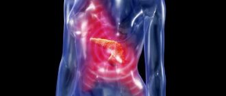
Causes
Doctors associate the pathogenesis of the disease with disruption of the normal circulation of bile, its stagnation and thickening. Subsequently, an infection occurs, provoking an inflammatory process. Chronic cholecystitis is characterized by slow development and sluggish course. In some cases, it can gradually move to the biliary tract from the walls of the gallbladder. A long-term course is characterized by: deformation of the bladder, adhesions, fusion with nearby organs, and the formation of fistulas.
The development of chronic cholecystitis can be triggered by the following factors:
- congenital disorders of the structure of the gallbladder, decreased tone (hypodynamia), prolapse of various abdominal organs, changes during pregnancy (contribute to stagnation of bile, caused mechanically);
- diet violations (obesity, alcoholism, overeating, regular consumption of spicy or fatty foods);
- hypotype biliary dyskinesias;
- the presence of intestinal parasites (giardia, amoeba, roundworm, opisthorchid);
- cholelithiasis (GSD).
Prevention
In order not to encounter gallbladder diseases, you just need to lead a healthy lifestyle and choose the right diet. It is necessary to eliminate all bad habits, especially alcohol, nicotine and fatty foods. It is also worth adjusting the amount of salt consumed. It should not be more than 5 grams per day. You also need to monitor your water balance.
Ultrasound of the gall bladder, prevention
As soon as you experience alarming symptoms, you should immediately consult a doctor. Only correct and timely diagnosis will significantly increase the chances of a quick recovery and prevent the occurrence of chronic inflammation of the gallbladder
.
Symptoms
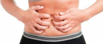
Pain syndrome. It is the main symptom of chronic cholecystitis. Painful sensations are localized in the right hypochondrium, sometimes in the epigastric region, they radiate to the right scapula, shoulder, collarbone, and left hypochondrium. The occurrence of pain and its intensification are often the result of a violation of diet, stress, physical activity, hypothermia, and concomitant infection. The pain can be different depending on the course of the inflammatory process (aching, intense, constant, nagging, etc.).
- Ker's sign. It manifests itself as pain when pressing in the projection of the gallbladder.
- Murphy's sign. It represents a significant increase in pain on inspiration during palpation of the gallbladder.
- Grekov-Ortner symptom. It is characterized by pain in the area of the gallbladder during tapping on the right along the costal arch.
- Georgievsky's sign - Mussi. Painful sensations when pressing on the right phrenic nerve, located between the legs of the sternocleidomastoid muscle.
Dyspeptic syndrome. Its manifestations are bitter belching or a constant bitter taste in the mouth. Patients often note a feeling of fullness in the upper abdomen, abnormal bowel movements, and bloating. Complaints of nausea and bitter vomiting are less common.
Increased body temperature. It is noted during exacerbation of cholecystitis. As a rule, the fever is low-grade; febrile levels are rarely achieved. Severe chills and severe sweating are always a consequence of purulent inflammation.
Jaundice. In chronic cholecystitis, it is rare, often due to obstruction of the outflow of bile due to the accumulation of epithelium, mucus or parasites in the bile duct or cholangitis.
Acute inflammation of the gallbladder is one of the most common complications of gallstone disease. Acute cholecystitis in cholelithiasis is called “stone” as opposed to “stoneless”, which occurs in the absence of stones in the gall bladder.
Etiology and pathogenesis. Calculous cholecystitis occurs in approximately 25% of patients with chronic calculous cholecystitis. Acalculous cholecystitis is rare, observed only in 5-10% of patients with acute cholecystitis. The main causes of the development of acute cholecystitis: impaired outflow of bile (most often due to blockage of the neck or cystic duct by a calculus), stretching of the walls of the bladder and associated ischemia of the wall, microflora in the lumen of the bladder. The microflora enters the gallbladder via the ascending route from the duodenum, descending via the bile flow from the liver, where the infection enters via the bloodstream, and, less commonly, through the lymphogenous and hematogenous routes.
In the vast majority of patients with chronic calculous cholecystitis, bile contains microflora. However, an acute inflammatory process occurs only when the outflow of bile is disrupted. Of secondary importance are ischemia of the bladder wall and the damaging effect of pancreatic juice on the mucous membrane of the bladder during pancreatobiliary reflux.
Clinical picture and diagnosis. The following clinical and morphological forms of acute cholecystitis are distinguished:
- catarrhal,
- phlegmonous,
- gangrenous (with or without perforation of the gallbladder).
Catarrhal cholecystitis is characterized by intense constant pain in the right hypochondrium, epigastric region with irradiation to the right shoulder blade, shoulder, and right half of the neck. At the onset of the disease, pain can be paroxysmal in nature due to increased contraction of the gallbladder wall, aimed at eliminating occlusion of the bladder neck or cystic duct. Vomiting of gastric and then duodenal contents often occurs, which does not bring relief to the patient. Body temperature rises to subfebrile levels. Moderate tachycardia develops up to 80-90 beats per minute, and sometimes a slight increase in blood pressure is observed. The tongue is moist and may be coated with a whitish coating. The abdomen participates in the act of breathing; there is only a slight lag in the upper parts of the right half of the abdominal wall in the act of breathing. With palpation and percussion of the abdomen, sharp pain occurs in the right hypochondrium, especially in the area of the projection of the gallbladder. Tension of the abdominal wall muscles is absent or slightly expressed. Symptoms of Ortner, Murphy, Georgievsky-Mussi are positive. In 20% of patients, an enlarged, moderately painful gallbladder can be felt. The blood test shows moderate leukocytosis (10-12 x109/l).
Catarrhal cholecystitis, like hepatic colic, in most patients is provoked by errors in diet. Unlike colic, an attack of acute catarrhal cholecystitis can be longer (up to several days) and is accompanied by nonspecific symptoms of the inflammatory process (hyperthermia, leukocytosis, increased ESR).
Phlegmonous cholecystitis has more pronounced clinical symptoms: the pain is much more intense than with the catarrhal form of inflammation, and intensifies with breathing, coughing, and changing body position. Nausea and repeated vomiting occur more often, the patient’s general condition worsens, body temperature reaches febrile levels, tachycardia increases to 100 beats per minute or more. The abdomen is somewhat swollen due to intestinal paresis; when breathing, the patient spares the right half of the abdominal wall, bowel sounds are weakened. Upon palpation and percussion of the abdomen, sharp pain occurs in the right hypochondrium, and pronounced muscle protection is also noted here; It is often possible to detect an inflammatory infiltrate or an enlarged, painful gallbladder. The examination reveals a positive Shchetkin-Blumberg symptom in the right upper quadrant of the abdomen, Ortner, Murphy, Georgievsky-Mussi symptoms, leukocytosis up to 12-18 x 109/l with a shift of the formula to the left, an increase in ESR.
A distinctive feature of the phlegmonous process is the transition of inflammation to the parietal peritoneum. There is an enlargement of the gallbladder: its wall is thickened, purple-bluish in color. There is a fibrinous coating on the peritoneum covering it, and purulent exudate in the lumen. If in the catarrhal form of acute cholecystitis, microscopic examination reveals only initial signs of inflammation (swelling of the bladder wall, hyperemia), then in phlegmonous cholecystitis, pronounced infiltration of the bladder wall with leukocytes is detected, tissue impregnation with purulent exudate, sometimes with the formation of small ulcers in the bladder wall.
Gangrenous cholecystitis is usually a continuation of the phlegmonous stage of inflammation, when the body’s natural defense mechanisms are unable to limit the spread of virulent microflora. Symptoms of severe intoxication with symptoms of local or general purulent peritonitis come to the fore, which is especially pronounced with perforation of the gallbladder wall. The gangrenous form of inflammation is observed more often in elderly and senile people with reduced regenerative abilities of tissues, decreased reactivity of the body and impaired blood supply to the gallbladder wall due to atherosclerotic damage to the abdominal part of the aorta and its branches.
When the inflammatory process transforms into a gangrenous form, there may be some reduction in pain and an apparent improvement in the general condition of the patient. This is due to the death of sensory nerve endings in the gallbladder. However, quite quickly this period of imaginary well-being is replaced by increasing intoxication and symptoms of widespread peritonitis. The condition of the patients becomes severe, they are lethargic and inhibited. The body temperature is febrile, severe tachycardia develops (up to 120 beats per minute or more), rapid and shallow breathing. The tongue is dry, the abdomen is swollen due to intestinal paresis, its right parts do not participate in the act of breathing, peristalsis is sharply suppressed, and in case of widespread peritonitis, it is absent. The protective tension of the muscles of the anterior abdominal wall becomes more pronounced, and symptoms of peritoneal irritation are revealed. Percussion sometimes detects dullness of sound over the right lateral canal of the abdomen. Blood and urine tests show high leukocytosis with a sharp shift in the leukocyte formula to the left, an increase in ESR, disturbances in the electrolyte composition of the blood and acid-base status, in the urine - proteinuria, cylindruria (signs of destructive inflammation and severe intoxication).
Acute cholecystitis in elderly and especially senile people with a decrease in the general reactivity of the body and the presence of concomitant diseases has a mild course: there is often no intense pain, protective tension in the muscles of the anterior abdominal wall is not expressed, and there is no high leukocytosis. In this regard, quite serious difficulties arise in diagnosing acute cholecystitis, assessing the condition and choosing a treatment method.
In typical cases, the diagnosis of acute cholecystitis does not present serious problems. However, a similar clinical picture can occur in acute appendicitis, acute pancreatitis, perforated gastric and duodenal ulcers, renal colic and some other acute diseases of the abdominal organs.
Among the instrumental methods for diagnosing acute cholecystitis, the leading role belongs to ultrasound, which can detect thickening of the gallbladder wall, stones in its lumen, and exudate in the subhepatic space. Among the invasive research methods, laparoscopy has become widespread, allowing visual assessment of the nature of morphological changes in the gallbladder. Both of these methods can also be used as therapeutic procedures in combination with puncture of the gallbladder and its external drainage.
Treatment. All patients with acute cholecystitis should be in the hospital under constant supervision of a surgeon. If there are symptoms of local or widespread peritonitis, emergency surgery is indicated. In other cases, conservative treatment is carried out. Limit food intake, allowing only alkaline drinks (acidic gastric contents, proteins and fats stimulate the release of intestinal hormones that enhance the motor activity of the gallbladder and the secretory activity of the pancreas). Non-narcotic analgesics are used to reduce pain.
It is not advisable to use narcotic analgesics, since the pronounced analgesic effect of the drugs can, by significantly reducing pain, obscure the objective signs of inflammation (peritoneal symptoms) and complicate diagnosis. In addition, narcotic analgesics, causing spasm of the sphincter of Oddi, contribute to the development of biliary hypertension and disruption of the outflow of pancreatic juice, which is extremely undesirable in acute cholecystitis.
Pain can be reduced through the use of anticholinergic antispasmodics (atropine, platifillin, baralgin, no-spa, etc.) agents. An ice pack is placed on the right hypochondrium to reduce blood flow to the inflamed organ. The use of a warm heating pad is absolutely unacceptable, since this significantly increases the blood supply to the gallbladder, which leads to further progression of the inflammatory process and the development of destructive changes. To suppress the activity of microflora, broad-spectrum antibiotics are prescribed, with the exception of tetracycline drugs that have hepatotoxic properties. For detoxification and parenteral nutrition, infusion therapy is prescribed in a total volume of at least 2-2.5 liters. solutions per day.
During treatment, the patient is constantly monitored: changes in subjective sensations and objective symptoms of the disease are recorded. It is advisable to keep an individual observation card, in which the pulse rate, blood pressure, body temperature, and the number of leukocytes in the blood are noted every 3-4 hours. Thus, the effectiveness of the treatment is assessed and the course of the inflammatory process is judged.
In acute cholecystopancreatitis, the complex of drug therapy should also include drugs used to treat acute pancreatitis.
In most patients, it is possible to relieve an attack of acute cholecystitis. During the process of observation and treatment, it is necessary to examine the patient; To identify stones in the gallbladder, perform an ultrasound. If they are detected and there are no contraindications (severe diseases of vital organs), it is advisable to operate the patient as planned 24-72 hours or 2-3 weeks after the acute attack subsides.
If, against the background of the treatment of acute cholecystitis, the patient’s condition does not improve within 48-72 hours, abdominal pain and protective tension of the abdominal wall continue or intensify, the pulse quickens, remains at a high level or the temperature rises, leukocytosis increases, then urgent surgical intervention is indicated for prevention of peritonitis and other severe complications.
In recent years, punctures and external drainage of the gallbladder have been successfully used to treat acute cholecystitis in patients with increased surgical risk. Under the control of a laparoscope or ultrasound, the gallbladder is punctured, its infected contents (bile, pus) are evacuated through the liver tissue, after which a flexible plastic catheter is installed in the lumen of the bladder for aspiration of the contents and local administration of antibiotics. This allows you to stop the development of the inflammatory process, destructive changes in the wall of the gallbladder, quickly achieve a positive clinical effect, avoid forced surgical interventions that are risky for the patient at the height of the process, and not perform surgery without proper preoperative preparation.
The situation becomes significantly more complicated with the development of obstructive jaundice against the background of acute cholecystitis. The disease can be complicated by cholangitis, damage to hepatocytes, further aggravation of intoxication, and the development of hepatic-renal failure. Obstructive jaundice often develops in elderly and senile people, whose compensatory capabilities of the body are very limited, and surgical intervention against the background of acute cholecystitis poses a great risk. In this situation, urgent endoscopic papillotomy is promising. A thin cannula is inserted through the biopsy channel of the duodenoscope into the major papilla of the duodenum, after which its upper wall is dissected using a special papillotome. In this case, the stones from the ducts either move away on their own, or they are removed with special tweezers using a Dormia loop (basket) or a Fogarty probe. This manipulation allows you to eliminate biliary and pancreatic hypertension, reduce jaundice and intoxication. Subsequently, gall bladder surgery is performed as planned.
Cholecystectomy is the main surgical intervention performed for acute cholecystitis. Removing the gallbladder can present significant difficulties due to severe inflammatory changes in the tissues surrounding it. Therefore, it is recommended to remove the bubble “from the bottom”. If indicated, cholecystectomy should be supplemented by intraoperative examination of the extrahepatic bile ducts (cholangiography). If choledocholithiasis or stenosis of the terminal part of the common bile duct is detected, the same manipulations are performed that are usually done in similar cases during planned operations in patients with chronic calculous cholecystitis (choledochotomy, T-shaped drainage, etc.). Drainage is left in the abdominal cavity to control blood and bile leakage.
Mortality after cholecystectomy performed for acute cholecystitis is 6-8%, reaching 15-20% in elderly and senile people.
Cholecystostomy with removal of stones and infected contents of the gallbladder is indicated in rare cases, as a necessary measure in the general serious condition of the patient and massive inflammatory infiltrate around the gallbladder, especially in elderly and senile people. This operation only eliminates acute inflammatory changes in the wall of the gallbladder. In the long term after surgery, as a rule, stones form again in the gallbladder and patients have to be operated on again.
Website illustrations: © 2011 Thinkstock.
Diagnostics
Blood analysis. The results of a blood sample during an exacerbation often reveal an increase in ESR, eosinophilia, neutrophilic leukocytosis, and a shift to the left in the leukocyte formula. Complicated forms are accompanied by an increase in blood levels of cholesterol, bilirubin, and transaminases.
Duodenal sounding. The severity of the inflammatory process is characterized by the results of analyzes of bile obtained from the body during duodenal intubation. As a rule, in this case, the bile is cloudy, it contains flakes, a significant admixture of mucus, cellular detritus, and columnar epithelium.
Bacteriological research. All portions of bile are analyzed. This makes it possible to determine the etiology of the inflammatory process and the susceptibility of the body's microflora to antibiotics.
Ultrasound and X-ray. X-ray examination can detect various signs of morphological or functional changes in the gallbladder or other digestive organs. Contrast examination of the gallbladder (cholangiography, cholecystography) makes it possible to identify disturbances in concentration ability, impaired motor function, and deformation.
Radioisotope method. Studying the state of the biliary tract and analyzing the absorption and excretion function of the liver can be carried out using the radioisotope method. More accurate diagnosis is achieved in combination with fractional multicomponent duodenal intubation. A more detailed study of the gallbladder and ducts is possible using the radio-X-ray chromodiagnostic method. Using ultrasound, it is possible to establish the absence of stones, as well as assess the contractility and condition of the gallbladder wall (a sign of chronic cholecystitis is its thickening by 4 mm or more).
What can you see on an ultrasound?
Using this research method, the following diseases can be diagnosed:
- different forms of cholecystitis,
- abnormalities in the development of the gallbladder during embryogenesis,
- gallbladder stones,
- tumor processes,
- agenesis,
- hydrocele of the gallbladder,
- organ enlargement.
what can be seen on an ultrasound
Only timely detection of the disease makes it possible to carry out full treatment. Most diseases can be cured with medications. In advanced cases, one can only hope for surgical intervention.
Usually the research takes about an hour, or even two. Therefore, it should only be carried out if prescribed by a doctor.
The person should lie down on the couch and remove clothes to the waist. To make the picture clearer, you need to lubricate the area with silicone gel. Using the sensor, the doctor can determine the structural features of the organ, its size, and more. A prerequisite is to carry out the procedure on an empty stomach.
Then you need to study the function of the organ. In this case, the patient should have breakfast before the procedure. Then the doctor will receive a picture before and after eating. It is best to use boiled chicken eggs for breakfast.
After the food has been digested a little, you need to conduct the study again. It is important to perform ultrasound in different positions of the patient.
Treatment
Medication. Treatment of chronic cholecystitis is almost always carried out conservatively by a gastroenterologist. During an exacerbation, it is aimed at eliminating acute symptoms, sanitizing the source of bacterial infection with the help of antibiotic therapy (broad-spectrum drugs are used for this, often belonging to the group of cephalosporins), detoxifying the body (introducing solutions of sodium chloride, glucose), restoring normal digestive function ( various enzyme preparations). Pain relief and inflammation relief during treatment are achieved with drugs from the group of non-steroidal anti-inflammatory drugs (NSAIDs), and spasm of the smooth muscles of the gallbladder and its ducts is relieved by antispasmodics. Elimination of bile stagnation is carried out with the help of drugs that enhance the peristalsis of the biliary tract (olive oil, sea buckthorn, magnesia). Choleretics are rarely used.
Operational. Chronic calculous cholecystitis is an indication for surgical removal of the gallbladder, which is the source of stone formation. The operation is not an emergency measure and is scheduled. Laparoscopic surgery or cholecystectomy are used. Percutaneous cholecystostomy is indicated for frail and elderly patients.
Non-surgical. If there are contraindications to surgery, non-surgical crushing of stones (extracorporeal shock wave lithotripsy) is used, but soon, as a rule, they re-form.
To make an appointment at the ABC-Medicine clinic for the treatment of chronic cholecystitis, call +7 (495) 223-38-83.
Surgery for cholecystitis
If the disease starts, surgical intervention cannot be avoided - cholecystectomy or laparoscopy. The latter method is used to treat calculous cholecystitis. The doctor removes stones and mucus from the cystic cavity. Classic surgery is performed for rupture of the gallbladder, destruction of the organ, or peritonitis - purulent inflammation of the organ.
Patients are prescribed diet No. 5, which excludes fatty foods, fried foods, smoked foods, carbonated drinks, baked goods, spicy seasonings and limits the consumption of eggs.