The article was prepared by a specialist for informational purposes only. We urge you not to self-medicate. When the first symptoms appear, consult a doctor.
The consequence of chronic inflammation of the mucous membrane of the stomach and duodenum can be atrophic gastroduodenitis. A characteristic feature of this disease is the destruction of the secretory glands that produce gastric juice. Instead of juice, the degenerated glands produce mucus. This form of gastroduodenitis is considered a precancerous condition and develops against a background of low acidity. Approximately 90% of all cases of atrophic gastroduodenitis are associated with the introduction of the Helicobacter pylori bacterium into the patient’s body.
After the body is infected with the Helicobacter bacterium, the concentration of gastric juice changes, protecting the gastrointestinal tract from dangerous infections. The pathological process from the stomach very quickly passes into the duodenum, which disrupts the process of digestion of the food bolus. During progressive inflammation of the gastric mucosa, secretory, or parietal, glands are lost, and metaplasia of individual areas occurs.
If the atrophic process occupies at least 20% of the total area of the stomach, we can say with absolute certainty that cancer is present. According to medical research, every eighth case of atrophic gastroduodenitis ends in cancer, while with other forms of gastritis this probability is 5 times lower.
With timely diagnosis of the disease, after 5 years of quality treatment, areas of metaplasia are significantly reduced, and the mucous membrane of the stomach and duodenum is restored.
Causes of atrophic gastroduodenitis
There are two main reasons leading to atrophy of the mucous membrane of the stomach and duodenum:
- An autoimmune process in which G cells of the secretory glands are damaged by their own immune antibodies;
- Long-term presence of the bacterium Helicobacter pylori in the gastrointestinal tract.
During the autoimmune process, antibodies mistake the cells of one's own secretory glands for foreign tissue. The acidity of gastric juice gradually decreases, and the parietal glands begin to produce mucus instead of hydrochloric acid. These processes lead to the impossibility of absorption of iron and vitamins by the walls of the stomach and duodenum, and the development of anemia. The addition of the Helicobacter bacterium accelerates the formation of areas of metaplasia.
The introduction of infection leads to cell damage, allowing free radicals to penetrate into them. The glands change their structure, their cells become precancerous. This is how intestinal metaplasia develops, when areas of the gastric mucosa acquire the properties of small intestinal and colonic epithelium. These transformations increase the likelihood of gastric adenocarcinoma.
Factors contributing to the development of the disease:
- Physical and mental stress;
- Hereditary predisposition to gastrointestinal diseases;
- Side effects of medications;
- Stress;
- Alcohol abuse;
- Occupational diseases;
- Somatic diseases in chronic form.
Diagnosis of gastroduodenitis
Treatment always begins with diagnostic procedures. For diagnostic purposes, several types of studies are used:
- endoscopic;
- histological;
- secretion assessment;
- antroduodenal manometry;
- fluoroscopy;
- Diagnosis of HP infection.
Endoscopic examination allows you to assess the condition of the walls. Using it, you can detect swelling, an increase in folds or, on the contrary, their smoothness, hyperemia (focal or diffuse), pallor of the mucous membrane, thinning.
Histology can confirm the conclusions that were made after examination with an endoscope. It assesses the degree of inflammatory and dystrophic processes - at what stage the disease is.
The intragastric pH-metry method is aimed at establishing the level of secretion. The pH level in the body of the stomach and its antrum, and the difference between these two indicators, is assessed. A normal reading indicates that the stomach sufficiently neutralizes the acid in the bolus of food before it enters the duodenum. Otherwise, acidification may occur. The indicators of both sections in the basal fraction (that is, normal) and after stimulation (histamine is administered) are also compared.
How is secretory function assessed?
- reduced if all indicators are reduced (in normal and stimulated states);
- increased if even one indicator is above normal.
In children, normal or increased secretory function is most often observed.
Since Hp infection is one of the main causes of gastroduodenitis, studies are carried out to search for it - histology and bacterioscopy. Antroduodenal manometry assesses gastric motor functions. For the same purpose, you can perform an EGG and ultrasound of the stomach filled with water. Fluoroscopy is usually used to distinguish gastroduodenitis from other diseases, such as pyloric stenosis or chronic duodenal obstruction.
Symptoms of atrophic gastroduodenitis
The disease develops slowly, starting from the bottom of the stomach, gradually moving to other areas of the mucous membrane. Vivid symptoms of the disease may not manifest themselves at first, which creates obstacles to early diagnosis and timely treatment.
Signs of anemic syndrome resulting from impaired absorption of vitamins and iron:
- Weakness;
- Fast fatiguability;
- Drowsiness;
- Paleness of the skin and mucous membranes;
- Burning and pain in the tongue;
- Varnished tongue surface;
- Dry hair;
- Brittle nails;
- Stitching pains in the heart;
- Shortness of breath with any exertion.
Signs of dyspeptic syndrome associated with digestive disorders:
- Heaviness in the stomach;
- Aching pain in the projection of the epigastrium;
- Heartburn;
- Belching;
- Nausea;
- Vomiting of recently eaten food, mucus and bile;
- Decreased appetite;
- Alternating constipation and diarrhea;
- Bad breath and unpleasant taste in the mouth in the morning;
- Gray coating on the tongue, imprint of teeth on it.
Digestive disorders lead to a sharp decrease in body weight, and in advanced cases – to dystrophy. Obstacles to the absorption of vitamins cause decreased immunity, frequent colds and infectious diseases.
Causes
There are a number of reasons why chronic gastroduodenitis may occur. If the cause is diseases of the internal organs, then they speak of a secondary form of the disease. If the development of the disease is influenced by external factors or poor nutrition, then doctors diagnose “primary gastroduodenitis.”
The secondary form is caused by the following factors:
- constant stress;
- dysfunction of the nervous system;
- low immunity;
- taking antibiotics;
- intestinal infections;
- diseases of the gallbladder and gastrointestinal tract.
Chronic infectious diseases also cause inflammation of the mucous membrane. Caries, tonsillitis and stomatitis must be treated promptly, because infections from the oral cavity enter the stomach. Sometimes the chronic form appears with reflux syndrome, when bile is thrown into the stomach.
The primary form is called:
- exposure to chemicals and poisons;
- poor nutrition and irregular diet (abuse of spicy, salty and smoked ingredients);
- bad habits, including drinking alcohol;
- eating hot or cold food.
Heredity also plays a big role. Genetic predisposition should be taken into account when making a diagnosis. Gastroduodenitis with high acidity often has an infectious etiology of origin. The inflammatory process is caused by Helicobacter pylori.
Diagnostics
The most informative modern method for determining the form and stage of gastroduodenitis is a hematological diagnostic panel. This study helps to establish the degree of damage to the secretory glands and the level of metaplasia of the mucosal epithelium.
Parameters determined by the study:
- Gastrin level-17;
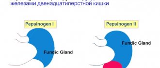
- Pepsinogen-1 and pepsinogen-2 levels;
- Histamine level-17;
- Indicator ratio.
Another informative study is FGDES with staining of the mucosa with methylene blue to assess the area of areas of epithelial metaplasia. During an endoscopic examination, a tissue biopsy of all altered areas of the mucous membrane is taken.
Additional methods for diagnosing atrophic gastritis:
- Gastrography;
- Intragastric pH-metry;
- Daily acidity measurement;
- MSCT (multispiral computed tomography) – if gastric cancer is suspected;
- Determination of the presence or absence of the bacterium Helicobacter pylori (breath test, ELISA test, PCR reaction).
Risk factors
An autoimmune case is caused by unfavorable heredity. Characterized by antibody activity against epithelial cells. It is registered by the specific presence of immunoglobulins in the blood serum. The disease resolves with increased levels of gastrin, which has a trophic effect on cells. This is how doctors explain the development of cancerous tumors.
Stomach cancer
A unified struggle strategy has not been developed. The role of Helicobacter in the development of inflammation of the antrum is beyond doubt. Additional research (biopsy) of the strain is required to determine sensitivity to antibiotics. Helicobacter is transmitted through the mouth.
Treatment of atrophic gastroduodenitis
The goal of treatment of atrophic gastroduodenitis is to prevent further development of intestinal metaplasia, destruction of the epithelium and its transformation into atypical cells (cancerous transformations). This goal can be achieved within 5 years of careful therapy.
A prerequisite for complete treatment is dietary nutrition. Food should be gentle in composition, temperature, and mechanical structure. After a short period of time, it is allowed to include low-concentrated lemon, cranberry, and cabbage juice in the diet. Bananas are the only acceptable fruit for this diet. Food should not be cold or hot, the diet should be frequent meals, small portions. Smoking and drinking alcohol in any dose are absolutely unacceptable during and after treatment.
Medicines for the treatment of atrophic gastroduodenitis:
- Antibiotics for eradication of the bacteria Helicobacter pylori;
- Proton pump inhibitors;
- Bismuth preparations;
- Glucocorticosteroids;
- Iron supplements;
- Vitamins;
- Enzymes;
- Mineral waters with high mineral content;
- Gastroprotectors;
- Antacids;
- Stimulators of cellular regeneration;
- Agents that stimulate peristalsis.
Additionally, physiotherapeutic treatment (electrophoresis, magnetotherapy, thermal procedures) and sanatorium treatment at a balneological resort are used.
Diet and nutrition rules
Proper nutrition is of utmost importance for gastroduodenitis. During remission, it is necessary to exclude the following foods from the diet:
- alcohol;
- strong broths;
- fatty meat, poultry and fish;
- spicy dishes (mustard, pepper, horseradish, garlic, etc.);
- strong coffee and tea;
- marinades, smoked meats, etc.
The basis of nutrition should be soups (with a second broth), lean meat, poultry, lean fish, cereals, vegetables and fruits. Products can be boiled, stewed, steamed or baked. In the acute stage, nutrition should be mechanically, chemically and thermally gentle. You can prepare liquid milk porridges, pureed soups with vegetable or weak meat broth, jelly, and compote. All dishes should be eaten warm.
Prevention and prognosis
To prevent atrophic gastroduodenitis, acute and chronic gastrointestinal diseases should be treated in a timely manner and the principles of rational nutrition should be followed. Early contact with a gastroenterologist and quality treatment will help prevent complications from occurring.
For elderly patients, the prognosis for the development of the disease is worse than for younger patients. At the age of over 50 years, mucosal atrophy most often ends in malignancy. If one course of treatment does not cure the disease, it should be repeated.
Author of the article:
Gorshenina Elena Ivanovna |
Gastroenterologist Education: Diploma in General Medicine received from the Russian State Medical University named after. N. I. Pirogova (2005). Postgraduate course in the specialty "Gastroenterology" - educational and scientific medical ]Our authors[/anchor]
Structure of the stomach
To better imagine the picture, let us describe the structure of the stomach and mucous membrane. The organ is hollow, with a volume of 0.5 liters, but can stretch 8 times more than usual. This is accompanied by unpleasant sensations.
The stomach is bean-shaped. In the left (front view) lower nook (antrum) there is an exit to the duodenum. This is where inflammation occurs when focal atrophic gastroduodenitis develops.
The entrance to the duodenum is guarded by the pylorus, a muscle formation similar to a sphincter. With the development of gastroduodenitis, this biological structure fails, and the intestinal contents penetrate into the stomach. The phenomenon of retrograde movement of chyme is called reflux. The enzymes it contains worsen the picture. The pH of the environment increases. This creates favorable conditions for the growth of bacteria.
An acidic stomach is a barrier to pathogenic flora. Microbes die here. But reflux sometimes disrupts the process, which leads to the introduction of dangerous strains into the intestines.
Gastroduodenitis becomes a prerequisite for the development of pathologies. Cancer is not caused by gastroduodenitis, but by dysbiosis that has developed against the background, the connection of which with carcinogenic processes has been proven. Symptoms make it difficult to establish a diagnosis. It is difficult to localize the genesis of pain.
Atrophic gastritis - a hidden threat: how to diagnose and help the patient
00:00
Oksana Mikhailovna Drapkina , professor:
- Let's move on to the next program. We will have a lecture by Associate Professor Tatyana Lvovna Lapina, dedicated to atrophic gastritis (AG).
Tatyana Lvovna Lapina , associate professor:
- Dear Colleagues! The topic of my lecture: “Atrophic gastritis - a hidden threat: how to diagnose and help the patient.”
Why do you need to make a diagnosis of atrophic gastritis?
The thing is that it has been clear for many decades that hypertension and such a change in the gastric mucosa, which is called intestinal metaplasia (this is a marker, a sign of hypertension), are precancerous diseases of the stomach.
Chronic gastritis and hypertension, as a rule, do not have their own symptoms. Having no obvious symptoms, gastritis is “silent” about itself. Atrophy of the mucous membrane does not make itself known. But this silence is very loud and threatening, because in the end we can get a stomach tumor.
The discovery of Helicobacter pyloricus in this chain of pathogenetic events from chronic gastritis to gastric cancer made it possible to very clearly show that this particular microorganism is the cause of chronic inflammation of the gastric mucosa.
Indeed, it has been absolutely clearly proven that for Helicobacter pyloric infection the connection with hypertension is obvious.
(Slide show).
Look here please. This is data from a recent meta-analysis that clearly links the presence of Helicobacter pyloric infection with the incidence of hypertension. The total relative risk is 5. Repeated presence of Helicobacter pyloric infection increases the risk of hypertension.
Naturally, Helicobacter pyloricus as such is associated with that stomach cancer, which has hypertension as its precancerous change. With non-cardiac gastric cancer, namely diffuse adenocarcinoma of the intestinal type according to the Lauren classification.
02:48
Look here please. I specifically present data from a meta-analysis on the relationship with Helicobacter pyloric infection of non-cardiac gastric cancer and cardia cancer. Cardia cancer is not associated with hypertension. It is clearly visible that there is no connection with Helicobacter pyloric infection.
Distal gastric cancer of the intestinal type has a specific stage of hypertension that precedes its development. It is clearly associated with Helicobacter pyloric infection.
Why can Helicobacter pylorus lead to the formation of stomach cancer? First of all, through inflammatory changes in the mucous membrane - through gastritis.
The bacterium itself is immediate. For example, thanks to the epigenetic changes that Helicobacter pyloric infection causes in the epithelial cells of the gastric mucosa.
This diagram, which is taken from a very good review (it’s called “Inflammation, atrophy and gastric cancer”) very clearly shows that at the molecular level it is clear what events in the gastric epithelial cell are triggered by Helicobacter pyloric infection. As a result, an imbalance in the processes of proliferation and apoptosis may occur, and genetic mutations may accumulate.
Of course, it is interesting to understand whether it is possible to prevent gastric cancer by eradicating Helicobacter pyloricus.
The most striking works are those on eradication therapy in patients diagnosed with early gastric cancer. These are Japanese works. You know that this country has a very good algorithm for managing such patients. Patients with early gastric cancer undergo endoscopic tumor resection.
Please look: 544 patients after endoscopic resection of early gastric cancer were observed for three years. Half of them underwent eradication of Helicobacter pyloric infection, half did not.
Three years later, in patients after eradication therapy, the incidence of new gastric tumors was 3%. Patients who remained infected with Helicobacter pylorus during 3 years of observation – almost 9%. A new stomach tumor was detected three times more often.
05:47
It is very easy to imagine everything in this model of endoscopic resection of gastric cancer. In fact, epidemiological studies on the prevention of gastric cancer through Helicobacter pyloric eradication are very difficult to implement, and it is very difficult to draw up a proper design for such studies.
However, it is clear that such research is being carried out. Indeed, from the analysis of these studies it is concluded that eradication of Helicobacter pyloricus statistically significantly reduces the incidence of gastric cancer by approximately 1/3.
Based on these calculations, some recommendations from certain medical communities include recommendations for possible screening for Helicobacter pyloric infection in the population. Especially in those populations where the incidence of Helicobacter pyloric infection is high.
Eradication of infection in seropositive individuals as a strategy and measure for the prevention of gastric cancer.
(Slide show).
From the point of view of developing such an algorithm for the prevention of stomach cancer in Japan, this article by very well-known researchers is interesting. Thanks to their calculations for Japan: the age of patients when eradication therapy for Helicobacter pyloric infection was carried out, and a possible decrease in the incidence of gastric cancer.
You see, the younger the age (depending on gender), the more effective eradication therapy is as a measure for the prevention of stomach cancer.
From the point of view of diagnosing atrophy, it is recommended to use the so-called level of serum pepsinogen I. This is a protein that is produced by the main cells of the glands of the body of the stomach. With hypertension, glands and chief cells are lost, so the level of serum pepsinogen significantly decreases. Certain threshold values may indicate the presence of atrophy of the gastric body.
08:47
Depending on the presence of Helicobacter pyloric infection and the presence of low titer serum pepsinogen as a marker of atrophy, certain groups of individuals in the population are identified that may (or may not) be subject to further observation.
Naturally, persons without Helicobacter pyloric infection and those with normal serum pepsinogen do not require further observation. But if we are talking about the presence of Helicobacter pyloric infection, we definitely mean eradication therapy.
Particularly severe group: Helicobacter pyloricus + and serum pepsinogen as a marker of atrophy is low. There is a special group of patients when the atrophy has gone so deep that pepsinogen is low. The antibody to Helicobacter pyloric is not even detected.
These patients should undergo endoscopic examination annually after eradication therapy.
The most interesting is the work that talks about preventive measures from the point of view of preventing stomach cancer. This is the work of BC Wahg et al. It was performed in a region with a high incidence of gastric cancer. A cohort of 1630 individuals was observed for many years. Moreover, almost 1000 of them at the time of inclusion in the study did not have hypertension and a marker of hypertension (intestinal metaplasia). They did not have dysplasia.
After eradication therapy and a comparison group that received placebo. Frequency of detection of stomach cancer after years of observation: 7 people and 11 people with stomach cancer.
There seems to be no difference. But when we looked at patients in whom eradication therapy was started before the formation of signs of hypertension, intestinal metaplasia, without dysplasia, the result of observation was zero cases of stomach tumors. Six cases of diagnosed gastric cancer in the absence of Helicobacter pyloric eradication.
Of course, there are independent risk factors: smoking, patient age. However, this study provides excellent evidence that the earlier eradication therapy is started (perhaps before the development of hypertension), the better this measure works in terms of prevention.
12:03
Who should receive treatment and when?
You see, a number of diseases and conditions are listed that can be considered indications for the eradication of Helicobacter pyloric infection.
Indeed, AG is present here. But will we conduct large-scale screening for serum pepsinogen and antibodies to Helicobacter pylori? There are still few regions in Russia in which such work is being carried out on a large scale.
What should a practitioner do?
A practitioner should use the recommendations of the Russian Gastroenterological Association for the diagnosis and treatment of Helicobacter pyloric infection in adults. They were developed by a group of experts as a result, in my opinion, of a very lively and lengthy discussion and published in No. 1 of the Russian journal “Gastroenterology, Hepatology, Coloproctology” for the current (2012) year.
Prescription of eradication therapy is desirable for chronic gastritis caused by Helicobacter pyloric infection. Including with hypertension.
A patient who has NSAID gastropathy, gastroesophageal reflux disease, functional dyspepsia, who came to the doctor with certain complaints, who is set up for an esophagogastroduodenoscopy, gastroenterological examination - of course, you should always keep in mind the diagnosis of Helicobacter pyloric infection. Whenever possible, use eradication therapy widely if indicated and if there are no contraindications.
How will we prescribe eradication therapy?
We will, of course, prescribe the well-proven standard triple therapy.
Proton pump inhibitor (PPI) in a standard dose 2 times a day. "Clarithromycin" - gram per day. "Amoxicillin" - 1 gram per day. A combination of Clarithromycin and Metronidazole (Metronidazolum) is possible.
Russian guidelines for the management of patients with Helicobacter pyloric infection recommend certain methods that can enhance the effect of standard triple therapy.
For example, prescribing a high dose of PPI twice daily, twice the standard dosage.
15:00
I would like to draw your attention to the point marked with an exclamation point. Adding Bismuthate tripotassium dicitrate to standard triple therapy.
PPI in a standard dose 2 times a day. "Clarithromycin", "Amoxicillin" and "Bismuth tripotassium dicitrate" 240 mg 2 times a day.
I must say that Bismuth salts are extremely interesting both from the point of view of their inclusion in eradication therapy, and from the point of view of possible use for chronic gastritis and hypertension.
One of the most reliable treatment regimens includes Bismuth salt. For example, "Bismuth tripotassium dicitrate." This is classic quadruple therapy with the drug “Bismuth”. It is recommended both as first-line therapy and as standard triple therapy.
In what situations?
For example, in a situation where you want to definitely achieve eradication, because quadruple therapy is a very powerful treatment.
When we encountered MALT lymphoma of the stomach associated with Helicobacter pyloric, we prescribed quadruple therapy to the patient, without any doubt in our decision.
Perhaps the patient cannot tolerate penicillin antibiotics, so standard triple therapy, which may include Amoxicillin, cannot be used in this situation. We turn to quad therapy.
If we carried out standard triple therapy, and still it did not turn out to be effective, we choose quadruple therapy with the drug “Bismuth” as the 2nd line of treatment.
This is indeed a very reliable treatment. Please look at the work from 2011. Quad therapy for 14 and 10 days. Even according to the very stringent Intention to treat (ITT) effectiveness criterion, 92% eradication is achieved, both in 10 and 14 days.
On the previous slide, in the recommendations of the Russian Gastroenterological Association, you saw the recommended duration of quadruple therapy - 10 days.
17:50
“Bismuth” preparations are drugs that have been used for the eradication of Helicobacter pyloricus from the very beginning of the “Helicobacter era”.
The discoverer of the H. pylori bacterium, Barry Marshall, came up with the idea of prescribing Bismuth drugs for eradication therapy of Helicobacter pyloric infection.
As a result of many years of research, a more or less clear algorithm for eradication therapy has been formed, which forms the basis for the recommendations of the Russian Gastroenterological Association on this issue.
Of course, there are different studies. There are studies that indicate the low effectiveness of triple therapy. Here is work that shows the low effectiveness of triple therapy and the not very high effectiveness of quadruple therapy.
Although for real clinical practice it is 77-78%. By and large, this is very close to the desired 80%. It is debatable to what extent this indicator in actual practice is an indicator of the failure of the scheme.
I would like to draw your attention to the search for the effectiveness of a combination of various drugs. I showed you what the Russian Gastroenterological Association recommends: strengthening standard triple therapy with Bismuth.
A recent study was conducted in a region where resistance to Metronidazole is 42% and to Clarithromycin 18%. High levels of resistance. It is not known what to prescribe.
Standard triple therapy with the drug "Bismuth" was prescribed. We obtained an excellent eradication percentage - 80% according to the strict criterion of calculating effectiveness according to the ITT (Intention to treat) criterion.
20:15
This approach - adding Bismuth to eradication therapy - is not new. This is an approach that, in different years of searching for optimal eradication therapy, takes one step or another. Either it is closer to 1st line therapy, or a little further from it.
(Slide show).
Huge meta-analysis. 93 randomized controlled trials. If you look at the last group of works, these are four-component therapies. This is the same therapy “Metranidazole” - “Tetracycline” - “Bismuth” and an antisecretory drug.
At the same time, there are various other options for adding Bismuth to standard triple therapy.
Such addition of this special anti-helicobacter drug, of course, can significantly increase the effectiveness of eradication.
How should we manage patients with gastritis, including hypertension.
These are the recommendations of the Russian Gastroenterological Association, based on the algorithm proposed by Vladimir Trofimovich Ivashkin in 2008.
From the point of view of preventing stomach cancer with gastritis, we recommend:
• balanced diet;
• possibly taking antioxidants;
• quitting smoking and concentrated alcohol.
Specific prevention in the presence of Helicobacter pyloric infection is Helicobacter pyloric eradication. But Bismuth drugs certainly occupy a certain niche here.
Due to what?
Please look at what the Russian Gastroenterological Association recommends. In case of chronic gastritis, including atrophic, after the end of eradication therapy, at the request of the doctor, in his opinion, it is possible to continue treatment with the drug “Bismuth” for up to 4-8 weeks to realize the protective properties of this drug.
You know that Bismuth preparations, in addition to anti-Helicobacter activity, have a very powerful potential to protect the cells of the gastric mucosa.
We will probably return to the cytoprotective effects of Bismuth in future lectures.
Thank you for your attention.
What is the doctor looking for?
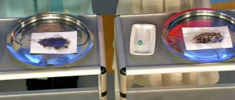
With atrophic gastroduodenitis, the mucous organs have a specific appearance: the diagnosis is made visually on the spot, using photographs or video recordings. The mucous membrane looks pale. Blood vessels are visible under the smooth relief (subatrophic gastroduodenitis). Atrophied mucous membrane is whitish, sometimes cyanotic in color.
There are no specific shapes or sizes. The doctor focuses on relief and transparency or changes in color. The name “atrophic” appeared against the background of severe dystrophy, observed visually. As research continued, the biological nature of the changes was established (see above).
Treatment methods for gastroduodenitis
When choosing treatment tactics, the specialist takes into account the results of tests and studies. Treatment is carried out in parallel with the appointment of a dietary table. If a Helicobacter pylori component is detected, antimicrobial therapy is necessarily prescribed. Also, drug treatment may include the use of antacids, enzyme therapy, vitamin therapy, and drugs that restore intestinal microflora. To relieve spasms, antispasmodic drugs are prescribed. If there is an increased stress load, use sedatives.
It is important to understand that only the complex effect on the body of diet and drug treatment allows us to talk about a favorable prognosis.
Research and diagnosis
The diagnosis is made using instrumental studies. The traditional method is esophagogastroduodenoscopy. Let's look at its etymology.
In 1868, Adolf Kussmaul figured out how to look inside the stomach through the esophagus. The name comes from two Greek words:
- “gaster” – stomach;
- "skopes" - to look.
We add here the Latin name of the duodenum - duodenum. The esophagus in Greek is called esophagos. Now the complex term makes it clear that the doctor:
- through the esophagus (esophagus) looks into...
- stomach (gaster), and then in...
- duodenum (duodenum).
By adding the words, you get the name of the procedure.
The submarine commander observes the maritime situation through the periscope eyepieces, remaining below the surface. Similarly, through the lenses, the doctor sees the picture in the stomach, highlighting the panorama.
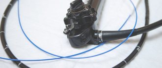
The difference between modern endoscopes is a flexible optical system that does not cause inconvenience to patients.
Treatment
Treatment of gastroduodenitis in adults is impossible without the patient refusing to drink alcohol, smoke, and comply with diet requirements.
Mode and nutrition
It is recommended to normalize mental and physical stress, and be more attentive to the organization of regular rest. You need to eat 5-6 times a day, in small portions and strictly following the schedule.
You will have to stop snacking “on the go” and dry food; food should be served warmed up, but not hot
Ice cream is excluded from the menu; cold temperatures are a stomach irritant. Among the prepared dishes, the diet does not allow everything that contributes to increased secretion of gastric juice. You should not eat anything that causes increased fermentation, mechanical and chemical effects on the mucous membrane. Raw vegetables are allowed only in pureed form.
Irritating foods include: fried fish and meat, smoked meats, pickled and canned vegetables, industrial juices (especially tomato), spices, hot seasonings, sauces, baked goods, fatty dishes, rich broths.
It is better to give up strong coffee and tea, chocolate, carbonated drinks, and choose low-fat dairy products. During exacerbations of gastroduodenitis, all dishes are prepared only in pureed form or from minced meat. The salt content is normal, unless there are contraindications due to other diseases.
Meat and fish are steamed, in the form of cutlets and meatballs. It is recommended to make stews from vegetables, and compotes and jelly from fruits and berries. If the disease occurs without intense pain or heartburn, there is no need for strict mechanical treatment in the diet, but fried foods remain contraindicated.
The diet is expanded as digestion improves. In case of acute gastroduodenitis, table No. 1 is sequentially expanded (first 1 “a”, then 1 “b”). In the chronic course of the disease, the patient will have to forever adapt to strict nutritional requirements.
Is it possible to cure gastroduodenitis only with medications without diet? This question worries people who are not accustomed to restrictions on food and catering at work. Our answer is that no matter how wonderful and expensive medications are prescribed, if there is no sparing of the stomach, a focus of continuous inflammation is created.
The process moves to deeper layers, causes ulcers, stimulates cancerous degeneration of cells
The use of medicinal methods
Since gastroduodenitis is most often accompanied by an increase in acidity, we will dwell in more detail on the treatment of this type of disease. To reduce acidity, drugs of different effects are used:
- Acid binders (antacids) - support the alkalization process, the group includes Almagel A, Phosphalugel, Maalox, De-Nol.
- Blocking synthesis by glandular cells of the antrum - Cimetidine, Omeprazole, Ranitidine.
- Increasing the activity of anti-acid-forming hormones - Gastal.
Antispasmodics - No-shpa, Platiphylline help reduce pain. Patients need to use a maximum of drugs by injection, unless there is a need for direct action on the mucous membrane.
In order to normalize nervous regulation, mild sedatives, infusions of plants, medicinal herbs (motherwort, valerian, ready-made drug Novopassit) are prescribed. To improve the protection of the mucous membrane, the recovery and healing processes, a course of biostimulants is prescribed (Aloe injectables, Actovegin, Solcoseryl).
If reduced acidity is detected in chronic gastroduodenitis, agents are used that stimulate the formation or replace gastric juice (ready-made preparation from gastric juice, Acidin-pepsin, Betacid.
For patients with identified Helicobacter pylori infection, therapy must get rid of the main cause. The treatment regimen includes antibiotics (Amoxicillin, Sumamed) in combination with Metronidazole and drugs based on bismuth compounds.
When identifying infection using the tank method. analysis for acute gastroduodenitis, a full course of antibacterial therapy is prescribed.
During periods of remission, physiotherapy, mud applications from healing springs, and mineral water intake are indicated. This is best done at resorts. Gastroduodenitis, complicating the postoperative period of gastric resection, needs to be monitored by the surgeon in terms of healing time and cause. Not all drugs are suitable for treatment with this option.