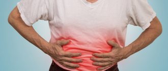
Ascites is an excess accumulation of fluid in the abdominal cavity.
This is usually a symptom of liver damage (cirrhosis, etc.) or the development of malignant neoplasms in the abdominal organs, as well as the pelvis. In most cases, ascites is accompanied by other clinical manifestations of the underlying disease.
There are conservative and surgical methods for treating ascites. Laparocentesis is a surgical method.
Laparocentesis (another name is paracentesis) is a procedure in which a catheter is inserted into the abdominal cavity through a puncture in the abdominal wall.
Laparocentesis can be therapeutic and diagnostic. Therapeutic laparocentesis involves the evacuation of a significant volume of fluid and is used for severe ascites that is not amenable to conservative treatment. Diagnostic laparocentesis is performed to take fluid samples for analysis to identify or cause ascites, often used when it reoccurs. As a rule, therapeutic laparocentesis also includes a diagnostic component.
Causes of ascites
Normally, fluid is present in the abdominal cavity, but in small quantities. The accumulation of excess fluid (ascites) can be caused by various reasons, among which three volume groups can be distinguished.
- Ascites can be caused by hypertension (high blood pressure) in the portal vein, which collects blood from the abdominal organs, due to cirrhosis of the liver, toxic or viral hepatitis. This type of ascites is called portal ascites.
- Ascites can develop due to a malignant tumor (primary or metastatic) in the liver or other abdominal organs, metastatic lesions of the peritoneum, or Hodgkin lymphoma. This is malignant ascites.
- Cardinal ascites is associated with decompensation of cardiovascular diseases and is often accompanied by congestive heart failure.
9500 patients annually
- We accept patients 24/7
- Stabilization, resuscitation, medical care
Call me back!
There are other, less common, causes of ascites:
- peritonitis;
- pancreatitis;
- kidney damage;
- congenital anomalies and vascular diseases of the abdominal cavity;
- nutritional disorders and protein deficiency.
In 65-75% of all cases (according to various sources), the cause of ascites is liver cirrhosis. In 15-20% it is associated with heart disease and the appearance of malignant tumors. Other reasons account for 5-10%.
Symptoms of fluid accumulation in the abdomen
In clinical practice, this anomaly is called “ascites.” It does not develop overnight and is diagnosed only when the volume of liquid substance in the peritoneal space reaches 1 liter. Rapid development of the disease is only possible if the portal vein is compressed. The main signs of pathology are:
- Changes in the size of the abdomen, its sagging or spreading depending on the position of the body.
- Obvious protrusion of the umbilical zone.
- The appearance of a noticeable vascular network around the navel.
- Difficulty breathing, the appearance of shortness of breath associated with the pressure of the filled peritoneum on the diaphragm and lungs.
- Feeling of fullness in the stomach, heaviness, bloating.
Ascites is not an independent disease; it is a complication of other pathologies. Depending on the underlying disease, other specific symptoms are present.
Mechanism of development of ascites
In liver cirrhosis, ascites occurs due to changes in the structure of the organ. Connective tissue replaces normal tissue, and in scarred areas the vascular network is transformed. Due to compression of the veins by connective tissue nodes, a number of pathological processes develop, leading, in particular, to an increase in pressure and resistance in the portal vein. As the pressure in the portal system increases, increased filtration and sweating of the liquid part of the blood into the liver tissue begins to occur. This increases the volume of lymphatic drainage from the liver - the lymphatic system thus responds to an increase in the volume of tissue fluid. However, this does not give the desired effect during progressive cirrhotic processes, and fluid begins to leak from the surface of the organ into the abdominal cavity. This is how the so-called “crying liver” appears. The peritoneal layers can absorb only part of the resulting fluid, and as a result, it accumulates in the abdominal cavity.
Our expert in this field:
Rudakova Maria Nikolaevna
Deputy Chief Physician, Oncologist-Surgeon, Doctor of Medical Sciences
Doctor of Medical Sciences
Experience: More than 30 years
Call the doctor
Call the doctor
Send documents by email The possibility of treatment will be reviewed by the chief physician of the clinic.
The process of ascites formation in heart diseases is very complex. The main role in this case is played by congestion in the systemic circulation and right ventricular failure.
Low stomach acidity
Stomach acid plays an important role in the human body - it is both a protective mechanism against ingested pathogens, and an activator of gastric juice enzymes and an assistant to enzymes in the digestion of proteins.
Low stomach acidity develops as a result of 3 main reasons:
- Gastric atrophy
- Taking medications that reduce gastric acidity (proton pump inhibitors, H2-histamine blockers)
- Removal of part of the stomach due to stomach cancer or as a bariatric treatment option (if the patient is severely obese, part of his stomach is removed for subsequent rapid weight loss)
I think it is worth covering in more detail the topic of atrophic gastritis as the reason for the decrease in the production of hydrochloric acid by the stomach.
With atrophy of the gastric mucosa, the number of glands gradually (over many years and decades) decreases and finally disappears completely. The production of hydrochloric acid first decreases and then stops altogether.
The main causes of gastric atrophy are Helicobacter pylori infection and autoimmune gastritis.
Helicobacter pylori is a bacterium that can lead to inflammation in the stomach; constant chronic inflammation contributes to gland atrophy. This process usually affects the antrum (outlet) of the stomach.
Autoimmune gastritis is a chronic disease that develops due to the fact that the human body, for unknown reasons, begins to perceive its own stomach cells as foreign and produces antibodies against them, which leads to inflammation and atrophy of the gastric glands. In autoimmune gastritis, the body and fundus of the stomach are mainly affected.
In the world, approximately 50% of people are infected with Helicobacter, and autoimmune gastritis occurs in approximately 1-2% of the population.
Of course, not all people infected with Helicobacter will develop such severe gastric atrophy that the secretion of hydrochloric acid will decrease. Moreover, with mild atrophy, the secretion of hydrochloric acid often remains normal.
In the presence of autoimmune gastritis, usually over the course of many years, atrophy becomes quite serious , and the presence of concomitant Helicobacter pylori infection aggravates the situation.
On average, the prevalence of atrophic gastritis worldwide is about 30%, most patients are people over 40 years of age.
About 2.5–5% of these people have progressive (moderate to severe) atrophic gastritis and an almost achlorid stomach in people over 50 years of age.
Reduced acidity and atrophy of the stomach leads to an increased risk of developing various infections , excessive growth of bacteria in the stomach and small intestine, deterioration of food digestion, and, accordingly, a deterioration in the absorption of vitamins and minerals. The absorption of vitamin B12 and iron slows down especially sharply, and the absorption of calcium, folic acid, and vitamin C is also worse.
Gastric atrophy and decreased secretion of hydrochloric acid can manifest as symptoms from the gastrointestinal tract, as well as from other organs, but you should know that in about half of the cases a person does not feel any symptoms for a long time.
Main symptoms of low stomach acidity
Main gastrointestinal symptoms include: epigastric pain, abdominal heaviness after eating, early satiety, heartburn (due to non-acid reflux), bloating, diarrhea or constipation.
Also clinically, atrophic gastritis and reduced gastric acidity are often manifested by anemia, which develops as a result of vitamin B12 and/or iron deficiency. The most common symptoms are the following: general weakness and fatigue, shortness of breath, pallor of the skin and mucous membranes, irritability and deterioration of mood, decreased cognitive abilities, perversion of taste - the desire to eat inedible objects (for example, soil or chalk), peripheral neuropathy and paresthesia (impaired sensitivity hands and feet - tingling and pinching sensation, numbness or, on the contrary, increased sensitivity of the skin), muscle weakness, lack of coordination. Decreased absorption of vitamin B12 and folic acid is accompanied by an increase in blood homocysteine levels, which can increase the risk of thrombosis, heart attacks and strokes.
Also, deterioration in calcium absorption can lead to osteoporosis (excessive bone fragility), which is manifested by pain in the bones/muscles, frequent bone fractures with minor trauma.
How to diagnose gastritis with reduced secretion of hydrochloric acid?
Quite a lot of methods for diagnosing reduced secretion of gastric acid are described in the literature, but most of them are either too complicated to perform and uncomfortable for the patient, or have very low sensitivity and insufficient evidence base.
I would like to note that quite often people over 50 years old come to the appointment and report with full confidence that their acidity is very low, their stomach “almost does not work.” And they gained confidence in this because once upon a time they were given an FGDS with a quick acidity test and were informed that their stomach was hypoacid, that is, there was very little acid. But I would like to note that the vast majority of these patients are fine with acidity (which is clarified during further examination), and the tests carried out in those days were extremely unreliable and showed hypoacidity . But currently there is a reverse trend - similar quick tests performed during FGDS, only now they diagnose high acidity in most patients. However, in foreign literature and recommendations, these tests are not mentioned as methods for diagnosing acidity levels, which means that these tests have not undergone reliable studies of sensitivity and specificity, therefore, the quality of these tests should also be doubted.
Therefore, in real clinical practice, it is worth relying on a blood test called “gastropanel” (the ratio of pepsinogen I/pepsinogen II, gastrin-17 and antibodies against H. pylori is studied), as well as an informative FGDS with multiple biopsies (according to OLGA or OLGA-M ) to determine the activity of inflammation in the stomach, the degree of atrophy and the presence of H. pylori - by the degree of atrophy one can indirectly judge the ability of the stomach to produce hydrochloric acid.
FGDS with biopsy according to OLGA or OLGA-M is the gold standard for diagnosing atrophic gastritis and identifying precancerous changes in the stomach.
If atrophic gastritis is detected, the patient is always tested for the presence of Helicobacter pylori infection. However, it must be remembered that in case of severe gastric atrophy with reduced secretion of hydrochloric acid, tests such as the C13-urease breath test and stool for antibodies to Helicobacter may be false negative. Therefore, in the case of serious atrophy, the best indicator of the presence of the microbe will be a biopsy of the gastric mucosa or detection of antibodies to Helicobacter IgG (antibodies in the blood - only if eradication of this microbe has not been carried out previously).
If the presence of Helicobacter pylori infection is detected, it must be eliminated with the help of antibiotics.
If there is B12 deficiency and/or atrophy of the body or fundus of the stomach, in addition to testing for Helicobacter, the doctor will always examine the patient for the presence of autoimmune gastritis. In this case, blood is taken for antibodies to gastric parietal cells and intrinsic Castle factor.
Currently, there is no specific treatment for autoimmune gastritis; only observation and elimination of vitamin and mineral deficiencies are indicated.
Patients with reduced gastric acidity, depending on the location of the atrophy, may be prescribed vitamin B12 for life (usually an intramuscular injection once a month), iron supplements, calcium supplements, folic acid, as well as other macro- and microelements, depending on their deficiency in a particular person. patient.
Patients with atrophic gastritis have an increased risk of developing stomach cancer and should undergo regular FGDS with biopsy (at least once every 3 years).
As for medications containing gastric enzymes (such as acidin, pepsin), the effectiveness of their use has not been proven, they are not mentioned in any recommendations, and their use refers rather to alternative medicine. Also, the effectiveness of any specific diet for low stomach acidity has not been proven, so meals should be regular, comply with modern ideas about proper nutrition, and also take into account the patient’s individual food tolerance (if any foods cause pain or other unpleasant sensations, it is recommended to avoid eating them).
Ascites in cancer
For cancer of the stomach, colon, pancreas, breast, uterus, and ovaries, especially in the later stages, ascites is a fairly common complication.
The immediate causes of development can be different:
- Blocking the outflow of the lymph nodes due to tumor damage to the lymph nodes;
- Damage by tumor cells to the peritoneum, a layer of connective tissue covering the internal organs and walls of the abdominal cavity;
- Damage to the liver, as a result of which albumin, a protein that maintains the oncotic pressure of the blood, ceases to be produced in the required amount;
- Increased pressure in the portal vein.
Ascites that develops against the background of oncology does not respond to conservative treatment. In these cases, only surgical methods can help - laparocentesis and peritoneal drainage. Sometimes additional surgical interventions are performed to prevent fluid from accumulating in the abdominal cavity in the future.
What does a stomach ultrasound show?
What does an ultrasound of the stomach show in adults:
- the presence (or absence) of gastritis or ulcers;
- malignant tumor;
- narrowing of the pylorus, if it is sufficiently pronounced;
- intestinal obstruction (this requires ultrasound examination of the entire gastrointestinal tract);
- any abnormal phenomena in the structure of the organ under study.
Decoding the result
When performing an ultrasound of the stomach, you can normally see the following:
- the organ in section usually looks like an oval ring-shaped formation with a rim, the nature of which is echo-negative, and the center is echo-positive;
- the walls of the stomach in the proximal sections have a thickness of about 5 mm, and in the pyloric section - about 7 mm;
- the presence of 5 layers in the wall of the stomach, differing in the degree of echogenicity;
- the serous membrane, which is located outside the stomach, is hyperechoic;
- the muscle sheath is hypoechoic, its thickness is about 2.5 mm;
- the submucosal membrane (with average echogenicity) measures about 3 mm;
- the mucosal plate, consisting of muscles, is usually low hypoechoic, its thickness is no more than 1 mm;
- the mucosa has a thickness of about 1.5 mm, its character is hyperechoic;
- A glass of liquid drunk by the patient is removed from the stomach in about 1/3 of an hour, and the primary withdrawal of liquid is normally about 180 seconds.
It is important to assess not only the thickness of the stomach walls, but also how uniform they are.
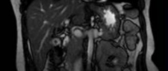
Can stomach cancer be seen on an ultrasound?
In the early stages, stomach cancer cannot be seen on ultrasound. This is due to the fact that at this stage of the disease, changes in the stomach are subtle and very difficult to identify.
In the later stages, changes in the body are already quite extensive. An ultrasound of the abdominal cavity will allow us to judge the presence of metastases and their size.
However, the sensitivity of the scanner is limited, so obvious tumors are detected only when they reach a large size.
Can a stomach ulcer be seen on an ultrasound?
Is a stomach ulcer visible on an ultrasound? The area where the ulcer is will not reflect ultrasound waves. Having noticed such a hole, the specialist will understand that the patient has an ulcer. However, such an indication on the disease screen is considered conditional. Additional examination will be required.
Types and clinical picture
There are minimal, moderate and severe ascites, or its three degrees.
Grade I ascites has no clinical manifestations. It can only be diagnosed using ultrasound, CT or laparoscopy. The amount of fluid in the cavity is slightly higher than normal.
Stage II ascites is characterized by the accumulation of a large amount of fluid and, accordingly, an increase in the abdomen in size, but no obvious stretching of the tissue is observed.
With grade III ascites, the stomach becomes huge and the figure becomes clearly disproportionate. There are difficulties in movement and breathing. The volume of fluid accumulating in the abdominal cavity can be 15-25 liters.
Methods for treating ascites
As mentioned at the beginning of the article, there are conservative or surgical treatments for ascites.
With conservative treatment, diuretics are prescribed to help remove excess fluid from the body. It is mandatory to monitor the amount of fluid you drink, daily urination and body weight. The patient should minimize salt intake, as it retains water in the body.
Among the methods of surgical treatment, laparocentesis is the most common. The operations of Kalb, Ruott and others are also used. The fluid outflow paths in their case are formed somewhat differently, but these procedures also imply the direct removal of accumulated fluid.
The choice of treatment method for ascites depends on its degree, cause, general condition of the patient, additional complications, etc.
How to prepare for an ultrasound of the stomach?
How reliable the results of the study are depends on whether gastric ultrasound is performed by specialists and on proper preparation for the gastric ultrasound procedure.
You are required to follow a diet for about two days before the procedure.
Avoid: foods that cause gas (rye bread, peas, beans, cabbage, kefir, carbonated mineral water, fresh fruits and vegetables).
- Questions often arise before an ultrasound: is it done on an empty stomach or not, is it possible to eat before an ultrasound of the stomach. The last meal on the day preceding the study should be no later than seven to eight o’clock in the evening.
- On the morning of the ultrasound of the stomach and intestines, do not eat, drink or smoke.
- Patients with severe hunger pains are allowed to drink half a glass of tea and eat a cracker.
How is laparocentesis performed?
Laparocentesis is performed under local anesthesia. The patient is in a semi-sitting or sitting position. Using a special instrument (trocar), which is a metal tube and a triangular needle inserted into it, a puncture is made in the abdominal wall. Then the needle is removed and the accumulated liquid is evacuated through the tube. The amount of fluid evacuated during one procedure is determined by the doctor.
To avoid injury to the intestines, laparocentesis is performed under ultrasound guidance. Or they use special devices with the help of which a space is created in the abdominal cavity free of intestinal loops.
Laparocentesis allows not only to remove fluid from the abdominal cavity, but also, if necessary, to find out the exact cause of the development of ascites through analysis of the composition of the fluid.
If long-term removal of fluid is necessary, then a drainage tube connected to a special container is installed for this purpose, but a more modern solution is peritoneal port systems. This is a titanium reservoir sewn under the skin and connected to the abdominal cavity with a catheter. One of the walls of the tank is a membrane made of a special material. To evacuate the liquid accumulated in the reservoir, it is enough to pierce the skin and the membrane under it with a needle. Thus, the port system creates much less inconvenience for the patient, being completely under the skin, and avoids regular laparocentesis procedures.
Preparation for laparocentesis
Laparocentesis is preceded by a standard set of studies, including:
- general chemical and biochemical blood test;
- coagulogram;
- general chemical urine analysis;
- tests for HIV, hepatitis B and C, syphilis;
- Ultrasound, which in this case is used to determine, among other things, the amount of fluid in the abdominal cavity.
A computed tomography scan may be prescribed. Immediately before the procedure, a cleansing enema is given, and it is also necessary to empty the bladder to avoid the risk of damaging it during the puncture of the abdominal wall.
If it is planned to remove a large volume of fluid, infusions of saline are given to fill the vascular bed with fluid.
Ultrasound process
The patient lies on his back or is in a semi-sitting position. The ultrasound probe is installed above the womb so that either both walls of the stomach or the curvature can be visualized on the screen at the same time.
It is considered normal if there is a small amount of fluid in the stomach. During an ultrasound, the doctor examines the shape and location of the stomach, the thickness of its walls, and existing pathologies.
As a rule, the stomach should be empty during the procedure. But in some cases a contrast agent may be needed.
Contraindications
Almost all contraindications for laparocentesis are relative. With the necessary preparation with the correction of the patient’s complications or compliance with conditions taking them into account, the procedure can be performed.
Such contraindications include:
- decreased platelet level (less than 20 × 103 / μl) (can be corrected by platelet transfusion before laparocentesis);
- bleeding disorders (corrected by infusions of fresh frozen plasma);
- adhesions in the abdominal cavity;
- cellulitis of the anterior abdominal wall;
- intestines dilated due to accumulation of gases;
- full bladder;
- pregnancy.
An absolute contraindication for laparocentesis is only an acute abdomen, which requires emergency surgery.
Possible complications
After laparocentesis, hematomas may remain on the abdominal wall and discharge from the puncture site may be observed. Possible in theory
- intestinal damage;
- infection;
- bleeding due to damage to a large blood vessel;
- decreased blood pressure, dizziness, fainting (with simultaneous evacuation of too much fluid);
- dysfunction of the liver and kidneys.
However, if the procedure is performed by experienced specialists with proper training, complications during laparocentesis are extremely rare.
+7
Call or leave a request
Request a call!