Bleeding stops on its own in approximately 80% of cases. Continued bleeding requires stopping it endoscopically as soon as possible. If this is not possible, then resort to active surgical tactics. In some cases, endovascular intervention or conservative treatment is performed.
The main tasks assigned to an anesthesiologist-resuscitator in the treatment of patients with gastrointestinal tract:
- Carrying out the prevention of recurrent bleeding after it has stopped;
- Restoration of systemic hemodynamics and other indicators of homeostasis. Naturally, the volume of assistance provided can vary widely: from resuscitation measures to simple dynamic monitoring of the patient;
- Providing assistance during endoscopic intervention or surgical intervention (if necessary);
- Timely detection of recurrent bleeding;
- In relatively rare cases, conservative treatment of bleeding is performed.
Sequence of assistance
If the patient received anticoagulants before bleeding occurred, they should, in most cases, be discontinued. Assess the severity of the condition and the estimated amount of blood loss based on clinical signs. Vomiting blood, loose stools with blood, melena, changes in hemodynamic parameters - these signs indicate ongoing bleeding. Arterial hypotension in the supine position indicates large blood loss (more than 20% of the blood volume). Orthostatic hypotension (a decrease in systolic blood pressure above 10 mm Hg and an increase in heart rate by more than 20 beats per minute when moving to a vertical position) indicates moderate blood loss (10-20% of blood volume);
In the most severe cases, tracheal intubation and mechanical ventilation may be required before endoscopic intervention. Provide venous access with a peripheral catheter of sufficient diameter (G14-18); in severe cases, install a second peripheral catheter or perform catheterization of the central vein.
Take a sufficient volume of blood (usually at least 20 ml) to determine the group and Rh factor, combine blood and conduct laboratory tests: general blood count, prothrombin and activated partial thromboplastin time, biochemical parameters.
Determining the cause and assessing the intensity of bleeding
Often the probable cause of bleeding can be determined by history.
Important things to consider:
- symptoms of dyspepsia (especially at night);
- symptoms of stomach ulcer;
- side effects of medications taken, especially those that inhibit blood clotting, such as non-steroidal anti-inflammatory drugs;
- likelihood of alcohol abuse;
- the presence of hepatitis B or C - these may indicate liver cirrhosis and portal hypertension as possible causes of bleeding.
Infusion therapy
Begin infusion therapy with the introduction of balanced salt solutions.
Important! If there are signs of ongoing bleeding or unstable hemostasis has been achieved, blood pressure should be maintained at the minimum acceptable level (SBP 80-100 mm Hg), i.e. infusion therapy should not be too aggressive. Blood transfusions are carried out if adequate infusion therapy fails to stabilize the patient’s hemodynamics (blood pressure, heart rate). Consider the need for blood transfusion:
– when the hemoglobin level decreases below 70 g/l. when bleeding has stopped;
– with ongoing bleeding, when hemoglobin is below 90-110 g/l.
In case of massive blood loss (more than 50-100% of the blood volume), transfusion treatment is carried out in accordance with the principles of “Hemostatic resuscitation”. It is believed that each dose of packed red blood cells (250-300 ml) increases hemoglobin levels by 10 g/l. Fresh frozen plasma is prescribed for clinically significant coagulopathy, including drug-induced coagulopathy (for example, the patient is receiving warfarin). And in case of massive blood loss (>50% of blood volume). If reliable hemostasis is achieved, there is no need to administer FFP even with significant blood loss (more than 30% of the blood volume). Dextrans (polyglucin, rheopolyglucin), hydroxyethyl starch (HES) solutions may increase bleeding and their use is not recommended.
How to make a diagnosis
The doctor examines the patient, assessing his external condition, the shade of the skin and mucous membranes. Then he measures blood pressure - often it is low.
In the clinic, the patient undergoes a general blood test. Using it you can quickly get an idea of the level of hemoglobin and the volume of other blood cells. Additionally, the diagnosis is made by biochemical analysis, but it is usually prescribed several days after the onset of blood loss, since the chemical composition of the blood changes only over time.
The main diagnosis concerns the detection of the very cause of the violation of the integrity of blood vessels. To do this, doctors use the following hardware examinations.
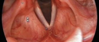
- Endoscopy - examination of the esophagus, stomach, duodenum using a flexible tube with a miniature camera allows you to quickly detect a problem area;
- Contrast radiography - an effective method for detecting bleeding in the gastrointestinal tract involves injecting a safe contrast solution into the organ, followed by an X-ray;
- Magnetic resonance imaging is a modern method that allows you to obtain comprehensive information about the condition of all tissues of a particular organ of the gastrointestinal tract.
Antisecretory therapy
Optimal conditions for the implementation of vascular-platelet and hemocoagulation components of hemostasis are created at pH > 4.0. Proton pump inhibitors and H2-histamine receptor blockers are used as antisecretory drugs.
Attention! It is not advisable to prescribe H2-histamine receptor blockers and proton pump inhibitors at the same time.
Medicines of both groups suppress the production of hydrochloric acid in the stomach and thereby create conditions for stable hemostasis of the bleeding vessel. But proton pump inhibitors show more consistent results in reducing gastric acidity and are significantly more effective in reducing the risk of recurrent bleeding. The antisecretory effect of proton pump inhibitors is dose-dependent. Therefore, the use of high doses of drugs is currently recommended, so the prescription regimens indicated below are not a typo by the author.
Patients are prescribed an intravenous infusion of one of the following proton pump inhibitors:
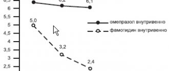
- Omeprazole (Losec) 80 mg IV as a loading dose, followed by 8 mg/hour.
- Pantoprazole (Controloc) 80 mg IV as a loading dose, followed by 8 mg/hour.
- Esomeprazole (Nexium) 80 mg IV as a loading dose, followed by 8 mg/hour.
The loading dose of the drug is administered in approximately half an hour. Intravenous administration of the drug is continued for 48-72 hours, using, depending on possibilities, a bolus or continuous route of administration. In the following days, they switch to oral administration of the drug at a daily dose of 40 mg (for all of the proton pump inhibitors listed in this paragraph). The approximate duration of the course is 4 weeks.
Attention. The administration of proton pump inhibitors should be started before endoscopic intervention, as this reduces the likelihood of recurrent bleeding.
In the absence of proton pump inhibitors, or patients intolerant of them, intravenous H2-histamine receptor blockers are prescribed:
- Ranitidine 50 mg IV every 6 hours or 50 mg IV followed by 6.25 mg/hour IV. After three days, 150-300 mg orally 2-3 times a day;
- Famotidine 20 mg IV drip every 12 hours. Orally for treatment use 10-20 mg 2 times/day or 40 mg 1 time/day.
Who is at risk?
Basically, diseases that lead to bleeding are observed in adults. Moreover, according to statistics, men are 2 times more likely than women to be diagnosed with problems with the gastrointestinal tract - the stomach, duodenum. As we noted above, ulcerative pathologies hold first place in terms of the number of diseases. The peak age for diseases is 40-45 years.
However, the problem is not limited to adults. The diagnosis associated with ulcerative lesions of the gastrointestinal tract is often made to adolescents who uncontrollably consume junk food and drinks. Cases of the formation of intestinal polyps are also common.
Gastric and intestinal bleeding is increasingly being detected even in newborns. Basically, they are caused by intestinal volvulus. In 3-year-old children, leakage can be caused by the formation of a diaphragmatic hernia, as well as abnormalities in the development of the organs of the lower gastrointestinal tract.
Preparation for gastroscopy
After relative stabilization of the patient’s condition (SBP more than 80-90 mm Hg), it is necessary to conduct an endoscopic examination, and, if possible, determine the source and stop the bleeding.
The following procedure can facilitate gastroscopy against the background of ongoing bleeding. 20 minutes before the intervention, the patient is given intravenous erythromycin by rapid infusion (250-300 mg of erythromycin is dissolved in 50 ml of 0.9% sodium chloride solution and administered over 5 minutes). Erythromycin promotes rapid evacuation of blood into the intestine, and thereby facilitates the identification of the source of bleeding. With relatively stable hemodynamics, 10 mg metoclopramide is used intravenously for the same purposes.
In patients with valvular heart disease, antibiotic prophylaxis is recommended before performing gastroscopy. Sometimes, to remove blood clots from the stomach (to facilitate endoscopic examination), a large-bore gastric tube (24 Fr or larger) must be inserted. It is recommended to lavage the stomach with water at room temperature. After the procedure is completed, the probe is removed.
Using a gastric tube for the purpose of diagnosing and controlling bleeding (if endoscopic examination is possible), in most cases, is considered inappropriate.
Treatment of acute ulcer bleeding
About the article
3919
0
Regular issues of "RMZh" No. 15 dated 07/09/2010 p. 933
Category: Gastroenterology
Authors: Gralnek IM, Barkun AN, Bardou M.
For quotation:
Gralnek IM, Barkun AN, Bardou M. Treatment of acute ulcer bleeding. RMJ. 2010;15:933.
Acute upper gastrointestinal (GI) bleeding, defined as bleeding originating proximal to the ligament of Treitz, is a common and clinically important condition with a significant impact on healthcare costs worldwide. Among the negative consequences of this phenomenon are recurrent bleeding and death, which in many cases is associated with decompensation of existing diseases caused by acute bleeding [1]. This review will focus on the treatment of patients with acute ulcer bleeding.
Acute upper gastrointestinal (GI) bleeding, defined as bleeding originating proximal to the ligament of Treitz, is a common and clinically important condition with a significant impact on healthcare costs worldwide. Among the negative consequences of this phenomenon are recurrent bleeding and death, which in many cases is associated with decompensation of existing diseases caused by acute bleeding [1]. This review will focus on the treatment of patients with acute ulcer bleeding. Epidemiology Every year in the United States of America, there are 160 hospitalizations due to acute gastrointestinal bleeding (GIB) per 100 thousand inhabitants, which corresponds to more than 400 thousand hospitalizations per year [2]. The majority (80–90%) of acute bleeding episodes are not associated with esophageal varices and are caused by gastric and duodenal ulcers [3]. Although several studies have suggested that the incidence of peptic ulcer bleeding may be declining worldwide [4], recently it was reported that the incidence is 60 per 100,000 population [5], with an increasing number of such bleeding associated with with taking acetylsalicinic acid and non-steroidal anti-inflammatory drugs (NSAIDs). In addition, in most cases, ulcer bleeding develops in the elderly, since 68% of cases are people over the age of 60 years, and 27% are over the age of 80 years [6]. Mortality from ulcer bleeding continues to be high, ranging from 5 to 10% [1,3]. It is estimated that the direct cost of inpatient care for patients with bleeding ulcers in the United States exceeds $2 billion per year [7]. Clinical presentation Initial management Haematemesis and melena are the most common initial manifestations of acute ulcer bleeding. Melena occurs in some cases in patients with lower gastrointestinal bleeding (i.e., distal small and large intestine), and in patients with upper gastrointestinal bleeding, blood may be present in the stool [8]. Hemodynamic assessment is important, including careful measurement of pulse and blood pressure (BP), including analysis of orthostatic changes to assess intravascular fluid volume and guide resuscitation measures. Patients with acute ulcer bleeding and a significant decrease in intravascular fluid volume have resting tachycardia (pulse greater than or equal to 100 beats per minute), hypotension (systolic blood pressure less than 100 mmHg), or significant orthostatic changes (pulse acceleration 20 mmHg). and more beats/min or a decrease in blood pressure by 20 or more mm Hg when standing up) [9,10]. The condition of the mucous membranes, neck veins and the volume of urine excreted should also be assessed, since they indirectly reflect the condition of the intravascular fluid volume [9]. The first goal of treatment is to restore fluid loss and hemodynamic stability. It is necessary to begin the administration of crystalloid solutions using large venous catheters (for example, two peripheral catheters of 16–18 gauge or a central catheter if it is impossible to install peripheral ones). In order to maintain adequate blood oxygen-carrying capacity, especially in older people with concomitant cardiovascular diseases, the use of oxygen and transfusions of blood plasma and red blood cells should be considered in the presence of tachycardia, hypotension, or a decrease in hemoglobin levels below 10 g/dL [9,11 ]. If indicated, coagulopathy should also be corrected [12]. Insertion of a nasogastric tube can be very useful during the initial evaluation of a patient (especially to determine the severity of the disease), although the value of this procedure remains controversial [10]. It has been suggested that the presence of scarlet blood in nasogastric tube aspirate is a poor prognostic sign indicating the need for urgent endoscopic evaluation [10,13]. At the same time, the absence of blood or material resembling coffee grounds does not guarantee the absence of bleeding, since 15% of such patients had high-risk lesions during endoscopic examination [10]. The use of large-diameter orogastric tubes for gastric lavage (with water at room temperature) improves visualization of the gastric fundus during endoscopy, but not treatment results [14]. Intravenous administration of erythromycin, which is a motilin receptor agonist, enhances gastric motility and significantly improves visualization of its mucosa during primary endoscopy. However, an increase in the diagnostic value of endoscopy or improvement in treatment outcomes as a result of the administration of erythromycin has not been proven (Table 1) [18]. Triage and risk determination Criteria based on clinical signs (i.e., not endoscopy) have been developed to speed up the triage of patients with acute gastrointestinal tract, identify those in need of urgent intervention, predict the risk of complications and determine the optimal method of treatment [19,20]. ]. The Blatchford score, a well-established risk classification method based on clinical and laboratory data, is used to predict the need for medical intervention in patients with upper gastrointestinal bleeding (Table 2) [16]. This score can range from 0 to 23, with higher scores indicating greater risk. The Rockall score is probably the best known risk assessment system for upper gastrointestinal bleeding and its effectiveness has been demonstrated in a variety of settings (Table 2) [17]. The Rockall clinical score (i.e., calculated before endoscopy) is derived solely from clinical data immediately upon admission. The full Rockall score includes both clinical and endoscopic data and predicts the risk of rebleeding and death, ranging from 0 to 11 points, with higher scores indicating higher risk. Both of these indices (Rockall and Blatchford) are useful in determining the prognosis of patients presenting with acute upper gastrointestinal bleeding and have common features, including data on hemodynamic status and comorbidities; the use of these scales makes it possible to reduce the number of emergency endoscopic interventions among low-risk patients [21]. Other methods for determining risk have been proposed [20]. Currently, the use of such scales is recommended as an addition to the clinical assessment of patients. The endoscopic appearance of a bleeding ulcer can be used to predict rebleeding based on the Forrest classification, which includes categories IA to III. High-risk changes include ulcers with visible heavy (Category IA) or mild bleeding (Category IB), a visible vessel described as a pigmented bump without bleeding (Category IIA), and an attached thrombus (defined as a red or black mass of amorphous consistency that cannot be dislodged by suction or pressurized water) (category IIB) (Figures 1A–1D). Low-risk lesions include flat, pigmented macules (category IIC) and clear-bottomed ulcers (category III) (Figures 1E and 1F) [8,22–24]. The dependence of the correctness of determining the category of ulcer on the qualifications of the specialist performing the study is low [25]. At initial endoscopic examination, high-risk lesions are detected in approximately one-third to one-half of cases [3], with rebleeding rates from these ulcers ranging from 22 to 55% if they are not treated endoscopically [8,22,24]. Additional research is needed to confirm the independent diagnostic value of endoscopic Doppler ultrasound examination of the bleeding area before and after intervention to stop bleeding in relation to risk determination [26]. Treatment approaches Joint treatment by various specialists with the timely involvement of a specially trained endoscopist is recommended [9,19,27]. Such tactics require round-the-clock availability of such specialists due to the fact that early endoscopy (performed within 24 hours of the patient’s presentation) is the cornerstone of treatment for patients with acute bleeding from the upper gastrointestinal tract and can improve individual treatment outcomes (amount of blood transfused and length of hospitalization ) in the case of patients classified as high risk. Early endoscopy also makes it possible to determine the indications for safe discharge of patients classified as low risk, thereby reducing the costs of their treatment [19,28]. The goals of early endoscopy are to determine the source of bleeding, prognosis, and provide endoscopic treatment when indicated. Treatment recommendations relate primarily to the first 72 hours after admission and endoscopic examination and treatment, since the risk of rebleeding is highest during this period (Table 1) [24,29]. High-risk patients High-risk patients should be hospitalized and undergo endoscopic examination and treatment. It is necessary to determine whether they require admission to the intensive care unit or sufficiently intensive observation during the first 24 hours, with a total hospital stay of approximately 72 hours. Patients with bleeding ulcers considered high risk on endoscopy (active bleeding or visible blood vessel), endoscopic hemostasis should be performed, which reduces the risk of rebleeding, emergency surgery and death [19,27,30]. Modern methods of endoscopic intervention include injections (for example, saline, vasoconstrictors, sclerosing agents, tissue adhesives, or combinations thereof), thermal effects (contact, such as multipolar electrocoagulation, and non-contact, such as coagulation using argon plasma) or mechanical influences (primarily endoscopic clipping). All methods of endoscopic interventions have been proven to be superior to the absence of any intervention [19,27]. At the same time, the addition of a second method of hemostasis (injection, thermal exposure) to the administration of adrenaline (in saline solution at a dilution of 1 to 10,000) further reduces the likelihood of rebleeding, surgical interventions and death [31] compared with the administration of adrenaline alone , which should be avoided [27,32]. Although the effectiveness of administering a sclerosing agent alone is considered questionable, it rarely results in tissue damage [33]. Existing recommendations consider combination treatment to be more preferable (administration of epinephrine to achieve local vasoconstriction and facilitate visualization of the bleeding vessel, followed by thermal treatment) [19], however, the advantages of combined treatment over thermal treatment alone have not been conclusively proven [32]. The feasibility of endoscopic treatment among high-risk patients with established thrombus remains controversial (Table 1) [15,19,34–36]. The use of mechanical impact methods, primarily endoscopic installation of clips, is considered very promising [19]. The exact role of this method is not fully defined, but existing data indicate similar efficacy to thermal treatment alone, a combination of injection and contact thermal treatment, and clip placement combined with injection [32,37]. This issue requires further study. It is possible that in the future, the optimal treatment method will depend on the location and appearance of the ulcer. Currently, endoscopists should use the methods of hemostasis that they know best, since all of these methods are quite effective. However, the use of epinephrine injection alone is unjustified. The exact role of new methods of endoscopic hemostasis, such as cryotherapy, suturing and the use of metal clips, needs to be established in future studies. Various clinical and endoscopic factors have been proposed to predict the effectiveness of endoscopic treatment in patients with ulcer bleeding. These include a history of peptic ulcer disease, a history of peptic ulcer bleeding, admission in shock, active bleeding during endoscopic examination, large ulcer size (more than 2 cm in diameter), large size of the vessel from which the bleeding occurs (
Possible complications
Bleeding from the upper gastrointestinal tract is dangerous due to aspiration of the respiratory tract. This means that bloody vomit can enter the nose or windpipe, causing pneumonia, which can be fatal. Prevention of aspiration usually involves inserting an endotracheal tube. This is especially necessary for patients who are unconscious.
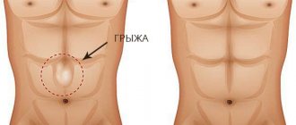
Also, gastrointestinal bleeding is fraught with general complications against the background of heavy blood loss (hypovolemia). If there is not enough blood circulating in the body, the risk of heart, kidney and brain failure increases. When more than 20% of the total blood volume is lost, a life-threatening condition develops - hemorrhagic shock. In this case, cellular metabolism is disrupted, oxygen starvation and tissue intoxication occur. Prognosis The bleeding episode does not affect the health of the digestive tract organs themselves. However, he points to the need for more careful monitoring of the course of the disease that provoked it. In the first few weeks, signs of posthemorrhagic anemia may be observed. This is caused by a drop in hemoglobin concentration against the background of heavy loss of red blood cells. Over the next six months, the patient needs to take iron supplements. A negative prognosis occurs when timely assistance is not provided during bleeding from the gastrointestinal tract. Moscow and other megacities with difficult traffic often do not allow a patient to be quickly transported to a hospital. Death occurs in 5-20% of cases (depending on the location of the damage and the general condition of the patient).
Endoscopic treatment of damaged vessels
For peptic ulcers, endoscopic vascular coagulation is usually used. The soldering of damaged walls can be carried out using electric currents or a laser. In case of bleeding from dilated veins of the gastrointestinal tract, the following is carried out: • Injection sclerotherapy - the introduction of sclerosants into the inner lining of the vein wall. These drugs destroy the damaged area, which eventually resolves. Subsequently, the vein sticks together and becomes hopeless. • Endoscopic ligation - mechanical clamping of the damaged area. • Application of a portosystemic shunt to redirect blood flow and reduce portal pressure on dilated veins.
The urgency of endoscopy depends on the patient's condition. If it is severe, treatment should begin no later than 24 hours from the moment of bleeding from the gastrointestinal tract.
How does the body cope with blood loss?
If blood loss is no more than 10%, the body is able to recover on its own. This is achieved through internal compensatory mechanisms: • Release of catecholamines by the adrenal glands immediately after the onset of bleeding from the digestive tract. Under the influence of these substances, blood vessels spasm, and the speed of blood flow adapts to new conditions. This prevents the blood supply to the organs from being cut off. • Release of tissue fluid into the vessels. On the third day, the body begins to use up red blood cell reserves from the spleen to restore normal blood volume. However, this occurs at the expense of cellular metabolism. • Restoration of blood composition by bone marrow. It will stabilize in a maximum of three weeks. However, this does not mean that treatment of gastrointestinal bleeding is not necessary. In practice, it is difficult for the body to cope with the consequences of such compensation. Therefore, he still requires medicinal support.