Neutral fat is a food material necessary for the human body. Thanks to it, the likelihood of hypothermia is significantly reduced, internal organs are protected from concussions, and vitamins are absorbed. In the body of a healthy person, this material is easily processed with the help of bile and pancreatic enzymes. If the digestive system malfunctions, feces contain breakdown products of neutral fats - fatty acids.
What are the main reasons?
There are several causes of steatorrhea in children. A small amount of neutral fats can be found as a result of dietary errors and consumption of increased amounts of fatty and fried foods. If, upon repeated examination of stool for coprogram, the fat level remains the same, it is necessary to exclude disturbances in the gastrointestinal tract - perhaps the child is developing the initial stage of pancreatitis, or the liver is damaged. Treatment depends on the type of pathology, existing symptoms, the age of the child and his well-being. Diagnosis when neutral fats are detected in feces can be extensive: this is necessary to identify the cause of the disorders and select an adequate treatment regimen.
Exocrine pancreatic insufficiency is most common in the first type of steatorrhea. It occurs against the background of a deficiency of enzymes involved in the breakdown of fats: lipase and colipase. Their secretion decreases in chronic pancreatitis, cystic fibrosis and after resection of the pancreas. In addition, the supply of digestive enzymes is reduced when the pancreatic ducts are blocked or compressed by mass formations in the abdominal cavity.
Clinical manifestations of steatorrhea:
- polyfecalia - the volume of feces is 2% greater than the volume of food eaten and liquid drunk;
- frequent bowel movements – 3-6 times a day;
- unformed, ointment-like stool with an oily sheen;
- gray stool;
- a large amount of neutral fat;
- undigested food particles and starch in stool;
- flatulence;
- abdominal pain without clear localization;
- weight loss.
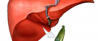
Another cause of impaired fat digestion is pathology of the liver, gall bladder and bile ducts. Bile converts the lipid mass found in the intestines into a fine-droplet solution. Thanks to this emulsification, the area of contact between fat droplets and lipases increases and their further absorption improves. In addition to participating in the digestion of fats, bile has a bactericidal effect and stimulates intestinal peristalsis. Insufficient flow of bile into the intestines (hypocholia) or its absence (acholia) disrupts the processes of fat emulsification and lipase activation.
Diagnostics
The inspection begins with the collection of complaints. On palpation, there is rumbling and splashing on the left side of the abdomen; you can feel the transfusion of the mass that is in the intestines. After the examination, the doctor prescribes a series of laboratory (stool, blood, urine tests) and instrumental (ultrasound, colonoscopy, MRI) studies.
Stool lipidogram
An important stage of diagnosis is a laboratory test of stool, in which stool is checked for the presence of fatty acids, neutral fat, and soap - the so-called stool lipid profile. This analysis is prescribed to assess the functions and identify pathologies of the digestive tract and pancreas. A stool lipidogram is indicated if the following diseases are suspected:
- pancreatitis;
- benign or malignant tumors of the gastrointestinal tract;
- intestinal tuberculosis;
- malabsorption syndrome;
- cholelithiasis;
- violation of the secretory function of the pancreas (for example, insufficient lipase activity);
- impaired lymphatic drainage due to damage to the mesenteric lymph nodes;
- increased peristalsis of the small intestine.
Compliance with the rules of preparation and collection technique affects the reliability of the lipid profile result. It is prohibited to collect material for research earlier than 2 days after an X-ray examination of the digestive tract using contrast. Three days before stool collection, it is recommended to exclude medications that affect peristalsis, composition and color of stool.
Protein metabolism disorders lead to a progressive decrease in body weight, a decrease in the amount of total protein and albumin, ascites, and hypoproteinemic edema.
Stool intended for analysis must be formed naturally; enemas or laxatives should not be used. On the eve of the test, it is necessary to exclude from the diet foods that contribute to the coloring of stool, excessive formation of gases in the intestines, and the occurrence of diarrhea or constipation.
Before collecting material, it is recommended to empty the bladder, wash the external genitalia with clean water, and pat dry with a towel. For hygiene procedures, it is better to use soap without foaming additives or fragrances.
Feces are collected in a plastic container with a special spatula built into the lid. You need to take care in advance about the container from which the feces will be collected for analysis. This can be a dry and clean vessel, or you can attach plastic film to the surface of the toilet. After defecation, about 20–25 ml of feces are collected in a prepared container. It is allowed to store the material in the refrigerator at a temperature of +3 to +7 °C for 24 hours. It is important not to forget to sign the container before submitting it for analysis.
There are several methods of analysis; a qualitative test is usually performed microscopically, and a quantitative test is usually performed using chromatography.
In a qualitative test, a stool sample is stained with Sudan dye and examined under a microscope, assessing the presence of fatty acids, soaps and drops of neutral fat, which have a bright orange-red color.
Quantitative detection of lipid levels is carried out on an automatic analyzer using chromatography. During the study, fats are extracted using a mixture of chloroform and ethanol. The extracts are subjected to chromatography on a layer of silica gel in the ether-acetone system (85:15). After staining with a solution of phosphomolybdic acid, all lipids contained in the test sample are detected. A stool lipidogram will be completed within one or two business days.
The main symptoms of steatorrhea are frequent urge to defecate, diarrhea with copious loose stools.
The norms for stool lipid profile parameters are shown in the table.
| Index | Norm |
| General lipids | 606–672 mg/dL |
| Non-esterified fatty acids | 17,1–20,5% |
| Monoglycerides | None |
| Diglycerides | 3,7–4,7% |
| Triglycerides | 10,2–13,3% |
| Phospholipids | 13,7–15,3% |
| Cholesterol | 33,2–35% |
| Coprosterol | Absent |
| Coprostanon | 18–20,9% |
Is it possible to recognize steatorrhea on your own?
The clinical symptoms of the pathology are quite specific, so you can suspect an increased content of neutral fat in the feces on your own. The main signs are associated with a change in the consistency of feces - they become sticky, oily, shiny, and can stain underwear. Such feces are difficult to wash off from the surface, sometimes light yellow spots remain on the laundry after washing. The color of the stool may remain the same, but in most cases the stool will lighten and become greyish. There is no strong odor with steatorrhea - on the contrary, such feces usually smell weakly or not at all.
Severe dyspeptic symptoms with steatorrhea are usually absent, but sometimes patients complain of bloating, a feeling of heaviness and fullness in the abdomen, dry mucous membranes of the oral cavity, and appetite disorders. If it lasts for a long time, the child may experience other signs not related to the functioning of the digestive tract. It can be:
- frequent headaches that turn into dizziness (in school-age children this is manifested by a decrease in academic performance and difficulties in mastering the material);
- joint pain (mainly in the back and lower back);
- bleeding gums;
- weakness and drowsiness.
In children of the first year of life, steatorrhea is manifested by increased excitability, frequent bowel movements, refusal of the breast or bottle, and poor weight gain. Feces are oily, difficult to remove from diapers and nappies, and have a stronger odor compared to children of the older age group.
One of the manifestations of pancreatic or hepatic steatorrhea may be a cough, which externally manifests itself as infrequent coughing. The cough is usually dry, not painful, goes away easily after drinking liquid, but can last up to 2-3 months. If a child suddenly begins to cough, but there is no wheezing in the lungs, and overall health remains satisfactory, a stool test should be taken for a coprogram to check the functioning of the digestive system.
Introduction
Acute intestinal infections (AI) rank second in frequency after acute respiratory infections.
Every year in Russia, more than 500 cases of acute intestinal infections per 100 thousand adult population are officially registered [1]. However, the real incidence of acute intestinal infections in Russia, according to experts, is 3–5 times higher than the officially registered one [2]. This is due to a significant proportion of erased and mild forms, the treatment of which is mainly carried out at home. In most patients with ACI of various etiologies, at the height of the disease, the thickness of the mucous membrane of the colon and the depth of its intestinal glands often increase, which is associated with interglandular edema of the lamina propria due to microcirculatory disorders and multiple hemorrhages [3]. Toxins of Shigella and Salmonella cause trophic changes in the tissues of the colon, which often persist for a long time. Thus, after 90–120 days, 39.1% of the examined salmonellosis convalescents showed signs of residual inflammation, and 13.1% showed signs of prolonged repair of the colon mucosa in the form of an enhanced macrophage reaction and changes in the linear parameters of the intestinal mucosa [4 ]. Solving the issues of diagnosis, prognosis, as well as early correction of dysbiosis in the initial period of recovery after acute intestinal infections will improve the quality of life of patients and achieve early and long-term remission. Continuing research in this direction will help improve understanding of the pathogenesis of gastroenterological pathology and its prevention. Despite the widespread prevalence of acute intestinal infections, many aspects of their pathogenesis in adults are extremely insufficiently studied. In particular, only a few studies are devoted to post-infectious irritable bowel syndrome (PIBS) and metabolic syndrome in disturbed microbiocenosis. Meanwhile, the development of PISCC is influenced not only by the intestinal microbiota, but also directly by the products of its metabolism, and in particular by short-chain fatty acids (SCFA), which are necessary to perform all functions of the large intestine and maintain physiological processes in the body.
How to accurately determine the cause of the disease
At the first visit, the doctor palpates the abdomen. When you press on it in the lower left part, you can hear a rumbling sound, the sound of iridescent liquid. After the examination, the following types of diagnostics are prescribed:
- Rectoscopy
is an examination of the rectum with an endoscope (up to 30 cm). Allows you to identify swelling, atrophy, ulceration of the intestinal mucosa. - Colonoscopy
is a complete examination of the intestines. Allows you to identify pathologies in all areas. - Biopsy
is the removal of affected organ cells for examination. Allows you to confirm or exclude cancer. - Coprogram
(stool microscopy) allows you to identify drops of fat and fatty acids in the stool and determine at what stage the disease is. - Ultrasound of the digestive organs.
Identifies pathologies of internal organs. - MRI
is used if ultrasound examination turns out to be uninformative. Allows you to identify any pathologies and neoplasms. - A blood test
shows an increase in leukocytes and ESR. This is typical for this disease.
The doctor determines what type of diagnosis to prescribe depending on the suspicion of a particular pathology.
Basic therapy
In order for the doctor to prescribe the correct treatment, it is necessary to find out which disease caused the disturbances in the digestive process. In most cases, children are diagnosed with pancreatic steatorrhea, which occurs as a result of a lack of lipase, so the basis of treatment in this case will be drugs containing digestive enzymes, for example:
- "Creon";
- "Festal";
- "Pancreatin";
- "Mezim."
Steatorrhea can be cured only after the root cause of the disease has been completely eliminated. The predominant method of treatment is medication. The doctor prescribes lipase drugs in high concentrations. They will help restore fat absorption and improve the functioning of the intestinal tract. Enzyme preparations are prescribed to restore good digestion. After diagnosing steatorrhea, doctors often suggest the use of antacids. In particularly difficult cases, these drugs are also accompanied by: adrenocorticotropic hormone, vitamin complexes, hydrochloric acid.
Table. Antacids for complex therapy of steatorrhea.
| Drug name | How to use | At what age should it be given? |
| 1-2 tablets up to 3 times a day (tablets should be dissolved until completely dissolved). | From 12 years old | |
| 1-2 packets 1.5 hours after meals. The maximum duration of treatment is 3 months. | From 15 years old | |
| 10-20 ml after meals and before bed. | From 12 years old | |
| The dosage regimen is selected individually. | From 10 years of age (can be used by children under 10 years of age, but strictly as prescribed by a doctor and under his supervision) | |
| 1-2 tablets 2 to 6 times a day (the exact dose depends on the age of the child). | From 6 years old |
Auxiliary therapy may include vitamin preparations and hormonal medications, but they should only be prescribed by a specialist after a thorough study of the clinical picture of the disease.
Treatment
Treatment of steatorrhea consists of eliminating the causes of insufficient absorption of fatty acids by the body, as well as correcting impaired metabolism. Drug therapy includes taking medications whose action is aimed at eliminating the consequences of steatorrhea: enzyme preparations with an increased content of lipases, antacids to neutralize gastric acid, cortisone, hydrochloric acid, vitamin complexes containing vitamins c. B, ascorbic and nicotinic acids, fat-soluble vitamins A, D, E and K.
An important condition for the treatment of steatorrhea is diet
Therapy also includes correction of diet and nutrition. It is recommended to eat small meals with a three-hour interval between meals, the weight of one serving should not exceed 200 g, and the daily amount of fat consumed should be 50–65 g. It is necessary to exclude fatty, fried and spicy foods, alcohol, and sweet carbonated drinks from the diet. Reduce carbohydrate intake. Preference should be given to dishes made from lean meats, lean fish, fermented milk products, low-fat cottage cheese, and fresh vegetables.
Video from YouTube on the topic of the article:
Additional measures
To normalize digestion and improve bowel movements, during treatment the child must follow a special diet with a high protein content. Before starting, you should take a urine test and evaluate your kidney function, since disturbances in the functioning of the renal system can lead to proteinuria. If the child is healthy, it is recommended to increase the proportion of protein foods in the diet by approximately 101005%. Boiled meat and fish, meat casseroles and souffles, eggs (it is better to choose quail eggs - they have less cholesterol), cottage cheese, and dairy products are useful for children with signs of steatorrhea.
If the child is not prone to intestinal disorders, you can give plant foods rich in proteins: chickpeas, beans, lentils. These products belong to legumes and contain a large amount of puric acid, which, when broken down, forms purines and causes bloating and flatulence, so it is better for children prone to colitis and enteritis to consume animal proteins.
Products that irritate the intestinal mucosa should be completely excluded from the diet.
Sample menu for children who have neutral fats in their stool
Breakfast:
- steamed scrambled eggs with herbs, tomatoes and pieces of boiled chicken;
- whole grain toast;
- tomato juice.
Lunch:
- cottage cheese casserole with apples.
Dinner:
- meat broth with meatballs and crackers;
- mashed potatoes with beef cutlet;
- compote.
Afternoon snack:
- natural thick yogurt without sugar;
- marshmallows;
- black tea.
Dinner:
- fish stew with a side dish of vegetables;
- lingonberry juice.
Before bedtime:
- a glass of milk or kefir.
Complications and consequences
Complications develop only if treatment is not started in a timely manner. There is a disruption in the absorption of nutrients in the intestinal tract. Against this background, hypo- and avitaminosis, protein deficiency and exhaustion of the body develop. Pathology of water and electrolyte balance is manifested by a constant feeling of thirst, edema, dehydration, and convulsive attacks.
The specialist diagnoses the appearance of oxaluria (excessive pathological excretion of oxalic acid salts from the body along with urine) and the formation of urinary stones of oxalate origin. This pathological condition occurs because, under normal conditions, oxalates do not enter the blood from the intestinal tract, since their combination with calcium makes them insoluble. With the development of steatorrhea, calcium is excreted in large quantities from the body along with feces. This leads to a significant release of oxalates from the intestines into the blood.
Physiological role of SCFAs
SCFAs are one of the products of the breakdown of proteins, fats and carbohydrates by microbial enzymes. They are produced predominantly by anaerobic microbes, and they can and should be considered as biochemical markers of changes in intestinal microecology. The main SCFAs are propionic acid, acetic acid, isobutyric acid, butyric acid and isovaleric acid [5]. Unbranched SCFAs (acetic, propionic and butyric) are formed as a result of anaerobic fermentation of carbohydrates, and fermentation of proteins and their breakdown products leads to the formation of already branched acids - isovaleric, isobutyric. Their determination allows for screening assessment of the state and activity of microflora and prescribing therapeutic correction using probiotics and other biological drugs [6]. The synthesis of SCFAs is one of the main factors of colonization resistance, ensuring the constancy of the composition of the intestinal microflora. An increase in the amount of SCFA is accompanied by a decrease in osmotic pressure in the lumen of the large intestine as a result of fermentation of polysaccharides [7]. The formation of a specific type of SCFA depends on the enzymes of which bacteria will cleave the substrate, and this makes it possible to assess the functional activity of certain representatives of the intestinal microbiota. Thus, if we consider anaerobic bacteria of the species Eubacterium rectale, Eubacterium ramulus, Eubacterium hallii, Roseburia cecicola, Roseburia faecis, Faecalibacterium prausnitzii
and
Coprococcus
, as well as fusobacteria and non-pathogenic species of clostridia, they play a major role in the production of butyrate.
And bacteria of the genera Bifidobacterium and Lactobacillus
are the main ones in the production of acetate [8].
SCFA is the main source of acetyl coenzyme A, as well as a respiratory substrate for colonocytes of the intestinal mucosa. SCFAs are involved in the proliferation of mucosal cells, the secretion of mucus and increased blood flow in the mucosa. The mechanism by which SCFAs exert their influence on cell proliferation has not yet been studied. Perhaps such a mechanism is their influence on the blood flow in the mucosa [9]. SCFAs maintain a slightly acidic environment. This allows butyrate-producing gram-positive bacteria to compete with gram-negative bacteria and thereby maintain balance in the intestinal microbiota. At a normal level of SCFA content, the development and increase in the number of pathogenic enterobacteria slows down, and they predominantly feed on protein components. This creates favorable conditions for suppressing decay processes and reduces the formation of ammonia, sulfides, endogenous carcinogens, and aromatic amines. A decrease in the SCFA content contributes to an increase in the number of gram-negative microbes and, accordingly, the content of lipopolysaccharides [10]. The highest concentration in the colon lumen is acetate (60%), to a lesser extent propionate (25%) and butyrate (15%) [11]. The anti-inflammatory effect of butyric acid has now been quite well studied, which is carried out mainly by reducing the activity of histone acetylase and suppressing the activation of the associated nuclear factor (NF-κB) in colon cells. Butyrate inhibits the activity of the NF-κB factor, which controls the expression of immune response genes and is responsible for the production of cytokines. As a result, there is a decrease in the production of pro-inflammatory cytokines, and the proliferation and activity of T cells decreases [12]. Butyrate is produced mainly by representatives of the gram-positive group Firmicutes
eg
E. rectale
,
Roseburia spp
.
and F. prausnitzii
[10, 13].
Studies conducted by H. Sokol et al. showed that F. prausnitzii
have the ability to inhibit inflammation in signaling systems, and the butyrate they produce blocks the activation of the transcription factor NF-κB [14]. Some studies have proven a significant decrease in the level of pro-inflammatory cytokines with increased consumption of dietary fiber or drugs containing SCFAs [14, 15]. Propionate and butyrate activate intestinal gluconeogenesis through the gut-brain neural circuit and promote metabolic control of body weight and glucose [16].
The main part (95%) of SCFAs released in the colon is reabsorbed. Absorption of SCFAs is carried out using active transport systems of intestinal cells [17]. In addition, SCFAs promote the absorption of calcium and magnesium. Today it has been proven that butyric acid plays a major role in the energy supply of colonocytes, which is necessary to improve metabolism, normal cell development and play a protective role in the prevention of diseases of the large intestine. It has been shown that SCFAs are also regulators of apoptosis and have an anticarcinogenic effect. Acetic and propionic acids that enter the colonocyte at the level of the colon participate in the regulation of its blood flow, improving blood supply in the mucous membrane, and thereby exhibit an anti-ischemic effect. The liver retains about half of the SCFAs that enter through the colonocyte, and another quarter is eliminated by peripheral tissues. In peripheral tissues, most of the acetate and propionate are used for glucose synthesis and only a small part (no more than 10%) is used for energy supply [17].
Recently published data from MC Kim et al. (2017) confirm the significant role of metabolites of symbiont microflora, including SCFA, in the formation of the humoral response; they have a direct and indirect immunomodulatory effect on B lymphocytes not only at the local but also at the systemic level and, therefore, enhance antibody formation [18]. It turned out that SCFAs, through their effect on coenzyme A, can also increase the activity of T cells [19]. Apolipoprotein-I (ApoA-I) is known to be the main protein component of high-density lipoproteins and removes excess cholesterol in the blood vessels. JZ Tayyeb et al. (2018) found that ApoA-I is significantly reduced during amoxicillin therapy due to the development of intestinal dysbiosis and, accordingly, changes in the composition and structure of SCFAs [20]. At the same time, H. Yu et al. (2018) concluded that disturbances in the quantitative and qualitative composition of SCFAs in the intestine can contribute to the accumulation of lipids and modulate the expression of metabolic enzymes [21].