Symptoms Causes Diagnostics Treatment Information block
Chronic gastritis associated with Helicobacter pylori (Chronic Helicobacter pylori gastritis) is an inflammation of the gastric mucosa that occurs under the influence of the bacterium Helicobacter Pylori.
Helicobacter pylori (H. pylori) is a gram-negative microaerophilic bacterium that colonizes the gastric mucosa and is associated with atrophic gastritis, gastric and duodenal ulcers, adenocarcinoma and extranodal B-cell MALT lymphoma of the stomach.
THE INSTITUTE OF ALLERGOLOGY AND CLINICAL IMMUNOLOGY has:
- range of services for accurate diagnosis of diseases
- special treatment programs
- consultations
- carried out by doctors with academic degrees.
When identifying concomitant diseases, consultations are held with specialists in related fields (otorhinolaryngologists, endocrinologists, neurologists, gastroenterologists, dermatovenerologists), consultations with Doctors of Medical Sciences.
Attention! Helicobacter pylori is a causative agent of cancer in the gastrointestinal tract.
Gastritis associated with Helicobacter Pylori: what is it?
Hp-associated gastritis (inflammation of the gastric mucosa) is a disease caused by the presence and activity of the bacterium Helicobacter Pylori in the stomach.
Hp-associated gastritis (inflammation of the gastric mucosa) is a disease caused by the presence and activity of the bacterium Helicobacter Pylori in the stomach. According to statistics, about 50% of the total population of the Earth is infected with this microorganism, and in some regions this figure reaches 80-90%.
In addition to the fact that inflammation of the gastric mucosa causes many unpleasant symptoms, it disrupts the digestive processes and absorption of nutrients. But the greatest danger is hp-associated gastritis with this bacterium, from the point of view of forecasts: according to various estimates, peptic ulcer of the stomach and/or duodenum develops in 15-24% of patients, and not much less often inflammation leads to atrophic processes in gastric mucosa - one of the predisposing factors for cancer.
Therefore, gastritis caused by Helicobacter Pylori requires timely and professional treatment - this will restore health and quality of life, and also protect against possible complications.
At the First Family Clinic of St. Petersburg, highly qualified specialists, modern diagnostic facilities and individual therapy programs are waiting for you, which together makes the treatment of gastritis the most comfortable and effective process.
Causes
Attention! Helicobacter pylori can be transmitted from one person to another due to violations of personal hygiene rules!
Helicobacter pylori produces highly active enzymes urease, catalase, and mucinase, which lead to changes in the qualitative characteristics of gastric juice. Helicobacter pylori, upon penetration into the pyloric part of the stomach, in which there is a high content of urea, creates an alkaline environment, converting urea into ammonia, which leads to the launch of the mechanism of infection pathogenesis: destructive processes at the molecular, cellular and tissue levels, cytotoxic effects, impaired secretion, compensatory reactions of the glands, an intense inflammatory reaction with a predominance of neutrophil infiltration and a large number of plasma cells producing IgA in the mucosa.
Predisposing factors:
- Dietary disorders and poor nutrition.
- Smoking.
- Alcoholism.
- Psychologically severe, stressful conditions.
Antral gastritis HP – associated (type B)
The primary treatment tactics are effective eradication of HP in the gastric mucosa, stabilization of the general status of the patient and prolongation (lengthening) of the remission period. The European Association of Gastroenterologists Maastricht II (2000) recommended that targeted eradication of HP be carried out only in patients with atrophic gastritis.
The average duration of eradication therapy is from 7 to 14 days and directly depends on the HP status:
- Degree of infection;
- Activity of urease test;
- The activity of the inflammatory process.
The combination of drugs is usually three-component and includes, in addition to a proton pump inhibitor (the main representative is omeprazole), two combined antibacterial drugs:
- clarithromycin and amoxicillin, or:
- furazolidone and clarithromycin.
The primary treatment tactics are effective eradication of HP in the gastric mucosa, stabilization of the general status of the patient and prolongation (lengthening) of the remission period.
Doses are selected individually depending on many factors: age, weight of the patient, life history, allergic disease. A sharply reduced secretory function in a patient predetermines the prescription of a bismuth drug instead of a secretion inhibitor - de-nol 240 mg twice a day. In addition, it has antioxidant and antibacterial properties. Evaluation of the effectiveness of the therapy should be carried out no earlier than a month to a month and a half after its completion.
If the patient suffers from herpesvirus infection type 4 in the acute stage, amoxicillin is not prescribed. It is possible to include an antiviral drug in the treatment regimen.
If the first line of therapy is ineffective, it is necessary to prescribe four-component therapy. Typically, tetracycline is added to the treatment regimen at a therapeutic dose. The ideal option is to prescribe antibacterial therapy after determining the sensitivity of HP to it.
The principles for prescribing symptomatic drugs are as follows:
- Prokinetics (Motilak).
- Preparations that adsorb excess gas in the intestines (silicon containing).
- Normalization of dysbiosis (probiotics and eubiotics).
Bifidumbacterin, subalin, bifiform, biosporin have proven themselves very well.
If the secretion of hydrochloric acid is reduced, the following is included in treatment:
- plantaglucide 1 g twice a day before meals;
- plantain juice 15 ml 2-3 times a day, it also has good reparative and anti-inflammatory properties;
- delicious bitters.
Among the herbal remedies that alleviate flatulence, you can prescribe romazulan or a special carminative mixture containing several herbs with a similar effect. Acidinpepsin, betacid, abomin or pepsidil are the first-line drugs prescribed for replacement therapy for achylia. For the same purpose, it is necessary to prescribe drugs containing a set of various pancreatic enzymes: digestal, pankurmen, solizym, pancitrate, etc.
A very effective and at the same time safe method of treating chronic atrophic gastritis, which is best used not as monotherapy, but in combination with other drugs prescribed by the attending physician, is the daily use of propolis for one month in a dose of approximately 10 grams. Necessary condition: thorough chewing of propolis.
Helicobacter-associated gastritis is a disease that requires a professional approach to treatment
Chronic gastritis is the most common disease of the gastrointestinal tract, affecting 50 to 80% of the world's adult population [1]. The main cause of chronic gastritis is infection of the gastric mucosa by Helicobacter pylori
.
According to numerous studies , H. pylori
-associated chronic gastritis accounts for approximately 80% of all forms of gastritis [2].
This infection causes a wide range of pathological changes in the gastric mucosa, including atrophic gastritis and intestinal metaplasia, which are precancerous conditions, against which precancerous changes in the mucous membrane (low- and high-grade dysplasia) and, in some cases, gastric cancer can develop [3-6 ]. Therefore, currently, diagnosis and timely treatment H. pylori
-associated gastritis are considered as a global strategy for the prevention of gastric cancer [7, 8].
Endoscopic examination with biopsy of the gastric mucosa is today the main method for diagnosing chronic gastritis [9-13]. However, white light endoscopy has limited capabilities in diagnosing H. pylori
-associated gastritis and associated precancerous conditions and changes in the gastric mucosa [14].
Only in a number of cases, with pronounced inflammatory and atrophic changes in the mucous membrane, specific endoscopic signs can be determined, such as multiple nodular formations [15, 16] with active chronic H. pylori
-associated gastritis, the disappearance of gastric folds and visualization of the vessels of the submucosal layer [17] with atrophy, as well as foci of ashen or whitish color [18, 19] with the development of intestinal metaplasia.
However, these signs have very low sensitivity, especially in patients with minimal inflammatory and initial atrophic changes [20]. With the introduction of new endoscopic technologies, such as magnifying and narrow-spectrum endoscopy, it has become possible to study in detail and in depth the minimal structural changes in the mucous membrane that occur under the influence of H. pylori
.
Narrow band endoscopy (NBI) is an optical endoscopic diagnostic technique based on the use of special optical filters that narrow the spectrum of the light wave and increase the contrast of the resulting image, which allows one to obtain a detailed picture of the vascular pattern of tissues [21]. Magnifying endoscopy (High-Magnification Endoscopy) is performed using special endoscopes that have an optical lens at the distal end and allow a detailed study of the mucous membrane with an optical magnification of more than 100 times. The combined use of magnifying and narrow-spectrum endoscopy makes it possible to evaluate in detail the surface of the epithelium and microvessels of the gastric mucosa [22].
Endoscopic examination with magnification of the gastric mucosa evaluates two main components [23]: the microvascular pattern and the microstructure of the epithelial surface. In the microvascular architecture of the superficial layers of the gastric mucosa, two main components are distinguished: the subepithelial capillary network (SECN) and collecting venules (CV). The microstructure of the surface of the gastric mucosa is represented by gastric pits and grooves of various shapes. The pits histologically correspond to the gastric glands, which have several main components: the mouth, the marginal pit epithelium, which represents the area surrounding the mouth of the gland, and the pars intermedium, the protruding zone between the mouths of the glands. Thus, the surface pattern of the epithelium is formed mainly by the intermediate part, bordered by the marginal pit epithelium.
The microanatomy of the mucous membrane has a different structure in different parts of the stomach [24]. According to K. Yao et al. [25], normally in the body of the stomach the microvascular architecture is represented by a subepithelial capillary network in the shape of a “honeycomb”, which are located on the periphery of the pits. Polygonal loops of true capillaries surround each gastric pit, forming a network under the epithelium, the branches of which converge into collecting venules that have the appearance of a “starfish”. The surface structure of the epithelium of the normal mucous membrane of the body of the stomach is represented by round-shaped pits, the mouths of which are presented in the form of points.
The mucous membrane of the antrum of the stomach has a different picture. The microvascular architecture of the normal mucous membrane of the gastric antrum is represented by coil-shaped microvessels that are located inside the epithelial structures. Collecting venules are visualized less frequently, since they lie in deeper parts of the lamina propria than in the body of the stomach and extend in an oblique direction. The surface pattern of the epithelium is represented by oval or linear pits; the mouths of the glands are not visualized, since they have an oblique direction.
An important feature of normal gastric mucosa that has not been infected with
H. pylori
is the presence of correctly located collecting venules (regular arrangement of collecting venules - RAC).
According to K. Yagi et al. [26], this sign has high sensitivity and specificity (93.8 and 96.2%, respectively). absence of collecting venules is a characteristic feature of H. pylori
-associated gastritis, which has sensitivity and specificity, according to G. Anagnostopoulos et al.
[27], 100 and 92.7%, respectively. , the very first sign of the onset of the inflammatory process induced by H. pylori
, is the disappearance of collecting venules while maintaining the round shape of the pits and capillary network. This is likely due to inflammatory infiltration, destruction of microvascular structure and changes in blood flow, as well as degeneration of the surface epithelium. Emphasizing the importance of describing collecting venules, S. Nakagawa et al. [28] identified several types of patterns. The correct type (regular, R-type) is determined by the presence of correctly located collecting venules, which is characteristic of normal mucosa. The hidden type (obscured, O-type) is characterized by the complete absence of collecting venules and is observed when gastritis occurs. Additionally, an irregular type (irregular, I-type) with chaotically located collecting venules was identified, which occurs in the case of the development of atrophic changes in the mucous membrane.
Further progression of inflammatory and degenerative changes in the gastric mucosa, which occurs under the influence of H. .
pylori
, is accompanied by characteristic changes in the microstructure of the gastric mucosa, observed when using magnifying endoscopy [29]. With active inflammation, the pits expand, are surrounded by a zone of erythema, dividing grooves appear, and the capillary network completely disappears. It is also noted that with an increase in the degree of the inflammatory process, the pits acquire a more expanded and elongated shape [30]. With the development of atrophic changes in the mucous membrane, the microstructure of its epithelial surface (pits and grooves) cannot be traced, the subepithelial capillary network is destroyed and cannot be determined, and the microvascular architecture is represented only by chaotically located collecting venules. These changes are described in detail in various classification systems.
In order to objectify and describe all changes in the mucous membrane that occur under the influence of H. pylori
, many specialists have created various classification systems.
One of the first works devoted to the study of the surface pattern of the mucous membrane in chronic gastritis was the work of N. Sakaki et al. [31]. Using a magnifying fiberscope FGS-ML II (Machida, Tokyo, Japan) with a magnification of 30 times, the microstructure of the gastric mucosa during various inflammatory and degenerative changes was described in detail for the first time. Four main types of pitting pattern of the gastric mucosa were identified, for which the most characteristic histological changes were subsequently determined: A - dotted pits (normal mucous membrane), B - short linear pits (chronic gastritis), C - striped furrows (atrophy and intestinal metaplasia ), D - cellular grooves (severe atrophy and intestinal metaplasia). The created classification is widely used by Japanese specialists to this day [32]. With the introduction of modern endoscopic equipment, changes in the microstructure of the mucous membrane that occur during chronic gastritis were described in detail in the so-called “Z-classification” created by K. Yagi et al. [33] (Table 1). Within this classification system, type Z-0 describes normal mucosa, types Z-1 and Z-2 are typical manifestations of
H. pylori gastritis, and type Z-3 is observed with the development of atrophic changes (see Table 1). Later, the same authors revised the “Z-classification” and added new types that describe in detail minimal inflammatory changes. The new classification system is called the “AB classification” [34], since it describes not only inflammatory changes in the body of the stomach (“body” - “B”), but also features of the mucous membrane of the antrum and atrophic changes (“atrophic mucosa and antral mucosa " - "A"). However, these criteria have not been validated in European countries; moreover, the criteria of various types are subjective and descriptive in nature and can be interpreted differently by different specialists. In this regard, leading European and Japanese experts developed a simplified classification system [35], describing four types of mucosal pattern, including the main types of histological changes (Table 2): normal mucous membrane (type 1), chronic gastritis (type 2 and type 3), atrophic changes (type 4). In the antrum of the stomach, the important and most characteristic sign of chronic inflammation is the disappearance of the capillary network [36].
Table 1. Z-classification of changes in surface microstructure and microvascular pattern in chronic gastritis according to Yagi
Table 2. Classification of changes in surface microstructure and microvascular pattern in chronic gastritis according to Anagnostopoulos.
A separate and very important task of endoscopic magnification is the diagnosis of intestinal metaplasia. According to a study by A. Bansal et al. [37], the villous and comb-like patterns correspond to the histological diagnosis of intestinal metaplasia with high sensitivity and specificity (80 and 100%, respectively). However, the most striking and characteristic sign of intestinal metaplasia is the detection of so-called light blue crests (LBC) - thin light blue lines on the ridges of the epithelium or grooves [38]. This phenomenon occurs only when viewed in a narrow spectrum of light due to the reflection of a short wavelength (400-430 nm) from the surface of tissue that has a ciliated surface, such as the brush border of intestinal metaplasia and duodenal cells. According to N. Uedo et al. [38], identifying light blue ridges as a predictor of intestinal metaplasia has high sensitivity and specificity - 89 and 93%, respectively. However, light blue ridges are not found in all areas of intestinal metaplasia, so it is necessary to use other signs during endoscopic examination. An alternative characteristic of intestinal metaplasia is the marginal turbid band - white, opaque stripes bordering the epithelial structures [39]. This phenomenon occurs due to the expansion and shortening of the intermediate part between the pits of the gastric glands. Typically, marginal dull streaks characterize recent development of intestinal metaplasia, while light blue ridges are more associated with long-standing lesions. In a study by J. An et al. [39] the sensitivity, specificity, and accuracy of marginal matte bands in the diagnosis of intestinal metaplasia were 100, 66.0, and 81.7%, respectively. This sign can be easily identified with the development of intestinal metaplasia in the body of the stomach, however, in the antrum of the stomach, the marginal matte stripes are sometimes difficult to differentiate from the marginal glandular epithelium of the structures of the normal mucous membrane.
Currently, eradication of
H. pylori
is a key tool in the therapeutic treatment of chronic gastritis and peptic ulcers, as well as the most promising strategy for the cancer prevention of gastric cancer. However, in the histological cascade of changes in the gastric mucosa, there is a so-called “point of no return”, upon reaching which eradication therapy cannot lead to regression of pathological changes and prevent the development of gastric cancer. That is why the earlier treatment begins, the more effective the restoration of the mucous membrane is [40].
Eradication of H. pylori
leads to cure and recovery in non-atrophic gastritis, however, with the development of atrophy of the gastric mucosa and intestinal metaplasia, the results of studies on the effectiveness of eradication are contradictory.
Studies show that eradication of H. pylori
eliminates inflammatory infiltration, improves the functional abilities of the gastric body, slows down or even stops the progression of atrophy, and in some cases, the atrophy process can be reversed.
However, there is currently no convincing evidence that eradication of H. pylori
can lead to regression of intestinal metaplasia [41].
Thus, eradication therapy for H . pylori
can improve the condition of the gastric mucosa, which is accompanied by changes in the microstructure of the mucous membrane, observed during magnifying endoscopy.
In a study by K. Yagi et al. [42], when observing patients 2 months after treatment, two signs of successful therapy were determined: the disappearance of erythema and edema between the gastric pits, as well as changes in the structure of the pits - enlarged white pits become dotted. Researchers also note a change in microvascular architecture in the form of a decrease in the density of the irregular capillary network. It is known that it takes several months for the mucous membrane to recover after successful elimination of the H .
pylori
, therefore a control study should be carried out no earlier than 2-3 months after eradication therapy. However, according to a study by T. Tahara et al. [30], even after 12 weeks, minor changes in the microstructure of the mucous membrane remain, manifested in minimal irregularity of the pits and capillary network. Therefore, complete restoration of the mucous membrane is a long process that requires endoscopic control and monitoring.
However, according to research, with moderate and severe atrophy, as well as in cases of intestinal metaplasia, changes in the microstructure of the mucous membrane were not observed after successful eradication therapy in a short time (3 months). It is likely that much longer observation intervals are required to monitor the dynamics of atrophic and metaplastic changes. Considering these facts, it is reliable to talk about the efficiency of H .
pylori
occurs only in patients without atrophic and metaplastic changes in the mucous membrane.
Thus, modern endoscopic technologies open up new prospects not only in the diagnosis of precancerous conditions and changes, but also in the detection of H. .
pylori
, as well as monitoring the effectiveness of eradication therapy.
and narrow-spectrum endoscopy in the complex diagnosis of patients with H. pylori
-associated gastritis will allow the diagnostic process to be elevated to a fundamentally different level, which in the future may lead to improved quality of monitoring of such patients and, as a consequence, the effectiveness of measures to prevent gastric cancer.
Helicobacter-associated gastritis: symptoms
Self-treatment of Helicobacter Pylori-associated chronic gastritis is impossible: to eliminate the infection and the damage it has caused to the stomach, the participation of qualified and experienced specialists and the use of the latest generation medical equipment is required. Therefore, if you have one or more of the following symptoms, be sure to consult your doctor:
- pain in the upper abdomen, which can occur both immediately after eating and 1-2 hours later;
- stool disorders, expressed in a tendency to constipation at the initial stage of the disease, and as it develops - in alternating constipation and diarrhea;
- heartburn and/or frequent belching with a sour taste;
- dyspeptic syndrome (a feeling of rapid satiety, a feeling of heaviness in the stomach, a bursting sensation in the epigastric region);
- appetite disorders.
Helicobacter-associated gastritis is a disease that requires a professional approach to treatment. This is the only way it becomes possible to eliminate the cause of the problem and prevent the development of gastric ulcers or atrophic processes in its mucous membrane.
Alarming symptoms of associated Helicobacter gastritis
Helicobacter pylori - gastritis, which occurs from the harmful effects of this microorganism, has 3 stages of development, each of which is accompanied by certain symptoms.
Initial, having increased or (which is quite rare) normal acidity. It is characterized by the following general symptoms:
- Epigastric pain that occurs 2 hours after eating;
- Frequent belching with a sour taste, turning into heartburn;
- Stool disorders (tendency to constipation).
Appetite is generally not affected, only occasionally an increase is noted. With the advanced stage of Helicobacter gastritis, the signs are the same, only their manifestation becomes more acute.
Helicobacter pylori - gastritis, which occurs from the harmful effects of this microorganism, has 3 stages of development, each of which is accompanied by certain symptoms
But the last, chronic, stage of this Helicobacter-associated gastritis will already be characterized by signs of atrophy, manifested in the mucous membrane of the digestive organ. Among them, the most common symptoms are:
- Indigestion (heaviness in the stomach after eating even a small amount of food);
- Lack of appetite;
- The presence of diarrhea due to the fact that the barrier function of hydrochloric acid is impaired;
- Rotten-tasting belching and metallic taste in the mouth;
- Significant reduction in body weight.
The complaints of patients with Helicobacter pylori gastritis are completely similar to the complaints of patients with stomach ulcers. These are hunger pains that appear in the morning (on an empty stomach), frequent constipation, sour belching, heartburn. If you do not pay timely attention to them and do not begin the necessary treatment, the disease begins to progress very quickly.
In this case, the following signs of bacterial gastritis are added - nausea, mostly turning into vomiting and containing bloody inclusions, as well as bloating, which appears due to excessive gas formation caused by the activity of Helicobakter pylori.
If a person has at least one of the above signs, you should not hesitate to contact a specialist, and not attribute it to the fact that perhaps you have eaten something of insufficient quality, and you need to drink no-shpa to prevent unpleasant symptoms stopped.
Only timely diagnosis of the gastrointestinal tract for the presence of Helicobacter gastritis will allow taking all necessary measures in a timely manner and preventing the disease from progressing to a more severe stage. Modern research methods suggest an accuracy of determining HP gastritis of up to 99%.
Erosive gastritis nr associated gastritis
The disease can occur in acute or chronic form.
Considering the factors that cause associated diseases in the chronic stage, primary and secondary types of associated diseases are distinguished. Diseases of the digestive tract disrupt the normal functioning of the body. The gastrointestinal tract is of great importance to the body, and inflammation can lead to complications if timely treatment is not started. It is extremely important to consult a doctor in time to carry out the necessary treatment complex. One of the most common diseases is associated gastroduodenitis, which is a type of gastroenteritis. Frequent diagnosis of associated gastroduodenitis is associated with constant contact of the duodenum and stomach. When an illness occurs, the patient's duodenal mucosa becomes inflamed.
The disease can occur in acute or chronic form. Taking into account the factors that cause associated diseases in the chronic stage, primary and secondary types of associated diseases are distinguished. The appearance of the primary type is characteristic of impaired digestion, which occurs due to an incorrect diet. The secondary type may appear as a consequence of other diseases, for example, gastritis, pancreatitis, etc.
There are several classifications of HP associated gastroduodenitis. In the presence of a certain level of acidity, types such as normal gastroduodenitis, an associated disease with a decreased or increased level of secretory function, are distinguished.
The appearance of gastroduodenitis is associated with the proliferation of the bacterium helicobacter pylori. Helicobacter is the cause of the disease in 80% of cases.
In advanced stages, it is impossible to determine the presence of Helicobacter, because the digestive environment becomes unsuitable for its existence. A properly formulated diet, an active lifestyle, and giving up bad habits are the main points necessary to maintain health and prevent the occurrence of inflammatory processes in the digestive tract. If unpleasant signs appear with associated gastroduodenitis, you should immediately consult a doctor to avoid complications.
Inflammatory processes at the initial stage can be characterized by such signs as:
- the appearance of heartburn, belching with a sour smell;
- increased appetite;
- pain in the area under the stomach that appears 2 hours after eating;
- Constipation may occur.
In advanced stages, it is impossible to determine the presence of Helicobacter, because the digestive environment becomes unsuitable for its existence.
The initial stage is most often characterized by an increased level of acidity. But in the last stages of the disease, the patient experiences atrophy of the gastric mucosa. This manifests itself in the development of symptoms such as:
- loss of appetite;
- a feeling of heaviness in the stomach area resulting from undigested food;
- dull pain in the gastric area;
- reduced barrier function of hydrochloric acid leads to diarrhea;
- the appearance of a metallic taste;
- the manifestation of belching, which may have the smell of consumed food, and sometimes has a rotten character;
- sudden weight loss;
- Cracks may appear in the corners of the mouth, which are called jams;
- the appearance of nausea.
If Helicobacter has spread to the duodenum, the patient begins to experience the following symptoms:
- a feeling of bitterness in the mouth, which may be accompanied by belching of bile;
- the appearance of nausea and vomiting;
- pain in the area of the right hypochondrium.
Treatment of gastroduodenitis associated with nr
The diet should be light; you can eat soups and cereals with water.
Any therapeutic complex aimed at treating the digestive system involves introducing a diet consisting of several points:
- The patient is allowed to eat soups and cereals cooked in water or meat broth, but without adding oil.
- Meat and fish products are recommended to be consumed boiled.
- The use of milk is allowed in the absence of diarrhea and allergic reactions. It is recommended to consume low-fat kefir, sour cream, cottage cheese and grated cheese that does not contain flavoring additives.
- You can eat stale types of bread and unpalatable crackers, but other rich foods are completely prohibited.
- Fruits and vegetables can be eaten boiled or raw, but the main thing is to grind them before eating.
- The use of natural juices, compotes, and unsweetened teas is allowed.
It must be remembered that the diet should consist of 5 meals, and the food should be at normal temperature so as not to burn the inflamed membrane. In addition, people who have been diagnosed with the development of associated gastroduodenitis should remain in bed for at least 8 days.
To destroy Helicobacter, the patient is prescribed a special medical treatment complex, including antibiotics. Such drugs are prescribed depending on the stage of development of the disease. Self-selection and treatment with antibiotics is prohibited, because the microflora of the digestive tract can be disrupted. Along with antibiotics, medications are prescribed that contain beneficial bacteria necessary for the normal functioning of the tract. In addition, the doctor may prescribe a complex of vitamins and minerals necessary to maintain body tone.
Features of the course of gastritis associated with HP
The main symptoms of chronic gastritis are dyspeptic disorders. Patients complain of nausea, heaviness in the stomach after eating, belching, heartburn, and rumbling in the stomach. A characteristic symptom of gastritis associated with HP is stool disorder: pasty stool, imperative urge to defecate. Abnormalities in bowel movements are intermittent, but they make themselves felt. Less common is bitterness and burning in the mouth, and belching of air. Chronic gastritis can cause symptoms such as weakness, irritability, increased fatigue, and loss of interest in what is happening. Such symptoms are called “general” and are characteristic of many infectious diseases.
Diagnosis of Helicobacter gastritis: gastroscopy, biopsy, helic test
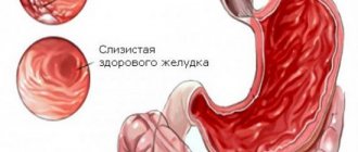
In addition to standard methods of examining the patient (blood tests, urine tests, stool tests, ultrasound of internal organs), if chronic gastritis of a bacterial nature is suspected, gastroscopy is mandatory. The stomach is examined through a probe with a lighted camera at the end. Changes in the gastric mucosa are assessed, its surface is examined for the presence of erosions, ulcers, and neoplasms.
During gastroscopy, a biopsy of tissue from different parts of the stomach is performed. The resulting samples are sent for microscopic examination. Using a special staining of the preparations, the doctor can determine the presence of Helicobacter pylori in the patient's stomach under a microscope. A simpler and more accessible method is to conduct a helic test (urease breath test). This is an absolutely painless test based on the ability of the HP bacterium to exhibit urease activity. The patient drinks a urea solution and then breathes through a special tube with an indicator. As a result of the hydrolysis reaction, gas is released, which is noted in the tube by a change in color.
Diagnostics
To make a diagnosis, the doctor should conduct a comprehensive diagnosis of the patient to exclude the development of other diseases. The diagnostic complex includes the following methods:
- conducting an endoscopic examination, which can be used to determine the swelling of the mucous membrane and determine the location of the source of inflammation;
- histological diagnosis, which allows you to determine the degree of the inflammatory process;
- electrogastrography and ultrasound make it possible to study motor function;
- X-rays can be used to evaluate the evacuation function, which is necessary for differentiation from other ailments with similar symptoms.