- Gallery
- Reviews
- Articles
- Licenses
- Vacancies
- Insurance partners
- Partners
- Controlling organizations
- Schedule for receiving citizens for personal requests
- Online consultation with a doctor
- Documentation
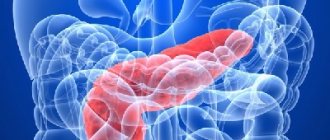
Pancreatitis is inflammation of the pancreas. There are 2 main forms of this inflammation: acute and chronic. These forms are more common in adults.
In recent years, it has become common to identify another one - reactive pancreatitis (correctly called reactive pancreatopathy) - which is more common in children.
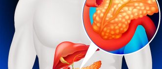
When the pancreas becomes inflamed, the enzymes secreted by the gland are not released into the duodenum, but begin to destroy it (self-digestion). The enzymes and toxins that are released often enter the bloodstream and can seriously damage other organs: the brain, lungs, heart, kidneys and liver.
In 97% of cases, the main cause of pancreatitis is:
- poor nutrition;
- monotonous food;
- regular overeating;
- acute poisoning;
- preference for fried, fatty, spicy foods and fast food instead of healthy foods.
Pancreatitis has the same symptoms regardless of the form of the disease: chronic or acute. The main symptom of the disease is acute pain in the abdominal area.
Diagnosis of “Pancreatitis”
True (primary) pancreatitis is a very rare diagnosis. Its cause in most cases is alcohol abuse. Frequent drinking of alcohol causes impaired motility in the sphincter of Oddi, because of this, protein plugs form in the pancreatic ducts, the outflow of enzymes is disrupted and pressure of pancreatic juice occurs on the tissue of the gland itself. This causes pain and inflammation. Long-term use of large amounts of medications is also a fairly common cause of pancreatitis. The disease can also be triggered by systematic poor nutrition, alcohol, frequent stress and hormonal imbalances.
More common is secondary (reactive) pancreatitis, which occurs against the background of disorders in the gastrointestinal tract. The main reason for the development of secondary pancreatitis is problems with the gallbladder (for example, cholecystitis - inflammation of the gallbladder, cholelithiasis, postcholecystectomy syndrome, bile duct dyskinesia and chronic gastroduodenitis). Thus, the development of pancreatitis is a consequence of other previous diseases of the stomach and gall bladder. In children, the disease may appear after viral infections and influenza. True, this happens quite rarely.
Emergency conditions. Acute pancreatitis
How to make a diagnosis? What complications pose a threat to the patient's life? Is it possible to stop the increase in incidence?
Acute pancreatitis is one of the most difficult to diagnose and at the same time widespread diseases of the digestive system. There is practically no specific therapy; manifestations are extremely varied and diagnosis is difficult even with the help of laboratory tests. It is accompanied by a number of life-threatening complications, and the clinical course and outcome are unpredictable.
This article is based on new British Gastroenterological Society guidelines for the management of acute pancreatitis. Patients unfamiliar with the symptoms and possible serious consequences of this disease may find it helpful to refer to a booklet produced by a pancreatitis support group [1].
- Definition, incidence and outcome
At the International Symposium held in Atlanta in 1972 [2], the following definition of acute pancreatitis was proposed: “acute inflammation of the pancreas, usually with an acute onset, severe pain, tension in the abdominal muscles, vomiting, and subsequent involvement of various organs and systems in the process " Objectively, the disease is confirmed by “increased levels of pancreatic enzymes in the blood or urine.” More accurate is the classification of complications, on which comparative analysis and scientific research are based (Table 1).
Table 1. Clinical classification proposed by the Atlanta International Symposium [1]
| Moderate attack Systemic disturbances are minimal, the recovery period is calm Severe attack One or more life-threatening or fatal complications develop Systemic complications
Kidney failure
|
Pancreatic manifestations
|
| *Cm. table 3. **Not to be confused with the more common “acute fluid collection,” which resolves on its own within four weeks and is not life-threatening. | |
Different diagnostic criteria make morbidity rates unreliable. However, it has been established that its frequency at a young age among the urban population significantly correlates with alcohol abuse, and among elderly rural residents - with cholelithiasis.
According to statistics in the UK, 200 out of a million people get acute pancreatitis every year, and about 1,500 die. On average, a general practitioner sees a new case once every three to four years. Over the past half century, the incidence has increased synchronously with the increase in alcohol consumption. At this time, hospital mortality remained within 10%, the total reached 20%, taking into account cases of post-mortem diagnosis.
Most attacks are limited to damage only to the pancreas, while others develop complications that are life-threatening or fatal. Usually this is an early developing organ failure, accompanied by damage to the pancreas in the form of necrosis, abscess or pseudocyst formation (Table 1). Recent reports from Yorkshire and western Scotland indicate that of 730 cases of acute pancreatitis, 73% of patients made a full recovery from a mild attack, 18% survived despite complications, and 9% died.
- Clinical manifestations, diagnosis and etiology
Sudden acute pain radiating to the back, nausea or vomiting is a typical onset of an attack. It is often preceded by food or alcohol intake
| Figure 1. The spread of inflammatory exudate along the retroperitoneal tissue causes bruising on the sides and in the umbilical region. Pigmentation disorders are caused by the destruction of hemoglobin; these spots disappear in a few days |
or a recent similar attack.
The examination reveals pallor, cold extremities, tachycardia, hypotension, indicating hypovolemia. Sometimes there is moderate fever, yellowness of the skin and mucous membranes; Abdominal symptoms range from mild muscle tension in the epigastrium to generalized symptoms of peritoneal irritation, which also develops with perforation of internal organs. In some cases, signs of shock and metabolic disorders are found in the absence of objective abdominal symptoms.
Bruising (Fig. 1) is pathognomonic only if it involves both the umbilical region (as in the case of termination of an ectopic pregnancy) and the side walls of the abdomen (as in the case of a ruptured aortic aneurysm). This sign indicates the severity of the attack, and the likelihood of death increases fourfold.
A four to sixfold increase in amylase levels in the blood is also diagnostically significant. Urine amylase testing using a dipstick test is used as a rapid screening method. Possible erroneous diagnoses that require urgent laparatomy rather than conservative treatment are given in Table. 2.
Table 2. Possible errors in determining hyperamylasemia
If the test result is positive
If the test result is negative
|
In doubtful cases, it is useless to carry out a conventional x-ray examination, since there are no specific signs of acute pancreatitis; ultrasound examination is usually uninformative due to flatulence, since gas accumulations hide the pancreas.
Computed tomography and magnetic resonance imaging provide reliable results, but they are not performed in obvious cases. Laparatomy does not provide a therapeutic effect, so people try to avoid it. According to Yorkshire/Scottish statistics, the majority of attacks in the world are provoked by cholelithiasis and alcohol - 41% and 25% respectively. But the cause of many of them remains unknown, although many etiological combinations have been proposed (Table 3) [3].
Since thiazides in 1959, more than a thousand drugs have been proposed that can provoke an attack, but in each specific case, to prove a medicinal etiology, it is necessary to exclude many other causes, which is practically very difficult.
- Patient management, complications and prognosis
Parenteral analgesia is usually required before hospitalization; This does not prevent an experienced surgeon from recognizing an acute abdomen. Intramuscular administration of pethidine is preferred over morphine, since the latter contracts the sphincter of Oddi, but this has little clinical effect.
The increased risk of renal dysfunction and gastrointestinal bleeding precludes the use of non-steroidal anti-inflammatory drugs.
The standard set of hospital tests includes a general clinical and biochemical blood test, determination of blood gases and a general urinalysis. In the first day, the course of the disease is unpredictable, and the patient requires constant monitoring and care, preferably in a specialized department.
Systemic complications include: respiratory failure (develops in 20% of attacks), renal failure (5%), cardiovascular collapse, the same as in septic shock (5% of patients).
Urgent retrograde cholangiopancreatography and endoscopic papillotomy are recommended for severe attacks. Although data on the results of such procedures vary, their effectiveness is confirmed by British studies [5]. During severe attacks, the intensity of catabolic processes increases, and with prolonged fasting (more than three to four days), there is a need for parenteral nutrition along with additional enteral food administration to preserve the barrier function of the intestinal mucosa. Diets are followed until the patient's fluid tolerance is restored and pain and muscle protection are subsided.
Pancreatic abscess manifests itself as symptoms of general intoxication in 2 - 3% of patients, usually several weeks after the attack. The encapsulated liquid pus is either aspirated or removed through percutaneous drainage. Acute pseudocyst complicates 2 - 3% of attacks; it contains pancreatic juice, delimited by mature fibrous or granulation tissue, in contrast to the epithelium of true cysts.
Pseudocysts develop due to damage to the pancreatic duct. The diagnosis is made with a routine X-ray examination, but sometimes cysts are detected even by palpation. The best methods for studying them are computed tomography and magnetic resonance imaging. The ultrasound method is used to monitor their development.
When ruptures occur, pancreatic ascites may occur, and erosions into the vessels lead to the development of inflammatory pseudoaneurysms or serious bleeding. Enlargement of the abscess causes pain and biliary or pyloric obstruction.
Table 3. Etiological factors, well established and possible
Congestion (with or without hypersecretion)
Toxic and metabolic disorders
|
Injuries
Mixed etiology
|
In the Yorkshire/Glasgow study, 90% of deaths were due to systemic organ failure, partially offset by the 50% of deaths due to fluid accumulation in the pancreas. Intensive treatment of early complications would seem to increase survival, but due to late, difficult-to-treat complications, overall mortality remains the same. It is also facilitated by depleted biological reserves of the body and aggravated premorbid background.
Thousands of patients have to fight for their lives, and those who survive usually make a full recovery. For young patients returning to work, more intensive treatment is recommended.
- Surveillance and prevention
Weight loss lasts about a month after the attack ends. At this time, many patients are depressed, and it is important to reassure them that it will return to normal. Stimulation of the pancreas is minimized through a diet low in fat and high in starchy substances. A short course of pancreatin helps restore weight, endocrine and exocrine functions. Regardless of the etiology of the disease, it is necessary to abstain from alcohol for three months. If alcoholism is suspected, it is necessary to carefully collect not only the patient’s life history, but also family history.
Prevention involves identifying and eliminating the cause of the attack. The national program to combat alcoholism in Scandinavia has stopped the increase in incidence, but otherwise prevention is essentially secondary. A transaminase level exceeding 60 IU/l makes it possible to suspect the presence of gallstones; if they are not detected by ultrasound, then RCP can identify them or establish another correctable cause.
| Figure 2. Worldwide, the majority of pancreatitis is caused by gallstones or alcohol |
Despite a low-fat diet, recurrent pancreatitis due to gallstones remains common. The best results in this case are achieved by cholecystectomy, but only if the patient is well prepared and liver function is normalized.
Cholecystectomy is also used in case of repeated idiopathic attacks if, according to RCP, the bile contains cholesterol crystals. It is pointless to determine the blood alcohol content, since an attack can develop several days after drinking alcohol. Advice to abstain from drinking alcohol is usually ignored.
Hypercalcemia and hyperlipoproteinemia detected after recovery require special correction. Despite all efforts to establish the etiology, about a quarter of attacks remain idiopathic.
- Are we making progress?
Numerous, mostly British studies have failed to show any increase in survival. The reason for this is the inability to clearly demonstrate “negative” results in control groups (type II error).
Patients in serious condition require constant care from experienced staff consisting of various specialists. If this is not possible, a patient with a severe attack should be transferred to a specialized center. Experience shows that an increase in the survival rate occurs if patients who are not yet dying are admitted there.
Be that as it may, mortality does not depend on the “art of management”, and a reduction in the number of such cases is achieved by a combination of drug therapy, radiological and surgical methods.
Literature
1.
Neoptolemos JP What's wrong with my pancreas?
Produced by the Pancreatitis Supporters Network. (Information leaflet available from Duphar Laboratories, Southampton). 2.
Braganza J. M, ed.
The Pathogenesis of Pancreatitis. Manchester: Manchester University Press, 1991. 3.
Bradley EL A clinically based classification for acute pancreatitis.
Arch. Surg, 1993; 128:586-90. 4.
Kingsnorth AN Role of cytokines and their inhibitors in acute pancreatitis.
Gut 1997; 40:1-4. 5.
Neoptolemus JP, Carr-Locke D. L, London NJ, Bailey IA, James DF, Ossard DP Controlled trial of urgent endoscopic cholangio-pancreatography and endoscopic sphincterotomy versus conservative treatment for acute pancreatitis due to gallstones.
Lancet ii:979-983. 6.
Larvin M., Chalmers AG, McMahon MJ Dynamic contrast enhanced computed tomography: a precise technique for identifying and localizing pancreatic necrosis.
Br. Med. J., 1990; 300:1425-1428. 7.
Saiffudin A., Ward J., Rigway L., Chalmers AG Comparison of MR and CT scanning in severe acute pancreatitis: initial experiences.
Clin. Radiol 1993;48:111-16. 8.
Larvin M., Chalmers AG, Robinson PJ, Mc Mahon MJ Debridement and closed cavity irrigation for the treatment of pancreatic necrosis. Br. J Surg 1989; 76:465-471.
Note!
- At a young age, the most common cause of acute pancreatitis is alcohol abuse; in the elderly - gallstones
- Overall mortality rate is 20%, including cases detected at autopsy
- A four to sixfold increase in amylase levels in the blood has diagnostic value.
- Do not prescribe non-steroidal anti-inflammatory drugs for acute pancreatitis - they increase the risk of renal complications and gastrointestinal bleeding
- British experience supports the usefulness of urgent RP and endoscopic papillotomy in severe attacks caused by gallstones.
- A diet low in fat and high in starchy substances is beneficial for pancreatitis. A short course of pancreatin restores weight loss and improves exocrine and endocrine functions of the gland. Regardless of the severity of the attack, you should abstain from alcohol for three months.
Symptoms of pancreatitis
Both forms of pancreatitis are accompanied by pain, disorders of the stomach and intestines, nausea and vomiting (these are the most characteristic symptoms of pancreatitis). However, it is worth taking a closer look at the symptoms of the acute and chronic forms of this disease.
Acute pancreatitis
Characterized by very severe pain. It is impossible to endure such pain, so an ambulance is immediately called for the patient and the patient is taken to the surgical department. The patient experiences pain in acute pancreatitis both in the upper abdomen and throughout the entire girth of the abdomen (girdling pain). Nausea, vomiting, and bloating appear. The patient complains of pain when palpating the abdomen. The heart rate increases and blood pressure may drop.
Chronic pancreatitis
Symptoms of chronic pancreatitis appear permanently and periodically worsen after exposure to various irritants. A person feels a constant dull pain in the epigastrium and both hypochondriums; due to a lack of enzymes for digesting food, diarrhea, bloating in the abdomen, and unstable stool may occur. During periods of exacerbation of pancreatitis, the patient should follow the instructions of his doctor or contact an ambulance service.
Constipation with pancreatitis
With reactive inflammation, constipation may occur. But its cause is not the disease itself, but a violation of the outflow of bile. Constipation needs to be treated, but the underlying cause must first be ruled out.
Why is pancreatitis feared?
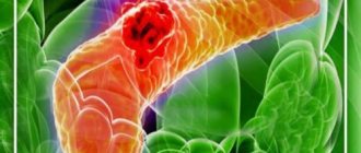
Clinical manifestations of chronic pancreatitis appear five years from the beginning of the process, its course differs from acute pancreatitis - the clinical symptoms are milder and the course is not so aggressive, there is no general inevitability of a quick and painful end of life. But chronic pancreatic disease is characterized by serious inflammation of the organ with gradual death of tissue or its replacement by scar tissue. And with each exacerbation, more and more of the gland atrophies, shutting down from active life. Scars disrupt the normal outflow of abundantly produced secretions, small ducts swell, forming cysts. Inside cysts of various sizes, dead gland tissue floats in a thick solution of enzymes. When an infection occurs, pus forms in the cysts. If the process takes a long time, almost no glandular tissue remains in the gland - a continuous scar.
During exacerbations, the quality of life deteriorates, and not only the quality, in the first decade of the disease, a fifth of patients die, because the gland, although small, is very important for the body, produces enzymes that, having escaped from cells and cysts during inflammation, themselves and digest. And after 20 years, half of the patients are already missing, dying from complications and acute processes initiated by the disease: vascular thrombosis, mucosal ulcers, hepatitis and pancreatic adenocarcinoma.
Treatment of pancreatitis in adults
In the chronic form, antispasmodics are prescribed, which improve the outflow of enzymes, antisecretory drugs that relieve the inflammatory process, and replacement therapy. If 90% of the pancreas does not work, enzyme preparations are prescribed. In the acute form of the disease, treatment can be described in three words: hunger, cold and rest. You need to adhere to this regime for three days. In addition, the patient is given a drip with antispasmodics and a drug that reduces secretion. In case of pancreatic necrosis, surgical intervention is necessary.
Can pancreatitis be cured?
The acute form of pancreatitis can end with only one attack; chronic pancreatitis cannot be completely cured, but remission can be achieved. To do this, you need to take medications on time, undergo tests (blood, feces for coprogram, ultrasound of the abdominal cavity, CT and MRI), engage in prevention, and follow a diet.
Hypertriglyceridemic pancreatitis
Hypertriglyceridemic pancreatitis accounts for 1-14% of cases of AP and 56% of cases of pancreatitis in pregnant women. Hypertriglyceridemia is defined as elevated fasting triglycerides (TG) (>1.7 mmol/L). Serum TG levels above 11.3 mmol/l can lead to the development of AP in 5% of cases. In cases where the TG concentration exceeds 22.6 mmol/l, the incidence of AP is 10–20%. The risk of AP increases with a TG concentration of 5.6 mmol/l: the higher it is, the higher the risk of AP and the more severe the pancreatitis.
Pancreatitis caused by hypertriglyceridemia is associated with both primary (genetic - dyslipidemia types I, IV, V) and secondary (acquired) disorders of lipoprotein metabolism. The main causes of acquired hypertriglyceridemia are obesity, diabetes, hypothyroidism, pregnancy and medication use - treatment with estrogens or tamoxifen, beta-blockers, antiretroviral drugs, thiazide diuretics.
During pregnancy, TG levels usually rise to 3.3 mmol/L in the third trimester and do not cause pancreatitis. Inheritance is most common during pregnancy due to hereditary familial dyslipidemia.
Inflammation of the pancreas is not caused by TGs themselves, but by toxic free fatty acids formed as a result of hydrolysis of the pancreas by pancreatic lipases. The clinical picture of the disease is usually no different from ordinary pancreatitis. In some cases, severe hypertriglyceridemia, xanthoma, xantheliosis, hepatosteatosis, etc. may be observed.
It is important to promptly diagnose hypertriglyceridemic pancreatitis and correct triglyceride levels. Standard treatment of pancreatitis is also prescribed - infusion therapy, analgesics.
The main methods of reducing hypertriglyceridemia are apheresis, especially plasmapheresis, and insulin therapy. However, there is no data from randomized studies on the effectiveness of these methods. Treatment is selected based on the severity of pancreatitis and warning signs, including hypocalcemia, lactic acidosis, exacerbation of systemic inflammatory response, and progressive organ dysfunction.
Intravenous insulin should be used if the patient does not tolerate or cannot undergo apheresis. Insulin lowers TG levels, but the goal of this treatment in severe hypertriglyceridemic pancreatitis is to prevent stress-induced release of fatty acids from adipocytes. The usual dose of insulin infusion is 0.1-0.3 U/kg/h. This requires hourly blood glucose measurements. To prevent hypoglycemia, a glucose solution is prescribed.
Hourly blood glucose measurements
For therapeutic apheresis, triglyceride levels should be monitored after each procedure. Plasmapheresis is continued until the TG concentration drops to 5.6 mmol/l. For insulin alone, TG levels are monitored every 12 hours. The insulin infusion is stopped when the TG concentration reaches 5.6 mmol/L (usually after a few days).
Further long-term correction of hypertriglyceridemia with TG <2.2 mmol/L is important to prevent recurrence of pancreatitis. There are medical and non-medical measures for this. The latter include diet, weight loss for obesity, increased physical activity, limiting added sugar, and good glycemic control for diabetes. You should also avoid medications that increase TG levels. Medicines include antilipid drugs, usually fibrates (fenofibrate).