01.05.2018
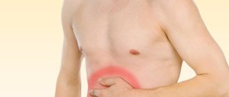
The appearance and development of a disease such as uterine fibroids is accompanied by a number of characteristic symptoms. One of them is an increase in the abdomen with uterine fibroids while maintaining the woman’s usual average weight and maintaining the size of the hips and breasts.
Yes, with uterine fibroids the abdomen grows - but this only happens when the tumor has reached such a large size that the peritoneum is stretched due to its volume. In addition, a tumor of this size puts pressure on neighboring internal organs, which can lead to malfunctions in their functioning. There are also cases in which an increase in the abdomen is expressed in the growth of the fat layer on it. The female sex hormone estrogen is to blame for this, exceeding the level of which can lead to the deposition of fat in the abdomen, hips and lower back. This material is intended for all those who ask questions like “Does the stomach grow with uterine fibroids?”, “Can the stomach grow with fibroids?” It will discuss the causes of this phenomenon, as well as the symptoms that arise in the process.
Causes of uterine fibroids
The main cause of the development of the disease is a malfunction of the ovaries when they begin to produce too much of the hormone estrogen. Contraceptives with a high concentration of this hormone can provoke fibroids and their intensive growth. Also, fibroids can occur after an abortion, complications during pregnancy, endometriosis, as a result of inflammation of the ovaries and fallopian tubes. At risk are nulliparous women over 30 years of age. Often the cause of the formation of nodes is obesity, low physical activity, disruption of the endocrine and immune systems, and a long period of insolation (stay under active sunlight). There is also a hereditary factor.
Factors promoting tumor formation
According to research results, the key factor in the occurrence of fibroids is an increase in estrogen with progesterone deficiency, which is associated with ovarian dysfunction. Estrogen promotes the growth of certain muscle groups of the uterus. An increase in the production of female hormones is provoked not only by inflammatory and infectious diseases of the reproductive system, abortions, contraceptives, but also by poor nutrition. Thus, foods high in refined carbohydrates, saturated fatty acids and insufficient fiber when consumed regularly increase the risk of developing tumors.
Fibroids can also appear as a result of trauma to the uterus - trauma during childbirth, curettage, diagnostic hysteroscopy, insertion of an IUD (intrauterine device). Women who are overweight are susceptible to tumor processes - fatty tissue transforms androgens into estrogens. Therefore, this disease occurs 3 times more often in patients weighing more than 70 kg. The distribution of fat mass affects the production of estrogen, so women with deposits in the upper torso and waist (apple body type) are most susceptible to fibroids. Hormonal levels are affected by hypertension, psycho-emotional stress, a sedentary lifestyle, regional factors, and incompatibility with a sexual partner.
Uterine fibroids and its types
Based on the sites of placement, the following types of tumors are distinguished:
- Subserous. It is located on the outside of the uterus and develops into the outer part of the pelvic cavity. It does not affect the functioning of the reproductive system and the menstrual cycle, but it compresses neighboring organs and tissues. Therefore, pain in the lumbar region, during bowel movements, and a feeling of incomplete bowel movement are possible.
- Intramuscular (intramural). It is formed in the middle muscle layers, causing an enlargement of the organ. This type of tumor is the most common among patients and is characterized by disruption of the menstrual cycle, pain, and heaviness in the pelvic area.
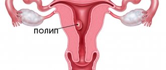
- Submucosal (submucosal). It develops under the mucous membrane lining the uterine cavity, growing deep into the organ and leading to acute symptoms.
- Intermuscular (interstitial). The nodes form inside the muscular layer of the uterus. This type of tumor is characterized by heavy menstrual flow, pain appears only with intensive growth of the fibroid, its necrosis or swelling, and neighboring organs - the rectum and bladder - also suffer. The enlargement of the uterus is uniform.
Symptoms
Myoma does not always have obvious symptoms, the most characteristic of which, occurring in most patients, are the following:
- disturbance, abnormal course of the cycle - increased duration (8 or more days), pain, blood clots;
- pain in the pelvic area;
- feeling of heaviness, pressure in the MT area (pelvis);
- pain in the legs and back;
- pain, discomfort during intimacy;
- frequent urination with little emptying of the bladder, which is associated with compression of the organ;
- constipation due to tight intestines;
- increase in abdominal volume.
If you have at least one symptom, you should urgently contact an antenatal clinic. If a visual examination and palpation are not enough, the doctor will prescribe an ultrasound, blood test, urine test, or smear test. Depending on the result, additional examination methods may be used.
Classification of nodes by size
The growth of the abdomen with fibroids directly depends on the size of the myomatous neoplasms. When classifying, it is customary to correlate their sizes with the weeks of pregnancy:
- small - sizes up to 20 mm (4-5 weeks);
- medium - sizes from 20 to 60 mm (5-11 weeks);
- large - sizes from 60 mm (12 weeks);
- giant - growths of gigantic size, deforming the uterus (from 16 weeks).
Most often, the neoplasm weighs no more than one kilogram, but it is known that the weight of the node was more than 60 kg.
Fibroids and pregnancy
Patients who have a tumor are concerned about the possibility of conception and pregnancy. This problem is very relevant, since the main age category of mothers is women over 30-35 years old, who are at risk.
Conception
The neoplasm is not the cause of infertility, but it does cause some difficulties: it compresses the fallopian tubes, disrupts ovulation, and reduces the permeability of sperm. Therefore, if the patient does not become pregnant and a large fibroid is detected, drug or surgical treatment is recommended. With hypertrophic growth of fibroids and significant enlargement and deformation of the uterus, restoring the ability to bear children is problematic, sometimes impossible.
1st trimester of pregnancy
Problems are fraught with contact of the tumor with the placenta, especially if the fibroids are large. With a small tumor, which occurs in 60% of women during pregnancy, there are no complications or symptoms, but early termination of pregnancy occurs. Causes:
- increase in the number of uterine contractions;
- impaired uterine circulation;
- problems in the functioning of the neuroendocrine system;
- chronic infections;
- proliferation of the uterine mucosa due to polyposis, hyperplasia.
2nd, 3rd trimesters
The risk of miscarriage increases as the space required for the fetus decreases and the intensity of uterine contractions increases. The number, size, location of neoplasms, and their contact with the placenta play an important role. If a miscarriage does not occur, fibroids can negatively affect the development of the fetus, causing deformations of the skull and other parts of the body, and infringement of internal organs.
Childbirth
The neoplasm is often the cause of protracted labor; doctors often decide on a caesarean section. The tumor itself does not affect the birth process, the reason is that due to their presence, there is an anomaly of presentation, the position of the child, when it is impossible to give birth naturally (pelvic, facial, transverse position). Also, during a caesarean section, the surgeon can immediately remove the tumor.
Placental abruption is a particular difficulty, especially if a tumor has formed behind it. During the birth process, doctors must take this into account.
Postpartum period
Myoma causes complications during the postpartum period. They can be early - causing bleeding, placenta accreta, and late. These include involution (the uterus does not return to its original size) and infections.
Robotic surgery
Robot-assisted surgery is a modern and promising type of laparoscopic surgery. It has a number of advantages over traditional laparoscopy, especially when working in confined spaces, such as the pelvic area, when there is involvement of vascular bundles, nerve endings, or complex localization of fibroid nodes along the posterior wall of the uterus.
Removal of uterine fibroids or the uterus itself is performed using a DaVinci robot. Access to the surgical field is achieved through several punctures on the anterior wall of the abdominal cavity. Through them, special surgical instruments are inserted, which the doctor controls remotely using manipulators. Essentially, this is an extension of the surgeon’s hands, allowing the operation to be performed with maximum precision with minimal trauma to the patient. This technology allows the surgeon to operate with even more precision than using a laparoscope.
The robotic assistant provides the surgeon with greater flexibility during surgery. It helps to identify the lesion as accurately as possible and remove it while preserving the uterus, which is important for patients who have not achieved reproductive function.
During the operation, modern anti-adhesion and hemostatic barriers (gels, meshes) are used. For large fibroids or the presence of anemia, the technique of temporary occlusion of the uterine vessels and modern blood-saving technologies (reinfusion of autoerythrocytes) are used, which allows such operations to be performed almost bloodlessly. All operations are performed on modern certified equipment using progressive and reliable suture material.
Pregnancy, childbirth and fibroids
During pregnancy, there is an increased production of estrogen and progesterone, which affects the condition of the tumor. Along with hormonal changes, physical changes occur - the myometrium stretches and increases in volume. Therefore, the location of the tumor and its size are important. The increase is typical for the 1st and 2nd trimesters, while in the 3rd trimester the fibroid decreases. In practice, most patients with this disease do not experience intensive tumor growth during pregnancy and childbirth, and it does not cause significant problems. The opposite effect is more common - destruction of the tumor, necrosis. This is a negative change that can cause bleeding, swelling, and the appearance of cysts. Necrosis of fibroid tissue is accompanied by an increase in body temperature, an increase in leukocytes in the blood, an increase in the level of ESR, and pain in the area where the node is located. If ultrasound and tests confirm the diagnosis, the pregnant woman is hospitalized and treated in a hospital. Surgical interventions are almost never prescribed.
Treatment
Therapy of neoplasms can be medicinal and surgical. For the treatment of patients of reproductive age, medicinal methods with the use of drugs are usually used; it is extremely rare that fibroids are removed surgically, as this jeopardizes the possibility of becoming pregnant in the future.
Drug therapy involves the use of iron supplements, vitamins B, E, A, which have a positive effect on the functioning of the endocrine and nervous systems, folic and ascorbic acids. Usually these are balanced complexes, dietary supplements. A good effect is observed after prescribing a special diet aimed at reducing body weight (often with fibroids, lipid metabolism is disrupted, which provokes weight gain). The patient's diet is adjusted, animal fats are replaced with vegetable fats, carbohydrate intake is limited, and fruit and vegetable juices are included. Hormonal drugs with a high content of progesterone, which inhibits the growth of tumors, are also prescribed. Thus, drug treatment is carried out in the following cases:
- childbearing age;
- if the fibroid is located in the muscle layer;
- the uterus is small in size and has not changed shape.
If the tumor is large and continues to grow, a decision is made about surgical intervention. Indications:
- uterine bleeding accompanied by anemia;
- large volume of the uterus;
- fibroids have formed under the mucosa;
- compression of the rectum, bladder;
- growth of neoplasm in postmenopause;
- the tumor is located in the cervix.
If there are indications, myomectomy (removal of fibroids) or embolization of the uterine arteries is performed in order to cut off the supply to the node, which leads to its reduction. Complete removal of the uterus can be practiced in exceptional cases, and also if the patient is over 45 years old.
Watch a video about fibroids
Original author's technique for performing bloodless myomectomy
During the operation, the vessels feeding the uterus are temporarily compressed, thus, due to the lack of blood flow, the intervention area remains dry, which allows you to visualize the boundaries of the node and the body of the uterus, qualitatively compare and reliably stitch the layers of the myometrium; later a full-fledged scar is formed here. At the end of the operation, which lasts no more than one hour, blood flow is restored.
Our clinic is equipped with modern high-quality equipment, for example, the use of the LigaSure electrothermal tissue ligation device during surgery makes it possible to simultaneously cut and seal the tissue, which eliminates the risk of bleeding. Ultrasonic scissors are used for minimal trauma to healthy tissue and better healing. In difficult cases, we use the V-lock suture system, which allows us to more carefully align the edges of the wound, which ensures better healing, while the suture is formed 3-4 times faster. To prevent the appearance of adhesions during the intervention, anti-adhesive substances are actively used, which minimizes the likelihood of adhesions.
I always strive to perform organ-conserving surgery - both in women of reproductive age and in patients who want to preserve the uterus as an organ. If a woman has concomitant diseases of the pelvic or abdominal organs that require surgical treatment, there is the possibility of performing simultaneous operations.
Organ-preserving operations using my method have been carried out in our clinic for more than 12 years. I personally have performed more than 4 thousand surgical interventions to remove fibroids. Today, the author’s technique, for which a patent has been received, is also actively used in leading clinics in Europe and America.
What will happen if left untreated?
Experts disagree about the need to treat fibroids. Some people prefer to watch and wait, as there are situations when the hormonal levels return to normal and the tumors resolve. This is especially true for young women of childbearing age and girls going through puberty. Vitamin complexes and proper nutrition are prescribed as aids.
Another category of specialists insists on immediate treatment, including hormonal agents, in order to promptly prevent further tumor growth and preserve the ability to bear children.
The decision is made after a series of analyses:
- blood,
- urine,
- for hormones
- smears for PPIs,
- Ultrasound diagnostics.
The fact is that if fibroids are not treated or folk methods are used, self-medication can lead to serious consequences - disruption of the functioning of other organs of the reproductive system, neighboring organs, and the development of other neoplasms. Considering that in the initial stages the disease occurs without symptoms, women are recommended to be checked annually, twice a year, by a gynecologist.
Physiotherapy
For small fibroids, especially when long-term observation is chosen, a woman can try alternative methods of therapy. Physical therapy exercises, massages, and acupuncture sessions will help eliminate unpleasant symptoms.
For painful menstruation, it is recommended to make compresses with castor oil. It is necessary to soak a clean flannel cloth with warm castor oil, wrap it in bandages and apply it to the lower abdomen. The cramps should go away within 10-15 minutes. You can also perform self-massage on a daily basis.
- Apply warm castor oil all over your belly.
- Make several circular stroking movements clockwise and counterclockwise.
- To relieve spasms, you can do a cycle of rubbing movements.
Aromatherapy enthusiasts can add a few drops of organic lavender essential oil to castor oil. The aroma of lavender activates blood circulation and promotes complete relaxation.
If you have uterine fibroids, you can also do yoga and perform simple physical therapy exercises.
Complications
After reducing or removing a tumor, it is necessary to pay attention to the body and see a doctor to prevent the development or appearance of a new tumor. This can be caused by various negative factors, hormonal imbalances, including those associated with the restructuring of the body during menopause. If surgical intervention was performed, you can plan a pregnancy no earlier than six months later, after consulting with your doctor. Otherwise, an unhealed postoperative scar on the uterus may cause it to rupture during childbirth.
During drug treatment, you should adhere to medical recommendations, lead a healthy lifestyle, play sports, regular intimacy and contraception prescribed by a gynecologist are important.
Hysteroresectoscopy
This is a minimally invasive endoscopic operation in which pathological formations are removed from the uterine cavity or cervical canal. The manipulation is performed through the vagina and does not require incisions, and the use of special optics to control the process allows for maximum preservation of healthy tissue.
To carry out the procedure, a hysteroresectoscope is used - an electrosurgical instrument equipped with an optical system. The tool guarantees the accuracy and efficiency of manipulations and avoids incisions, which is important for patients planning a pregnancy.
If a submucosal (submucosal) myomatous node is detected, its removal during hysteroresectoscopy is possible if the diameter of the node does not exceed 4-5 cm.