16 June 2020
54743
2
4.1 out of 5
Hemangiomas are vascular tumors that can occur in the skin and any internal organs that contain a network of blood vessels. Thus, such formations cannot form only in the articular surfaces and optically transparent environments of the eyes. The spine is one of the places where it can form. In such cases, a hemangioma of the spine, or more correctly, a vertebra, is diagnosed.
A tumor of this kind in 100% of cases is a benign neoplasm and never becomes malignant, i.e., does not turn into malignant. Vertebral hemangioma is localized in its thickness, which is due to the peculiarities of the blood supply to the structural elements of the spine, and is, so to speak, an advantageous location for a tumor of this type. Growing inside the spongy tissue of the vertebra, the vascular formation is reliably protected from external influences by the bone, which reduces the risk of injury to a minimum. At the same time, hemangiomas formed in the parenchyma of internal organs are easily damaged and can become a source of severe bleeding.
In recent years, the frequency of diagnosing vertebral hemangiomas has increased significantly, but not due to an increase in the number of cases of their formation, but due to the introduction of modern diagnostic methods: CT and MRI.
What is hemangioma
Hemangioma in children (ICD 10 code: D18) is a benign formation that appears as a result of proliferation of the vascular wall at the cellular level (hyperplasia). The pathology can be congenital or can develop in the first months of a baby’s life.
In adults, abnormal proliferation of vascular epithelium is rarely observed, since it can spontaneously resolve in childhood. In the case when a growth appears on clean skin in adulthood for no apparent reason, you need to contact a specialist for histological examination.
If in young years the pathology is expressed insignificantly, then in the process of growing up, under the influence of a number of factors, its growth and increase can be observed.
hemangioma on a child's head
Hemangioma in infants, formed as a result of vascular malformation, appears as a birthmark.
The detection rate of a vascular tumor in newborns is 3%. In the first year of life, pathology develops in 9% of babies.
In pediatric practice, this is the most common benign neoplasm. In adulthood, the manifestation of hemangioma on the face and head is observed in rare cases (if full treatment was not carried out in childhood).
Tumor formation in most cases occurs on the skin of the neck, scalp and face. The torso, legs and arms are affected much less frequently. On internal organs it is most often localized on the lungs, bone tissue and liver.
Hemangioma is expressed predominantly in solitary formations, but there are exceptions. If more than 5 tumors are observed on the skin, most likely the internal organs are also affected by the disease.
Prevention
Since hemangiomas most often form in the embryonic period, the pregnant woman must take responsibility for preventing this phenomenon. It is recommended to avoid viral infections, contact with toxic chemicals, and taking potent medications during pregnancy, especially in the early stages.
For a favorable outcome of any pathology, its early detection is very important; for this purpose, a diagnostic examination must be carried out annually, regardless of whether there are or are not any undesirable symptoms.
For patients with liver hemangioma, experts advise sticking to a healthy diet and avoiding excessive physical activity, which can cause tumor rupture and intraperitoneal hemorrhage. Women who are deciding on a method of contraception should also be careful: contraceptives containing estrogen are contraindicated for women with a hemangiomatous node in the liver, since it is this hormone, according to one assumption, that activates the growth of tumor formations.
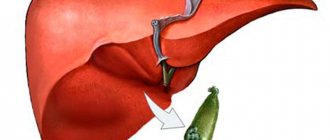
Hemangioma of the liver is a pathological vascular formation of a benign nature, which most often does not bother the patient and in very rare cases develops into a malignant tumor. However, this does not mean at all that the dynamics of this phenomenon do not need to be monitored. People with hemangiomatous nodes in the liver should be extremely attentive to their pathology; with regular examinations, proper nutrition, and normalization of physical activity, they may not even need drug treatment. Otherwise, there is a risk of further growth of the tumor or the development of various undesirable complications.
For what reasons does hemangioma appear?
Modern science has not fully elucidated the causes of pathological proliferation of endothelial cells. According to one version, hemangioma begins to grow during intrauterine development of the fetus under conditions of chronic oxygen starvation. According to another, the overgrown cells are residual embryonic cells. Another theory suggests that the neoplasm is formed under the influence of maternal infections in the 1st trimester of pregnancy. But these are just guesses, since the formation of hemangioma also occurs in babies who were born during a normal pregnancy, not complicated by infectious diseases.
It has been noted that pathology is observed more often in multiple pregnancies and in premature newborns.
Risk factors for the development of a benign tumor of this type include the mother’s age exceeding 40 years, as well as medical indicators: preeclampsia (protein in the urine, high blood pressure, swelling), threat of miscarriage, entanglement of the umbilical cord, inflammatory processes of the placenta, its abruption and presentation (when the embryonic organ is located as low as possible in the uterus, which makes childbirth difficult).
hemangioma on a child's face
Possible complications
Progressive liver hemangiomas are dangerous for the patient due to the undesirable consequences that they can entail. The greatest danger is rupture of the tumor, followed by hemorrhage and massive blood loss. This usually occurs as a result of excessive physical exertion, trauma to the abdominal area, or sudden movements. Such complications may be indicated by pain in the abdominal area that does not go away within two hours; in such a situation, it is necessary to urgently seek medical help.
There are known cases of gastrointestinal bleeding, which begin due to increased pressure in the portal system of the organ. When a hemangioma becomes infected or undergoes a necrotic process, the likelihood of developing sepsis increases, and this is a consequence of another complication - thrombosis of the hemangiomatous node. Another aggravating circumstance is the occurrence of liver failure, as well as cirrhosis, which is most often observed with total damage to the organ by hemangiomas.
How is pathology classified?
Infantile hemangioma is divided into congenital (formed, reaching its maximum size in the womb) and infantile.
A child is born with a vascular pathology, which can significantly decrease in size during the first 1.5 years, but not disappear completely, or spontaneously involute or remain unchanged. If a congenital hemangioma grows, it occurs in proportion to the physical growth of the baby. For infants of different sexes it occurs in equal percentages. Large neoplasms can be detected during ultrasound screening.
The development of infantile hemangioma occurs in the first weeks of a child’s life. It is located both under the skin and on its surface. A combined type is also found. The tumor forms from a small red spot and continues to grow until six months of age. This is followed by a fading phase, lasting up to 2 months, followed by a slow decrease in size of the formation, lasting up to 10 years. In girls, the development of infantile vascular anomaly is observed 5 times more often.
In accordance with the caliber and histology of the vessels, hemangioma is divided into several forms.
Capillary
Another name for such a hemangioma is superficial. It occurs most often (in 95% of cases). This is a small tumor consisting of densely woven capillaries covered with overgrown epithelial cells. Located on the surface of the skin, it looks like a small port-wine stain. A characteristic difference of this form is the intensity of growth, therefore it is considered to be the initial stage of the formation of pathology.
Venous
The venous form develops as a result of the proliferation of cells of medium and small caliber veins. This happens both above the surface and deep into the skin.
Cavernous
This is the next stage of the formation of venous hemangioma. The tumor is characterized by a cavernous structure, consisting of cavities separated by partitions from the vascular epithelium. The formation of pathology occurs when defective capillaries are excessively filled with blood, leading to rupture of the wall, the formation of caves and hemorrhage, expressed as a hematoma. Increased coagulation of blood in the cavities leads to the formation of small clots.
Combined
This is a neoplasm, which is characterized by signs of several types of hemangioma simultaneously, differing in varying degrees of severity.
Definition
Liver hemangioma (code in ICD-10, or International Classification of Diseases D18.0) is a vascular neoplasm that is considered a nonepithelial benign tumor. It may not change size for a long time or grow quickly, and is characterized by a predominantly asymptomatic course.
Liver hemangiomas are visually described as red formations with a bluish tint, flat, with clear edges. The surface is moderately bumpy or, conversely, smooth. From the inside, the tumor consists of numerous vessels, which are separated from each other by partitions of fibrous tissue and may contain liquid blood and blood clots. Sometimes part of the lesion is located outside the boundaries of the liver and is connected to its parenchyma (tissue) leg.
Hemangioma is a tumor that is not prone to malignancy (malignancy). This means that it does not develop into cancer.
How does hemangioma manifest?
A capillary-type formation, which is a growth in color from burgundy to faint pink, grows into the surrounding tissue. After pressing, it becomes pale, quickly returning to its original appearance. Hemangioma on a child’s head , localized on the skin of the frontal part, is called “angel’s kiss”, on the back of the head - “stork bite”. Rising somewhat above the skin, a superficial hemangioma has smooth, clearly defined outlines (when growth has stopped and a phase of reduction begins) or vague (in the process of active growth).
so-called “strawberry” hemangioma
The venous type is characterized by a dark red, slightly bluish color. The development of this form is rarely observed, but the growths are quite large in size.
The difference between the cavernous form will be:
- roughness of the surface that shows through the capillaries,
- elasticity,
- clear delineation from surrounding tissues,
- pronounced crimson-violet color.
The pathological growth mostly penetrates deep into the tissues, affecting the subcutaneous tissue, muscle fibers and only slightly rising above the surface of the skin. If you press on the formation and release it, it will turn pale and wrinkle, reducing its volume, but will immediately return to its normal state.
In combined hemangiomas, the signs of the subcutaneous and superficial type are combined (the part located under the skin has the largest volume).
Classification
Formations in the liver are divided according to size:
- Less than 3 cm – small.
- From 3 to 10 cm – large
- More than 10 cm – gigantic.
Single or multiple tumors can be located in the parenchyma or tissue of an organ. They are formed from venous vessels and are fed from the arterial network. Histologically (according to the type of structure), hemangiomas are distinguished:
- capillary (characterized by the narrow lumen of the vessels);
- scirrhous (has a supporting structure of connective fibrous tissue);
- cavernous (characterized by large cavities containing blood and blood clots).
If there are different signs of the histological structure, they speak of a mixed variant of the tumor structure.
Hemangioma of internal organs
Minor tumors that have developed on internal organs, as a rule, do not appear. They are discovered by chance, during a medical examination carried out for another violation.
For an internal anomaly to manifest itself, its size must be impressive (up to 5–10 cm). If the formation arose inside the spinal column, it will not have manifestations. But as soon as the growth touches the ligaments or the periosteal membrane, pain will appear. By compressing the nerve roots, it can cause sensory disturbances in the upper and lower extremities.
In some cases, the development of pathology in internal organs may immediately manifest itself as symptoms. It depends on the location of the tumor. Its growth can quickly lead to disruption of the organ. A large hemangioma poses a danger to the eyes, throat and trachea. Liver hemangioma in a child rarely causes negative symptoms.
Symptoms
Spinal hemangioma does not manifest itself in any way until the integrity of the vertebral body is compromised and the above-described disorders occur. Only in very rare cases is radicular syndrome observed, i.e., pinching of the nerves passing through the affected vertebra and the occurrence of:
- sharp, shooting pains along its course, caused by various movements, coughing, laughter;
- sensations of numbness, crawling;
- limited movement.
If the hemangioma is located in the cervical vertebra, cerebral circulation may be impaired, which will lead to headaches, dizziness, hearing, vision and sleep disturbances. When it is localized in the thoracic vertebra, disturbances in the functioning of the pelvic organs, heart, and digestive organs may be observed, and when a tumor forms in the lumbar vertebrae, there are frequent cases of pain radiating from the lower back to the groin and hips.
This is only possible when the hemangioma reaches a large size and leads to a compression fracture of the spine without the formation of individual bone fragments, which occurs only in 0.1% of patients with vertebral hemangioma. In such situations, the vertebra seems to be evenly flattened. As a result of a decrease in its height, the size of the natural openings through which the nerves pass also decreases, which leads to their compression and the development of radicular syndrome.
In the vast majority of cases, radicular syndrome occurs due to intervertebral hernia.
The patient must clearly understand that hemangioma, although it is a tumor, has nothing to do with oncology and is not capable of degenerating into a malignant neoplasm and giving metastases. This is very important to reduce stress levels and avoid the development of anxiety, since negative experiences weaken the body and false back pain may occur, which significantly reduces the quality of life.
Treatment and removal
Treatment of hemangiomas in children includes the use of one of the modern methods (treatment with liquid nitrogen, laser, etc.), drug therapy with the prescription of antitumor and hormonal drugs, or surgical excision.
The initial stage of diagnosing vascular wall pathology involves asking the patient (in the case of a child, his parents) about the manifestation of symptoms and the dynamics of the development of the defect. Next, the doctor examines the tumor in detail. If necessary, (depending on its location) it is referred to a specialist for a more in-depth examination (ENT specialist, ophthalmologist, etc.).
The structure, size and depth of growth of the anomaly are clarified during an ultrasound examination. During an ultrasound, blood flow in the affected area is studied.
Important! The diagnostic method of ultrasound examination will not be informative for all forms of hemangioma.
Removal of hemangioma in children is mandatory if it bleeds, itches, causes discomfort, or is located in areas rubbed by clothing.
Diet
An integral part of the treatment process is dietary nutrition. For liver hemangiomas, gastroenterology prescribes table No. 5, which includes limited consumption of fats, acidic foods, coarse fiber, as well as foods that stimulate excessive production of digestive enzymes. Dishes should be served boiled; steaming is encouraged. The central place should be given to vegetable soups and slimy porridges, lean meat and fish, vegetables and non-acidic fruits, dried bread, weak tea, compote from dried fruits and mineral water.
Patients with liver hemangioma are prohibited from consuming the following foods and dishes:
- Fried food;
- Marinades and preservation;
- Borscht, cabbage soup, soups with strong meat broth;
- Sausages, smoked meats;
- Meat and fish of fatty varieties;
- Dairy products;
- Mushrooms;
- Cabbage, legumes, radishes, ginger, garlic, spinach and sorrel, horseradish;
- Pearl barley and corn porridge;
- Pastries, chocolate;
- Ice cream;
- Coffee, cocoa, kvass, strong tea, alcoholic and carbonated drinks, concentrated juices.
Dietary table No. 5 provides for fractional but frequent meals. One of the important recommendations of this diet is to avoid excessively hot or cold foods, as well as to drink enough water - it should be about 2 liters per day.
Medicinal methods
Depending on whether the patient has a particular complication, treatment tactics will be different - for example, topical corticosteroids may be prescribed. At the same time, the clinical response to even the most powerful drugs develops quite slowly—over several weeks. Therefore, this method is not suitable for vision-threatening conditions.
Injectable corticosteroids provide a faster effect: blanching of hemangiomas is observed already on days 2–3, and involution becomes noticeable after 2–4 weeks. The success rate of this therapy is about 75%.
Systemic corticosteroids are used for amblyogenic life-threatening lesions. A marked response is usually observed in 30% of patients, a moderate response in 40%, and no response in 30%.
Alpha-2a interferon is prescribed when hemangioma is resistant to corticosteroid therapy. Despite its good effectiveness, its use is associated with undesirable side effects: fever, arthralgia, retinal angiopathy.
Propranolol can be used to treat hemangiomas , although the evidence base is relatively weak and mainly consists of anecdotal cases. However, some studies indicate that propranolol may prevent loss of visual acuity in the case of periocular hemangioma.
For some superficial (and in some cases deeply located) neoplasms of a limited area, timolol .
Clinical case
As I emphasized earlier, hemangioma in newborns and adults is a silent pathology that is often disguised as other diseases.
To prove my words, I can give an example from practice. A 55-year-old patient came to me with complaints of rapid satiety when eating, abdominal discomfort, flatulence and periodic vomiting. The man admitted that he sometimes drinks alcohol. In addition to the symptoms already mentioned, heartburn (after episodes of vomiting) was bothersome, the abdomen was sensitive to palpation and, although there was no significant pain, pressing on the epigastric area caused severe discomfort. To establish a diagnosis, an examination was required; Based on the results of ultrasound and tomography of the abdominal organs, a space-occupying formation with clear contours was identified. Further diagnostics confirmed the presence of hemangioma in the liver parenchyma.
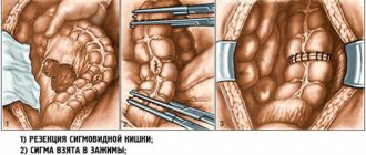
The patient was sent to the vascular surgery department for additional examination and endovascular embolization of the tumor arteries. A few days after the intervention, he was discharged with improvement. The most striking symptoms with which he came to me - a feeling of fullness in the stomach and vomiting - disappeared.