Very often, during the next act of defecation, a person may feel severe pain, discomfort and burning in the anus. There can be many reasons for this. Among them there is a disease such as sphincteritis of the rectum (inflammation of its membranes).
In proctology, this is a common disease that is not life-threatening for the patient, but if not treated in a timely manner can cause many unpleasant pathologies. Therefore, it is very important to contact a specialist at the first unpleasant symptoms who will prescribe effective treatment. In this article we will look at what sphincteritis of the rectum is, symptoms, and treatment of this disease.
What is a sphincter
There are about 35 sphincters throughout the human digestive system. What is it? These are special muscle valves that perform an obturator function in the human body . It is thanks to them that food moves throughout the body, smoothly passing from one organ to another.
Rectum
Among the many sphincters, we will consider the anal one in detail. This valve is responsible for the movement of feces through the rectum and is responsible for the processes of emptying . It has two parts:
- external , which consists of striated muscles. A person can control their reduction on a subconscious level. The structure of this part is ring-shaped. It is about 10 cm in length and 2.5 cm in width, located in the coccygeal area;
- internal , which is formed by smooth muscles. It also has a ring shape. This part of the sphincter originates inside the rectum and is located directly at the exit from the sphincter of the colon. Contractions of the muscles of this sphincter must occur mechanically; a person cannot influence them in any way. Thanks to it, feces and excess gases are retained in the rectal area, which can come out during efforts made by a person.
Bladder sphincter
More than 30% of pathologies of the sphincter apparatus in women of different ages are associated with the functioning of the bladder sphincter, which regulates urination and prevents involuntary release of urine. In women, there are two sphincters in the bladder. One of them is located in the cervical part of the organ and compresses the walls of the bladder, causing urination. The second sphincter is part of the muscular apparatus of the pelvic day and is located in the middle part of the urethra (urethra). It narrows the opening of the urinary canal and prevents involuntary urination.
If the muscles that form the urethral sphincter are weakened, they cannot squeeze the opening of the urethra with sufficient force, causing the woman to experience incontinence (enuresis). This pathology is called secondary, since it is acquired in nature and develops against the background of concomitant diseases and disorders. Urinary incontinence is very common in women over 50 years of age, since at this age there is a natural weakening of muscle fibers, including the muscles of the sphincter apparatus. Frequent urge to urinate due to the release of a small amount of urinary fluid is also typical for patients with diabetes, so treatment in this case should also include drugs that normalize blood sugar levels.
Other causes of weakened urethral sphincter muscle tissue include:
- chronic infectious diseases of the urinary system (cystitis, pyelonephritis, glomerulonephritis, urethritis);
- childbirth and complicated pregnancies;
- stress and a state of chronic emotional tension;
- pathologies of the digestive system, accompanied by prolonged constipation (gastritis, colitis);
- chronic heart failure.
In some cases, the cause of weakening of the pelvic muscles, which includes the sphincter of the bladder and urethra, may be obesity. If a woman does not take timely measures to correct her body weight, incontinence can become chronic.
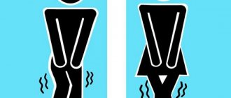
How to strengthen the sphincter during incontinence?
Special gymnastics can help cope with the problem at an early stage. It is very useful to do the following exercise daily:
- squeeze the muscles of the perineum and anus;
- count to 10;
- relax.
The exercise must be repeated 5-6 times a day, 101005 times. During urination, you must try to stop the process by tensing the perineum. With regular training, you can cope with the problem in 1-2 months. During this period, it is useful to follow a diet that limits the consumption of spicy and fatty foods, spices, marinades, and vinegar. Alcohol negatively affects the functioning of muscle valves, so women suffering from various forms of enuresis should avoid any drinks that contain ethyl alcohol.
You can also increase the tone of the pelvic muscles with the help of physiotherapeutic treatment, for example, electrophoresis or magnetic therapy. If conservative methods are ineffective, surgical treatment is indicated.
Sphincter diseases
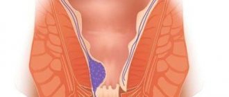
The most common diseases of the rectal sphincter are: spasm and sphincteritis. A photo of inflammation of the rectal sphincter can be seen below.
In the first case, this is a chronic form of the disease, in which a person experiences constant pain and discomfort in the anal area. This disease develops over a long period of time and causes severe discomfort to the patient’s life. Therefore, it is recommended not to delay the treatment of this problem.
Sphincteritis is an inflammatory process in which its muscles become inflamed . This disease is characterized by wave-like exacerbation; treatment takes a long period of time. Below is a photo of rectal sphincteritis.
What is rectal sphincteritis
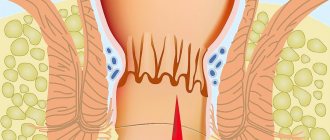
This disease is one of the most common in proctology. Sphincteritis is an inflammation of the sphincter muscles. In this case, the full functioning of the latter is disrupted, and if treatment is not timely, the patient can delay the process to the point of severe complications. With advanced sphincteritis, the muscles completely relax and the patient loses the ability to retain feces and gases in the body, thus an unexpected act of defecation may occur .
This is a rather delicate problem, so it is recommended to treat it promptly. The development of the disease occurs as follows: if there are wounds or cracks in the anus, pathogenic microorganisms can get there, they begin to actively multiply and provoke the formation of purulent masses. Next, inflammation of the affected area occurs and acute unpleasant symptoms of sphincteritis appear.
Sphincter of the rectum
The internal sphincter of the rectum is one of two sphincters of the human anus. According to physiological characteristics, it is considered a continuation of the inner layer of muscle in the rectum. On one side, this sphincter connects to the skin in the anus. The total thickness of the internal sphincter is up to 5 mm. Its length can reach 30 mm. The rectal sphincter muscles run at right angles along the intestine.
The internal sphincter of the rectum is constantly contracting. It performs a peculiar function of preventing involuntary bowel movements in humans. When feces appear in the rectum, the sphincter reflexively relaxes, which makes it possible to defecate. The “work” of the internal sphincter is realized through three separate mechanisms:
- The nervous system that regulates the contraction and relaxation of the internal sphincter.
- Rectal plexuses that control local reflexes.
- Incoming substances that control myogenic tone.
If the coordination of the sphincter muscle fibers is disrupted, this can become a serious reason for the development of chronic constipation in a person.
How does constipation affect the functioning of the anal sphincters?
If a woman suffers from chronic constipation, weakening of the rectal muscles may occur. The pathology is accompanied by impaired defecation, pain during bowel movements, unpleasant sensations and discomfort during the movement of feces through the large intestine.
To improve gastrointestinal motility, you should follow the following tips.
- Compliance with the drinking regime is of great importance. This is especially true for pregnant women, elderly and senile patients, children and adolescents. The drinking water consumption rate per day for an adult is 1.5 liters.
- The amount of fresh vegetables, fruits and berries in the diet should be at least 600 g. The norm of greens consumption for an adult is 20-30 g per day.
- A good way to normalize intestinal motility is physical therapy. If a person cannot engage in sports or gymnastics for health reasons, walking in the fresh air is useful.
In order for the anal sphincters to work normally, it is necessary to treat diseases of the stomach and intestines in a timely manner. This is especially true for inflammatory processes in the rectum, for example, hemorrhoids and proctitis. If the inflammation is severe or the disease is in an advanced stage, contraction of the sphincter muscles can cause pain and intensify the pathological symptoms of the underlying pathology.
Causes of the disease
Oddly enough, there are many causes of sphincteritis. The development of inflammation of the sphincter muscles can be caused by hemorrhoids, fissures in the anus, frequent constipation or other pathologies.
The independent development of sphincteritis is impossible; it is the result of other diseases in the anal area.
Other factors that can trigger the development of inflammation of the sphincter muscles include:
- bacterial anal infections;
- disorders of the gastrointestinal tract;
- spicy food;
- tumor formations in the rectum;
- injury to the anus during anal sex;
- frequent spastic constipation (when hard-formed feces pass through the sphincter, the muscles of which are in good shape);
- intestinal disorders that cause severe irritation of the anal mucosa
Anal incontinence (anal sphincter insufficiency)
Insufficiency of the anal sphincter (anal incontinence) is a partial or complete violation of the voluntary or involuntary retention of the contents of the colon.
Normally, the rectal closure apparatus is capable of retaining solid, liquid and gaseous intestinal contents not only in various body positions, but also during exercise, coughing, sneezing, etc. The ability to retain rectal contents depends on circumstances such as quantity and quality intestinal contents, the state of the closing apparatus of the rectum and pelvic floor muscles, the integrity of the reflex arc and the autonomous innervation of the colon and anal sphincter.
Causes
According to the causes of occurrence, the following types of incontinence are distinguished: post-traumatic, postpartum, functional and congenital.
The most common cause of incontinence is injury to the obturator apparatus of the rectum, most often associated with obstetric or surgical trauma. Then in frequency comes functional insufficiency of the anal sphincter, associated with diseases of the peripheral or central nervous system. In third place are various anorectal malformations, which in most cases are complicated by insufficiency of the anal sphincter.
The most common cause of anal incontinence is traumatic injury to the obturator apparatus. Among the damaging factors leading to anal sphincter insufficiency, the most common is surgical trauma to the sphincter muscle fibers during interventions for various diseases of the distal rectum and perineum. The risk of damage to the sphincter is especially high during operations for paraproctitis. In more than half of the patients in this group, insufficiency develops after surgery for chronic paraproctitis. Incontinence of intestinal contents occurs in 10% of cases after operations for acute paraproctitis, in 6% - after operations for rectovaginal fistula and anal fissures, in 7% - after hemorrhoidectomy and in 3% of cases - after operations for caudal teratomas of the pararectal fiber. Other causes of anal sphincter insufficiency include domestic trauma such as “falling on a stake,” traumatic ruptures of the rectum by foreign bodies, etc. Anal sphincter incontinence due to traumatic injury to the rectum and perineum accounts for 11% of cases.
The cause of postpartum anal sphincter insufficiency in 20% of patients is birth trauma. III degree perineal lacerations during childbirth are the most common cause of anal incontinence. This fact is due to the fact that suturing postpartum defects of the rectovaginal septum is often accompanied by wound suppuration, suture dehiscence, and the development of scar tissue, which quite often leads to anal sphincter insufficiency.
Functional disorders of the obturator apparatus of the rectum are caused by neuro-reflex disorders and pronounced local changes in the muscular structures of the pelvic floor and anal canal. The cause of these disorders is most often concomitant diseases of the rectum and anal canal. It is known that atony of the anal sphincter occurs with proctitis, proctosigmoiditis, and colitis. In these diseases, as a result of the inflammatory process, the state of the receptor apparatus is disrupted, and the motor function of the colon suffers. Constant stretching of the anal sphincter, which occurs with prolapse of hemorrhoids and prolonged prolapse of the rectum, leads to a decrease in the contractility of the obturator apparatus of the rectum.
Congenital insufficiency of the anal sphincter is caused by two reasons: 1) congenital disorders of the central or peripheral innervation of the rectum, which occur with non-fusion of the arches of the sacral vertebrae, spinal cord herniation; 2) atresia of the anal canal with complete or partial absence of the rectal closure apparatus. Congenital insufficiency of the anal sphincter is quite rare.
Retention of intestinal contents is the key to successful treatment of any rectal disease. When studying the causes of anal sphincter incontinence, it is necessary to understand how intestinal contents are retained in the rectum. It is ensured by the interaction of the sensitive zone of the receptor apparatus of the distal rectum and anal canal, the nerve pathways of the sacral plexus, spinal cord and brain with the muscular structures of the internal and external sphincters, supporting tonic and volitional continence. In addition, factors such as the closure of the anus, its slit-like shape, anorectal angle, and coordinated motor-evacuation activity of the colon must be taken into account when diagnosing and choosing a treatment method for anal sphincter incontinence.
In the development of anal incontinence with clinical and functional changes in the obturator apparatus of the rectum, disorders of muscle structures and neuro-reflex pathology are distinguished. Organic changes in the muscular structures of the anal sphincter without pronounced neuro-reflex defects are characterized by the development of a scar process in the area of the anal sphincter.
Switching off the reflex or neuromuscular link in the complex system of anal continence causes various clinical manifestations of anal insufficiency. Loss of the function of the external sphincter leads to incontinence of intestinal contents at the time of filling of the rectum. In this case, the patient, with a persistent urge to defecate, cannot retain the intestinal contents when the rectum is filled. If the innervation of the internal sphincter is disrupted, incontinence occurs when the conscious control of the sphincter function during sleep and emotional stress is turned off. When the receptor apparatus of the distal rectum is damaged, there is no urge to defecate and the presence of intestinal contents in it is perceived only from the perianal skin. When the central nervous system is damaged, there is a disruption in communication and coordination of the external and internal sphincters. It should be remembered that if the receptor apparatus of the pathways or the central nervous system is disrupted, any surgical intervention will be ineffective.
Symptoms, clinical course
There are three degrees of clinical manifestation of anal sphincter insufficiency. In grade I, patients cannot retain gases; in grade II, this symptom is accompanied by incontinence of liquid feces; in grade III, patients cannot retain all elements of intestinal contents. In addition to subjective sensations, when determining the degree of insufficiency, an objective characteristic of the contractility of the obturator apparatus of the rectum is of great importance. Normally, the muscle tone of the sphincter, according to sphincterometry, is on average 410 g; with maximum contraction of the anal sphincter, it increases to an average of 650 g. With degree I of anal sphincter insufficiency, sphincterometry indicators decrease to 260-360 g, with degree II - to 130-300 g, with III - up to 0-180 g.
Classification and types
In the literature there are various classifications of anal sphincter insufficiency. In practical work, the classification described above is most often used, taking into account the degrees of deficiency. Different types of treatment are used for each degree of deficiency. In case of I degree of insufficiency with a sphincter defect of less than 25%, the main method of treatment is conservative. In case of anal sphincter insufficiency of II-III degree, surgical treatment is indicated.
It is recommended to use a classification that subdivides anal sphincter insufficiency according to form, etiology, degree of retention of intestinal contents, and clinical, functional and morphological changes in the anal sphincter.
Classification of anal sphincter insufficiency
I. Form: 1. Organic. 2. Inorganic. 3. Mixed.
II. By etiology: 1. Congenital (associated with developmental defects). 2. Traumatic: - after operations on the rectum and perineum; - postpartum; - actually post-traumatic.
III. According to the degree of retention of intestinal contents: 1. I degree. 2. II degree. 3. III degree.
IV. According to clinical and functional changes in the obturator apparatus of the rectum: 1. With a violation of the muscle structures: - internal sphincter; - external sphincter; - pelvic floor muscles. 2. With neuro-reflex disorders: - receptor apparatus; — conducting paths; - central nervous system.
V. According to morphological changes in the obturator apparatus of the rectum. 1. With localization of the muscle defect around the circumference of the anal canal: - on the anterior wall; - on the back wall; - on the side wall; — on several walls (combination of defects); - around the entire circumference. 2. According to the length of the muscle defect around the circumference of the anal canal: - up to a quarter of the circumference; - quarter circle; - up to half the circle; - half a circle; - three quarters of a circle; - lack of sphincter.
Complications
As a complicated form of anal sphincter insufficiency, it is advisable to distinguish its combination with chronic paraproctitis, rectovaginal fistulas, and strictures of the anal canal. Patients with this form of the disease account for 17% of all patients with anal sphincter weakness. The difficulties of treatment are aggravated by the presence of a purulent process, as is observed in patients with chronic paraproctitis or the presence of a pronounced cicatricial process after repeated operations.
Diagnostics
The diagnosis of anal sphincter insufficiency is primarily based on the patient's complaints of gas and fecal incontinence. The patient is examined on a gynecological chair in the position as for hemorrhoidectomy. At the same time, the closure of the anus and its location, the presence of cicatricial deformation of the perineum and anus, the condition of the skin of the perianal region, sacrococcygeal region and buttocks are assessed. Sometimes, when examining the perineum and anus, it is possible to identify such concomitant diseases of this area as anal fissure, hemorrhoids, fistulas or rectal prolapse. Palpation of the perianal area helps to determine the presence of a scar process and the condition of the subcutaneous portion of the external sphincter.
Of great importance is a digital examination of the rectum, which is used to determine the presence and extent of the scar process, its distribution within the wall of the anal canal, the elasticity and extent of the sphincter, the safety and condition of the pelvic floor muscles. The anatomical relationships of the muscle and bone structures of the pelvic ring are also determined. The doctor notes the tone of the anal sphincter, the nature of its contraction, and the presence of a gap after the finger is removed.
Anoscopy makes it possible to visually examine the walls of the anal canal and distal rectum and determine the extent of the scarring process. During sigmoidoscopy, the mucous membrane of the rectum and distal sigmoid colon is examined. Proctography determines the relief of the rectal mucosa, the size of the anorectal angle, and the condition of the pelvic floor. In addition, patients undergo irrigoscopy with double contrast. This study allows you to assess the condition of the colon, identify the presence of narrowed and dilated areas, fecal stones, and abnormal location of sections of the colon. Often, patients with incontinence of intestinal contents experience unstable stools, bloating, and increased gas formation. When examining their intestinal flora, dysbiosis is often detected in them, so the examination includes a bacteriological examination of feces with inoculation on selective aerobic and anaerobic nutrient media. According to indications, in patients with postpartum trauma and rectovaginal fistula, it is imperative to conduct a study of the degree of vaginal cleanliness.
Physiological research methods are of great importance in diagnosing and assessing the extent of the scarring process and the severity of anal sphincter insufficiency. The most common method for assessing the functional state of the obturator apparatus of the rectum is sphincterometry, which determines the contractile function of the external and internal sphincters. The magnitude of tonic tension largely characterizes the state of the internal sphincter, and volitional contraction characterizes the contractility of the external sphincter. The study of the contractility of the muscles of the obturator apparatus made it possible to establish average normal values for individuals of both sexes. It has been established that with the traumatic nature of incontinence, tonic and voluntary pressure in the area of the external sphincter decreases, and with congenital insufficiency of the anal sphincter, the reflex activity of the external and internal sphincters often changes, the total pressure in the anal canal and the nature of the pressure in the projection of the internal sphincter decrease.
Electromyography has a certain significance in the study of the obturator apparatus of the rectum. It has been established that the external sphincter and pelvic floor muscles have continuous electrical activity, the magnitude of which changes with voluntary and reflex influences.
An important component in determining the condition of the rectal obturator apparatus is the assessment of the anal reflex. When studying the contractility of the sphincter muscles and the severity of the anal reflex, a direct relationship was noted between them. The anal reflex study is carried out by streak irritation of the perianal skin with a button probe. The reflex response is assessed as alive (or normal) when a full contraction of the external sphincter occurs in response to irritation; increased - when simultaneously with the sphincter there is a contraction of the muscles of the perineum, sometimes the buttocks, and adduction of the hips; weakened - if the reaction of the external sphincter is hardly noticeable.
The most complete picture of the functional state of the anal sphincter is provided by profilometry, a method for assessing the geometric model of intracavitary pressure. Using the appropriate computer program, you can record the pressure throughout its entire length and have a clear idea of the distribution of the scar process and the degree of dysfunction of the anal sphincter. These research methods make it possible to determine the functional state of the obturator apparatus of the rectum, assess the basic properties of the muscular frame and neuroreceptor apparatus of the rectum, and establish the boundaries of the functionally intact muscles of the anal sphincter and pelvic floor. In persons with impaired continence function, this complex makes it possible to determine the degree, nature and extent of the lesion, which determines the choice of treatment method and the type of surgical intervention aimed at correcting the obturator apparatus of the rectum.
Treatment
With a mixed form of incontinence with damage to nerve and muscle structures, it is necessary to carry out complex treatment both in the pre- and postoperative period.
Conservative therapy as the main and only type of treatment is indicated for patients with inorganic insufficiency of the anal sphincter, in particular those developed as a result of rectal prolapse or hemorrhoids. This type of treatment is used in patients with I-II degree of weakness of the obturator apparatus of the rectum, as well as in cases of disruption of neuro-reflex connections at different levels, atrophy of the muscle fibers of the anal sphincter associated with changes in the central nervous system. In addition, patients with organic damage to the sphincter with a linear defect along one quarter of the circumference of the anal canal at the mucocutaneous level, involving the superficial layers of the sphincter muscles and the absence of deformation of the walls of the anal canal, are subject to conservative treatment. Conservative treatment includes electrical stimulation of the muscles of the anal sphincter and perineum, a complex of physical therapy and drug therapy. Electrical stimulation actively affects the obturator apparatus of the rectum and increases tonic muscle tension. Therapeutic exercise is aimed at increasing strength and improving muscle contractility. Drug treatment is aimed at improving excitation at neuromuscular synapses and muscle tissue activity.
Surgical treatment is performed for most patients with organic weakness of the anal sphincter. Indications for surgical correction include sphincter defects measuring one quarter of a circle or more, the spread of scarring to the muscles of the obturator apparatus of the rectum, and deformation of the walls of the anal canal. Surgical treatment is indicated for patients with degree II-III weakness of the anal sphincter, which has developed as a result of rectal prolapse with the presence of atrophy of the pelvic floor muscles and disruption of the anatomical relationships of the obturator apparatus muscles. A contraindication to surgical correction is damage to the central and peripheral nervous system innervating the pelvic organs.
Organic damage to the muscular structures of the obturator apparatus without pronounced neuro-reflex disorders are subject to surgical treatment. The nature of the surgical intervention is determined by the localization of the muscle defect around the circumference of the anal canal, its length and the level of spread of the scar process.
Stone's operation - relocation of the distal rectum into the preserved obturator apparatus - can be performed in patients with congenital impairment of the function of continence and the location of the anus outside the anal sphincter.
In patients with organic insufficiency of the anal sphincter of I-II degree, with a defect extending up to one quarter of the circumference of the anal canal, the spread of a scar process at the level of the perianal skin, mucous membrane and sphincter muscle, any localization of the defect along the circumference of the canal, deformation of the anal opening, sphincteroplasty is performed . For more pronounced changes in the obturator apparatus of the rectum, sphincterolevatoroplasty is performed. Indications for it are organic insufficiency of the sphincter of II-III degree, the presence of a defect up to one quarter of its circumference along the anterior or posterior semicircle of the anal canal, spread of the scar process to the anal sphincter muscle, as well as insufficiency of II-III degree, developed as a result of rectal prolapse after the liquidation of the latter.
In case of II-III degree deficiency with a sphincter defect of up to one third of its circumference and localization along the lateral or anteroposterior semicircles, the spread of the scar process to the sphincter and pelvic floor muscles, it is necessary to form the obturator apparatus of the rectum and strengthen the pelvic floor. For this purpose, sphincterogluteoplasty is performed - replacing the sphincter defect with a short flap of the gluteus maximus muscle.
The greatest difficulties arise in the treatment of patients with extensive defects of the anal sphincter or its absence, both acquired and congenital. These may be patients after various sphincter injuries or with congenital absence of the muscles of the obturator apparatus of the rectum. In this case, there is a need to form an almost new obturator apparatus.
In patients with extensive damage to the anal sphincter, it is optimal to create an artificial obturator apparatus of the distal rectum and form the pelvic floor from long flaps of one or two gluteal major muscles. The operation can be performed in 1 or 2 stages, alternately using the right and left gluteal muscles. The feasibility of this technique is explained by the fact that the gluteus maximus muscles, compared to others, are closest to the rectum. They are large and have long muscle fibers. Their innervation, like the muscles of the external sphincter, comes from the sacral plexus. The gluteus maximus muscles, contracting when necessary, assist the external sphincter in retaining intestinal contents.
The conducted anatomical, topographical and experimental studies make it possible to introduce into clinical practice an original operation - the formation of the obturator apparatus of the rectum with a fascial-muscular flap of the tender thigh muscle. In addition to muscle plastic, a device used in clinical practice is an elastic air-filled balloon placed around the distal colon in the form of a circular cuff. This type of plastic surgery is accompanied by a high incidence of complications caused by implantation of the device.
Muscle plastic surgery remains the most promising method, and to solve this problem it is necessary to study the methods and results of forming the obturator apparatus of the rectum from the tender thigh muscle.
Forecast. In general, the use of conservative and surgical treatment makes it possible to achieve recovery or improve continence function in the vast majority of patients.
Main symptoms
Characteristic signs of inflammation of the anal sphincter are:
- itching and burning in the anus;
- bloating and discomfort;
- stool disorder in the form of constant diarrhea or constipation;
- sharp pain during defecation;
- lower abdominal pain;
- loss of appetite, insomnia;
- pain symptoms in the lower back or coccygeal region;
- frequent urge to defecate, often false;
- mucous or bloody discharge in the stool;
- high body temperature;
- nausea, vomiting, severe malaise.
It is important to note that the main symptoms will be directly related to the concomitant proctological disease. Therefore, if any of the unpleasant symptoms appear, you should immediately see a doctor.
Damage to a woman's sphincter can occur during childbirth . It is then that anal fissures and hemorrhoids may appear, which give impetus to the onset of the inflammatory process of the anal sphincter. How to understand that the rectal sphincter is damaged in women? It is impossible to do this yourself from a photo; an examination by a specialist is required.
Treatment options
First of all, to clarify the diagnosis, you need to undergo a comprehensive diagnosis, which includes:
- examination of the patient by a proctologist using palpation;
- blood tests for biochemical, immunological and cytological parameters;
- stool analysis;
- performing rectoscopy of the anus.
Only after receiving all the results can the doctor determine the form of the disease and prescribe effective treatment.
Treatment with suppositories for symptoms of rectal sphincteritis is carried out in acute forms of the disease. They use rectal suppositories such as Posterizan, Relief, Proctoglivenol, or others. They quickly help relieve pain and heal the affected areas.
How to relax the sphincter? For this, a special blockade is used, which includes relieving pain and relaxing the sphincter muscles .
Thanks to this procedure, the patient’s process of natural emptying is simplified.
It is performed as follows: a syringe with an anesthetic substance is inserted into the anus and the anus is closed with a tampon with glucosteroid ointment. The tampon is in the anus until the first urge to defecate. Sphincteritis is also treated with various creams and ointments for rectal administration .
The course depends on the form and degree of the disease and is selected individually by the doctor. In some cases, complex concomitant disease may require surgery followed by antibiotics.
A prerequisite for increasing the effectiveness of treatment is adherence to a strict diet and moderate physical activity . Examples include “Proctosan”, “Bezornil”, “Aurobin”, “Heparin ointment”, etc.
A very popular method of treating the inflammatory process in the sphincter is sphincterotomy . This operation is performed under general anesthesia. The doctor removes a small area of skin on the anus and makes a small incision in the sphincter. This helps the muscles relax and normalize the process of natural emptying.
Pylorus of the stomach - what is it?
The pylorus is one of the most important sphincters of the digestive system. It consists of muscle tissue and is located immediately after the stomach. The main function of the gastric pylorus is to regulate gastric contents treated with hydrochloric acid into the duodenum. If the pyloric muscles are weakened, there may be a constant reflux of acidic contents into the small intestine, which can lead to the formation of local ulcerative defects on the surface of the duodenal mucosa. In severe cases, ulcers can also occur in the submucosal layer, increasing the risk of perforation.
To assess the functioning of the pylorus, the doctor may prescribe a comprehensive examination to the woman. The choice of methods for diagnosing pathologies of the digestive system is quite large, but most often combined diagnostic schemes are used to clarify or confirm a preliminary diagnosis.
Table. Diagnosis of pyloric pathologies.
| Method | Characteristic |
| Intragastric pH-metry | Determination of the acidity of the gastrointestinal environment and assessment of quantitative indicators of hydrochloric acid production. |
| Stool analysis | Helps to identify hidden blood and suggest the presence of ulcerative defects in the intestines. |
| Bacteriological research | The presence and degree of contamination of the mucous membranes of the duodenum with causative agents of the infectious form of gastritis and duodenitis - spiral-shaped bacteria Helicobacter pylori - is revealed. |
| Biopsy | Allows you to exclude malignant formations that have the clinical appearance of peptic ulcer disease. |
| X-ray examination | Detects disorders of gastroduodenal motility, which is directly related to the functioning of the gastric sphincter. X-rays can also be used to identify signs of cicatricial and ulcerative deformities in the duodenal bulb. |
| Blood test (biochemical and general) | Increased hemoglobin and red blood cells, leukocytosis and accelerated ESR in combination can be manifestations of duodenal ulcer. If the patient is anemic, the doctor may suspect an ulcer with bleeding. |
One of the causes of pathologies of the duodenum, which is the initial part of the small intestine, is weakness of the pyloric muscles. The pathology is corrected by a therapeutic diet, physical therapy, and lifestyle changes. Drug therapy is selected individually depending on the existing diagnoses and the results of combined diagnostics.