Where is the pancreas located?
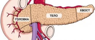
The weight of the pancreas in a person reaches 80-90 g; it is located, as you can easily guess from the name, under the stomach if the person lies on his back and behind the stomach on the back wall of the abdominal cavity if the person is in a standing position. This is a lobular organ about 15-20 cm long, grayish-pink in color, which is closely adjacent to the duodenum and is located at the level of the I-II lumbar vertebrae. The components of the pancreas are the head, body and tail. A groove with the portal vein separates the head and body of the gland, which has a triangular-prismatic structure and three surfaces: anterior, inferior and posterior.
What functions does the pancreas perform?
The pancreas is involved in the regulation of metabolic processes in the body and performs exocrine and intrasecretory functions. Its cells produce enzymes, without which digestion processes in the lumen of the small intestine would be impossible. Moreover, unlike other substances required for normal digestion of food, pancreatic enzymes begin to be produced a couple of minutes after food enters the stomach and this process continues for about 10-14 hours after consuming the last foods. The functions of the pancreas can only be performed if there is a sufficient amount of bile.
Pancreas – structure and function, role in the development of diabetes
The pancreas (Latin: páncreas) is a palm-sized organ in the abdominal cavity, located between the stomach and the spine.
Rice. 1. The structure of the pancreas. Adapt. from Wikipedia
- Gallbladder
- Pancreatic lobes
- Pancreatic duct
- Common bile duct
- Accessory duct of the pancreas
- Major duodenal papilla
- Duodenum
The pancreas has three sections: head, body, tail (Fig. 1). All parts of the pancreas perform the same functions, namely:
- produce enzymes that help digest food;
- produce hormones such as insulin and glucagon that control blood glucose levels.
Digestive enzymes from the pancreas enter the intestine through the pancreatic duct [3]. The pancreatic duct[3] connects with the common bile duct[4], which carries bile from the gallbladder[1] and liver, and they empty into the duodenum[7] in the area of the major duodenal papilla[6]. This function of the pancreas is also called exocrine, that is, directed outward. The bulk of the pancreas performs this function.
The second function of the pancreas is the endocrine function, that is, directed inward, is the production of hormones that control blood glucose levels. This function is performed by separate groups of cells, they are called pancreatic islets or islets of Langerhans. There are about 1 million islets in the entire pancreas, representing 1-2% of the total mass of the pancreas. They are located diffusely throughout the entire volume of the pancreas. Unlike enzymes that are synthesized in the pancreatic duct and drain into the intestine, cells in the islets of Langerhans produce hormones directly into the blood, namely into the small blood vessels passing through the pancreas.
Fig.2. Islet of Langerhans. Adapt. from Anatomy, Physiology, and Pathology of the Digestive System. Introduction to Medical Science - Duke University TIP
If you look at the islet of Langerhans through a microscope (Fig. 2), you will find in it:
- Beta cells that produce insulin
- Alpha cells that produce glucagon
- Delta cells that produce somatostatin
- PP cells, which produce pancreatic polypeptide (the function of which is still unclear)
Beta cells contain a kind of “built-in” glucose meter. If blood glucose levels rise, they release insulin. If blood glucose levels decrease, insulin secretion stops. If glucose levels fall below normal levels, alpha cells release glucagon. Other hormones produced by the cells of the islets of Langerhans are necessary for the islet cells to communicate with each other. The islets of Langerhans are very small, approximately 0.1 mm in diameter. All adult human islets contain approximately 200 units of insulin. The volume of all of them combined is no more than the tip of a finger. Insulin is a hormone that helps the body absorb and use glucose and other nutrients. It is like a “key that opens the door” for glucose to enter the cell. Without insulin, blood glucose levels rise (more about insulin in the section Insulin and its importance for the body).
The role of the pancreas in the development of diabetes mellitus
In order to understand this issue, we will consider each type of diabetes separately.
Diabetes mellitus type 1
In type 1 diabetes, beta cells die and the pancreas produces little or no insulin. Type 1 diabetes usually develops when the immune system destroys beta cells in the pancreas. This is called an autoimmune response. The body's own immune system perceives beta cells as foreign objects, such as bacteria or viruses, and begins to attack and destroy them. When more than 90% of beta cells are destroyed (a process that takes anywhere from several months to a maximum of several years), the body begins to feel a lack of insulin and blood glucose levels rise. Then the person experiences “big” symptoms of diabetes, such as thirst, frequent urination, and weight loss. Previously, this type of diabetes was called insulin-dependent diabetes mellitus. This means that treatment requires insulin as soon as the diagnosis is made.
At this time, it remains unknown why this autoimmune response occurs. Genetically, a person can be transmitted a tendency to autoimmune diseases (type 1 diabetes mellitus is only one of many autoimmune diseases), but what exactly serves as a trigger, a trigger for type 1 diabetes mellitus, is not yet clear enough. (You can learn more about this in the section Causes of type 1 diabetes).
Diabetes mellitus type 2
In type 2 diabetes, the ability of the pancreas to produce insulin does not disappear completely. But the body becomes increasingly resistant to insulin. That is, a situation is created when the normal level of blood insulin cannot “open the door to the cell for glucose.” Therefore, if the body does not respond to normal blood insulin levels, the pancreas has to produce more and more insulin. And if this process is not influenced in any way, it will lead to depletion of the pancreas and absolute insulin deficiency.
The causes of type 2 diabetes are a complex of genetic factors and environmental conditions. Genetically, a person inherits a group of genes for susceptibility to type 2 diabetes, and most often a person with type 2 diabetes has relatives with type 2 diabetes. Also, some ethnic groups have a higher predisposition to this disease. But whether type 2 diabetes mellitus develops or not depends on the person himself and his lifestyle. This is influenced by the nature of nutrition, level of physical activity, etc. (You can learn more about this in the section Causes of type 2 diabetes).
The tablets used to treat type 2 diabetes do not contain insulin and work either by increasing the body's sensitivity to insulin or by increasing insulin secretion from the pancreas. Diet and loss of excess weight (if any) are also the main components of the treatment of type 2 diabetes. It is rare that insulin injections are necessary in the early stages of type 2 diabetes. But when the pancreas is depleted, insulin therapy may become a necessary component in the treatment of type 2 diabetes.
Gestational diabetes mellitus
This form of diabetes occurs during pregnancy and in most cases goes away after the baby is born.
Throughout pregnancy, the placenta produces hormones that interfere with the normal functioning of insulin (they increase insulin resistance). Typically, the pancreas can simply increase the amount of insulin it produces, and the woman will have normal blood glucose levels. Sometimes, however, the pancreas cannot compensate for the body's need for insulin, and then the woman's blood glucose levels rise and develop gestational diabetes.
All pregnant women should be screened for gestational diabetes. It is important to recognize and treat gestational diabetes as soon as possible to minimize the risk of complications in the baby. (You can find out more about this in the section Gestational diabetes mellitus).
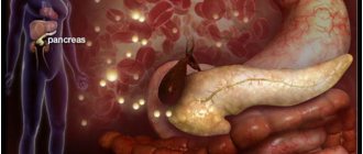
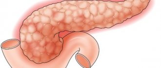
Pancreatic diseases and their symptoms
Diseases of the pancreas arise due to its insufficient functioning, and can also be caused by eating fatty foods, especially when combined with alcohol. Alcohol alone also has a destructive effect on the pancreas. In older people, disorders occur due to circulatory disorders of the pancreas, atherosclerotic processes in blood vessels, and thrombosis. Sudden inflammation of the pancreas is called acute pancreatitis, it can be caused by both the above factors and diseases of the duodenum, gall bladder, and drug allergies. Depending on the severity of the disease, the symptoms may differ slightly, but any disturbance is always accompanied by pain, which manifests itself in the abdomen or epigastric region; often a person feels pain in the left hypochondrium, which can be constant or paroxysmal in nature or encircle. The pain increases significantly with overeating, consuming fatty, spicy and fried foods, and alcohol.
Inflammation always manifests itself acutely; it is very difficult to relieve pain; you can only reduce it by sitting down and bending forward or lying on your side. In acute pancreatitis, hemorrhage occurs in the tissues, swelling occurs, sometimes even leading to necrosis of the pancreas. The condition is accompanied by nausea and vomiting, low blood pressure, and rapid pulse.
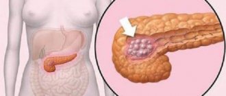
If the functions of the pancreas are slightly impaired, a person may experience a decrease in appetite and weight loss. More accurate results are achieved through ultrasound examination, computed tomography, and x-ray examinations, since there are tumors (benign and malignant) and pancreatic cysts.
Blood supply to the pancreas
The pancreas has three parts: head, body, and tail. The head is located in the bend of the duodenum, and sometimes surrounds it with a ring. The body lies along the lower part of the posterior wall of the stomach, the tail rests against the gate of the spleen. In close proximity are the liver, mesentery, and intestinal omentum.
From the abdominal aorta, which is part of the systemic circulation, nutrients and oxygen enter the arterial vessels. Arterial blood from the left ventricle is pumped under high pressure into the aorta. Some of it flows to the head and upper limbs, and some goes down to the organs of the chest, abdominal cavity, and lower limbs.
Pancreaticoduodenal arterial network
The celiac trunk departs from the abdominal part of the aorta, giving rise to the vessels that nourish the pancreas tissue.
Arterial branches carrying blood to the lobules and glandular islets, in descending order of vascular caliber:
- celiac trunk;
- common hepatic and splenic arteries;
- superior anterior and posterior vessels of the pancreas and duodenum;
- the inferior pancreaticoduodenal artery with anterior and posterior branches, which arises from the superior omental arterial trunk;
- dorsal pancreatic artery;
- large artery of the pancreas;
- tail artery;
- inferior pancreatic artery.
Next, the blood flows through the arterioles to the capillaries that open in each lobe of the gland. It brings oxygen and nutrients to the tissues. Hormones enter the bloodstream from the pancreatic islets through the arterial system.
Angiography is used to diagnose pancreatic diseases. The essence of the method is to study the external and internal vascular pattern in order to identify pathological changes in the arteries, the formation of a new network of capillaries, and identify areas with excess blood supply (hypervascularization) or its deficiency (hypovascularization).
A contrast agent is injected into the celiac trunk and superior mesenteric artery from the femoral approach. A series of radiographs is taken, which allows one to determine the nature of the course of the vessels and the degree of their filling outside and inside the organ. In this way, the phase of chronic pancreatitis, cysts, benign and malignant tumors can be diagnosed.
Venous drainage
Pathways of blood flow towards the portal vein
The venous collectors receive “processed” blood from the glandular lobules and islets. Outflow occurs through the system of the portal and inferior vena cava through a dense vascular network.
Blood moves through the venules in the following direction:
- superior, inferior mesenteric veins;
- splenic veins;
- left gastric vein;
- portal vein.
After passing through the liver, the blood enters the inferior vena cava. Then it moves towards the opening of the right atrium. Once in the cavity of the heart, the blood flow ends the systemic circulation.
Innervation of the pancreas
The innervation and blood supply of the pancreas ensure the functioning of the complex process of neurohumoral work of the endocrine part of the organ. Together with the arteriovenous network, sensory fibers form a neurovascular bundle.
The innervation of all parts of the gland is carried out by the autonomic nervous system. The main task of this department of vital activity regulation is to maintain the functional activity of all organs and the constancy of the internal environment. The autonomic system includes the central nuclei and pathways located in the brain, the spinal cord and individual pancreatic nerve ganglia.
Conducting paths and nodes (designations in the text)
Neurohumoral regulation of the endocrine and exocrine pancreatic apparatus is carried out by the following structures:
- celiac plexus (2), located on the abdominal aorta (1) next to the inferior vena cava (16);
- large branches of the vagus nerve;
- right and left branches of the posterior vagus trunk;
- fibers accompanying the gastroepiploic artery (12);
- nerves located next to the pancreaticoduodenal anterior arteries - lower (9) and upper (11), feeding the duodenum (10);
- branches passing through the thickness of the gland (6);
- superior mesenteric plexus (7), accompanying the vein and artery of the same name (8);
- fibers of the hepatic plexus (13), located along the native artery of the liver (14) and the portal vein (15);
- nerves of the splenic plexus (3), running along the splenic artery (4) to the hilum of the spleen (5).
The nerve trunks approach the gland along with the vessels, forming a single pancreatic plexus on its surface and inside. A dense network of afferent fibers provides continuous neurohumoral regulation, sensitive to the slightest fluctuations in the internal environment.
The receptor apparatus of the enzyme-forming part of the gland reacts to the entry of a bolus of food into the lumen of the duodenum. Thanks to complex physicochemical interactions, pancreatic juice begins to be produced, which enters the intestinal cavity through the main duct and promotes the breakdown of proteins, fats, and carbohydrates.
Receptors of the endocrine apparatus of the pancreas are sensitive to fluctuations in glucose levels in the bloodstream. When it is in excess, neurohumoral regulation activates the production of insulin, which promotes the utilization of sugar molecules. When there is a deficiency, glucagon is released, which induces liver cells to synthesize glucose from glycogen stores.
The complex formation of the pancreatic neurovascular plexus ensures the functioning of the organ itself, its active participation in digestion and regulation of metabolism. The connection with large arterial and venous collectors determines the involvement of the gland in the pathological process when they are damaged.