Diseases of the esophagus, stomach and duodenum
Recommendations of the Russian Gastroenterological Association for the diagnosis and treatment of gastroesophageal reflux disease 2022
Authors : V.T. Ivashkin, I.V. Maev, A.S. Trukhmanov, T.L. Lapina, O.A. Storonova, O.V. Zairatyants, O.B. Dronova, Yu.A. Kucheryavyi, S.S. Pirogov, R.G. Sayfutdinov, Yu.P. Uspensky, A.A. Sheptulin, D.N. Andreev, D.E. Rumyantseva
The prevalence of GERD in the Russian Federation varies from 11.3 to 23.6%. The diagnosis of GERD is established on the basis of anamnestic data and the results of instrumental examination. Esophagogastroduodenoscopy (EGD) can detect signs of reflux esophagitis of varying severity and identify columnar cell metaplasia of the esophageal epithelium. Patients with a refractory course of the disease (in the absence of convincing clinical and endoscopic remission within 4–8 weeks of therapy with a standard dose of PPI) or in the presence of strictures, Barrett’s esophagus, need to undergo endoscopy with a biopsy of the esophagus and histological examination of the biopsy samples. According to indications, patients should undergo intraesophageal 24-hour pH-metry or pH-impedansometry, high-resolution manometry, which allows a complete study of the functional state of the esophagus and esophagogastric junction, to predict the variants of the course of the disease and the results of its treatment.
Clinical guidelines of the Russian Gastroenterological Association for the diagnosis and treatment of gastroesophageal reflux disease 2022
Authors : V.T Ivashkin, I.V Maev, A.S. Trukhmanov, E.K. Baranskaya, O.B. Dronova, O.V. Zairatyants, R.G. Sayfutdinov, A.A. Sheptulin, T.L. Lapina, S.S. Pirogov, Yu.A. Kucheryavyi, O.A. Storonova, D.N. Andreev
Treatment of patients with GERD should be individualized according to the clinical manifestations of the disease and the severity of symptoms. The goal of treatment is to eliminate symptoms, for erosive esophagitis - to heal erosions and prevent complications, for Barrett's esophagus - to prevent the progression and development of dysplasia and adenocarcinoma of the esophagus.
Clinical guidelines of the Russian Gastroenterological Association for the diagnosis and treatment of peptic ulcer 2016
Authors : V.T. Ivashkin, A.A. Sheptulin, I.V. Maev, E.K. Baranskaya, A.S. Trukhmanov, T.L. Lapina, S.G. Burkov, A.V. Kalinin, A.V. Tkachev
Peptic ulcer continues to be one of the most common diseases of the digestive system. Despite the trend towards a decrease in the frequency of hospitalizations of patients with uncomplicated peptic ulcer disease, an increase in the frequency of complicated forms of the disease has been noted, which is mainly associated with the increasing use of non-steroidal anti-inflammatory drugs (NSAIDs). The leading role in the etiology of peptic ulcer disease belongs to Helicobacter pylori infection.
Clinical recommendations of the Russian Gastroenterological Association for the diagnosis and treatment of achalasia cardia and cardiospasm 2016
Authors : V.T. Ivashkin, A.S. Trukhmanov, E.A. Gogello, I.V. Maev, Yu.V. Evsyutina, T.L. Lapina, O.A. Storonova
Achalasia cardia is a primary disorder of the motor function of the esophagus, manifested by impaired relaxation of the lower esophageal sphincter and defects in peristalsis of the thoracic region. The etiology remains unknown. Clinical recommendations contain a detailed description of all currently existing methods of treating cardia achalasia and cardiospasm (conservative, endoscopic, surgical), which are aimed at expanding the cardia and reducing its tone to improve the passage of food through the area of the esophagogastric junction.
Clinical guidelines of the Russian Gastroenterological Association for the diagnosis and treatment of dysphagia 2015
Authors : V.T. Ivashkin, I.V. Maev, A.S. Trukhmanov, T.L. Lapina, A.A. Sheptulin, O.A. Storonova, D.N. Andreev.
Dysphagia is considered as difficulty in starting to swallow (defined as oropharyngeal dysphagia) or as a sensation of obstruction in the passage of food or liquid from the mouth to the stomach (defined as esophageal dysphagia). Thus, dysphagia is a feeling of obstruction of the normal passage of ingested food.
Clinical guidelines of the Russian Gastroenterological Association for the diagnosis and treatment of infectious esophagitis 2015
Authors : V.T. Ivashkin, N.D. Yushchuk, I.V. Maev, A.S. Trukhmanov, O.P. Alekseeva, T.L. Lapina, S.S. Pirogov, A.V. Tkachev, A.A. Sheptulin.
Infectious esophagitis (ICD K20) can be caused by fungal, viral, bacterial or even parasitic agents. Risk factors include: use of antibiotics and steroids, chemotherapy and/or radiation therapy, malignancies and immunodeficiency syndromes, including acquired immunodeficiency syndrome. Characterized by an acute onset with the appearance of symptoms such as dysphagia and odynophagia.
Clinical recommendations of the Russian Gastroenterological Association for the diagnosis and treatment of erosive and ulcerative lesions of the stomach and duodenum caused by non-steroidal anti-inflammatory drugs, 2014
Authors : V. T. Ivashkin, A. A. Sheptulin, I. V. Maev, E. K. Baranskaya, A. S. Trukhmanov, T. L. Lapina.
The relevance of the problem of erosive and ulcerative lesions of the stomach and duodenum associated with the use of NSAIDs (NSAID-associated gastropathy) is determined, first of all, by the fact that the use of drugs in this group is extremely common among the population. NSAIDs are over-the-counter drugs; many patients take them without prior consultation with their doctor. The consequence of such uncontrolled use is a high incidence of gastroduodenal ulcers and erosions, which in such cases increases 5 times.
Clinical guidelines of the Russian Gastroenterological Association for the diagnosis and treatment of gastroesophageal reflux disease 2014
Authors : V.T. Ivashkin, I.V. Maev, A.S. Trukhmanov, E.K. Baranskaya, O.B. Dronova, O.V. Zairatyants, V.D. Pasechnikov, R.G. Sayfutdinov, A.A. Sheptulin, Yu.A. Curly, T.L. Lapina, O.A. Storonova, V.O. Kaibysheva
The relevance of the problem of GERD is determined by a number of circumstances. Epidemiological studies in recent years have shown that, in terms of prevalence, GERD takes a leading position among other gastroenterological diseases. Heartburn, the leading symptom of GERD, is detected in 20-40% of the population of developed countries.
Clinical guidelines of the Russian Gastroenterological Association for the diagnosis and treatment of Barrett's esophagus 2014
Authors : V.T.Ivashkin, I.V.Maev, A.S.Trukhmanov, V.V.Sokolov, S.S.Pirogov, O.V.Zayratyants, A.A.Sheptulin, T.L.Lapina, G.O.Zayratyants, V.O.Kaibysheva
Growing attention to the problem of Barrett's esophagus (BE) is caused by the high incidence of esophageal adenocarcinoma (AEC) against the background of BRE and the emergence of AEC in first place among malignant tumors of the esophagus.
Clinical Guidelines of the Russian Gastroenterological Association for the diagnosis and treatment of peptic ulcer disease 2013
Peptic ulcer disease is a chronic relapsing disease that occurs with alternating periods of exacerbation and remission, the leading manifestation of which is the formation of a defect (ulcer) in the wall of the stomach and duodenum.
Clinical guidelines of the Russian Gastroenterological Association for the diagnosis and treatment of functional dyspepsia 2013
Authors : V.T. Ivashkin, A.A. Sheptulin, T.L. Lapina, I.M. Kartavenko, V.A. Kiprianis, O.Z. Okhlobystina.
In accordance with the recommendations of the consensus meeting of the International Working Group on Improving Diagnostic Criteria for Functional Diseases of the Gastrointestinal Tract (Rome III Criteria, 2006), FD is understood as a set of complaints, including pain and a burning sensation in the epigastric region, a feeling of fullness in the epigastrium after food and early satiety, which have been observed in the patient over the last 3 months (with a total duration of at least 6 months) and which cannot be explained by organic diseases.
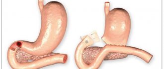
Treatment algorithm for patients with peptic ulcer
P
Recent decades have been characterized by significant changes in the epidemiology of some common gastroenterological diseases.
Along with the increase in the incidence of gastroesophageal reflux disease, there has been a noticeable decrease in the incidence of peptic ulcer disease. Thus, if back in the 70–80s of the last century it was generally accepted that every tenth person may develop a peptic ulcer in his life [2], now the prevalence of peptic ulcer has decreased several times and is, for example, currently in the USA 2.5% [7]. At the same time, while the frequency of ulcer perforation remained at the same level, the frequency of ulcer bleeding
(due to gastric ulcers) has increased significantly, which is caused by the increasing use of nonsteroidal anti-inflammatory drugs (NSAIDs) [8]. The costs associated with the treatment of patients with peptic ulcer disease remain significant, which in the USA, for example, amount to $3.1 billion, ranking 4th after the costs associated with the treatment of gastroesophageal reflux disease, cholelithiasis, and colorectal cancer [7 ].
Such positive dynamics are largely due to changes in our ideas about the pathogenesis of peptic ulcer disease (primarily, with the discovery of the role of Helicobacter pylori
(NR) in the development of peptic ulcer and its relapses) and revision of approaches to its treatment. Previously, a variety of different treatments could be recommended for the treatment of this disease, incl. “exotic” products (for example, quail eggs, “living” and “dead” water, etc.), which served as V.Kh. Vasilenko [1] as the basis for formulating his famous aphorism (“gastroduodenal ulcers heal with treatment, without treatment, and even in spite of treatment”), then already in the 90s of the last century a protocol for the treatment of peptic ulcer disease was created, based on modern approaches and principles of pharmacotherapy for this diseases.
Principles of pharmacotherapy for peptic ulcer disease:
- the same approach to the treatment of stomach and duodenal ulcers;
- mandatory basic antisecretory therapy;
- choosing an antisecretory drug that maintains intragastric pH>3 for about 18 hours a day;
- prescribing an antisecretory drug in a strictly defined dose;
- endoscopic control at 2-week intervals;
- duration of antisecretory therapy depending on the timing of ulcer healing;
- eradication therapy against Helicobacter pylori in HP-positive patients;
- mandatory monitoring of the effectiveness of anti-Helicobacter therapy after 4–6 weeks;
- repeated courses of anti-Helicobacter therapy if it is ineffective;
- maintenance anti-relapse therapy with an antisecretory drug in HP-negative patients;
- influence on risk factors for poor response to therapy (replacement of NSAIDs with paracetamol, selective COX-2 inhibitors, combination of NSAIDs with misoprostol, ensuring patient compliance, etc.).
The treatment protocol for peptic ulcer disease involves, first of all, basic antisecretory therapy.
, the purpose of which is to relieve pain and dyspeptic disorders, as well as achieve scarring of the ulcerative defect in the shortest possible time.
In 1990, W. Burget et al. [6]. Neither H2 blockers, nor selective anticholinergics, much less antacids, can fulfill this condition. Only proton pump blockers satisfy this requirement, which explains why drugs of the latter group are currently the most effective in the treatment of peptic ulcers.
The pharmacotherapy protocol involves prescribing a basic antisecretory drug in a strictly defined dose. The duration of treatment depends on the results of endoscopic monitoring, which is carried out at two-week intervals (i.e. after 4, 6, 8 weeks). To assess the effectiveness of a particular antiulcer drug, one uses the calculation not of the average time of scarring, but of the frequency of healed ulcers in 4, 6, 8, etc. weeks of treatment.
Although the mechanism of action of various proton pump blockers is the same (blocking the activity of H+, K+–ATPase of the parietal cell), their effectiveness may not be the same. The time at which these drugs begin to act depends on how quickly they are converted from their inactive form to their active (sulfenamide) form. For example, rabeprazole is converted to its active form more quickly than omeprazole, lansoprazole and pantoprazole, which results in a more rapid onset of its inhibitory effect compared to other proton pump blockers [5]. This makes the prescription of rabeprazole during basic antisecretory therapy more preferable. In addition, rabeprazole, to a lesser extent than other proton pump blockers (in particular, omeprazole), interacts with the cytochrome P450 enzyme system in the liver, as a result of which, firstly, its antisecretory effect is more stable, and secondly, metabolism taking other medications does not interfere with its use.
An important principle of modern pharmacotherapy for peptic ulcer disease is the absence of fundamental differences in approaches to the treatment of gastric ulcers and duodenal ulcers
. For a long time it was believed that duodenal ulcers require the use of antisecretory drugs, and gastric ulcers require drugs that stimulate regeneration processes. It is now generally accepted that after confirmation of the benign nature of gastric ulcers, the treatment of such patients is carried out in the same way as the treatment of patients with duodenal ulcers (but only for a longer time, given the somewhat slower scarring of gastric ulcers).
The “Achilles heel” of conservative treatment of peptic ulcer disease is, as is known, the high rate of relapse of ulcers after stopping the course of treatment for exacerbation of the disease, which reaches 70% during the first year. maintenance pharmacotherapy after completion of
. The most common practice for this purpose is to take half doses of proton pump blockers daily, which reduces the incidence of recurrent ulcers within a year by up to 15%. The effectiveness of other methods of supportive antisecretory therapy - “yourself treatment” or “on demand” therapy, when patients themselves determine the need to use drugs based on their well-being, turns out to be less high: the frequency of exacerbations of peptic ulcer disease during the year with this treatment is 30–35%. Currently, when anti-Helicobacter therapy has begun to play a major role in anti-relapse treatment, the indications for maintenance therapy with antisecretory drugs have narrowed significantly. It is considered necessary in HP-negative patients with peptic ulcer disease (15–20% of patients with gastric ulcers and about 5% of patients with duodenal ulcers), as well as in patients in whom several attempts at anti-helicobacter treatment using different eradication regimens have been unsuccessful.
Each patient with peptic ulcer in whom HP is detected in the gastric mucosa by one method or another (rapid urease test, morphological method, DNA determination of HP by polymerase chain reaction, etc.) undergoes eradication therapy
.
This therapy involves a combination of several antibacterial agents. Most anti-helicobacter therapy regimens include proton pump blockers, which, by increasing the pH of the stomach contents, create unfavorable conditions for the life of HP and, in addition, increase the effectiveness of antibiotics. At the same time, the use of rabeprazole in eradication therapy regimens should be considered more preferable compared to other proton pump blockers, given the faster onset of its antisecretory action and more pronounced direct anti-Helicobacter activity (in vitro), which favors and potentiates the effect of antibiotics. This makes it possible to avoid the preliminary administration of antisecretory drugs (as, for example, in the case of omeprazole) before eradication.
By the decision of the Maastricht Consensus Conference of the European Group for the Study of Helicobacter Pyloricus (1996), the use of 3 possible 1st-line regimens was recommended for eradication therapy, each of which necessarily included the prescription of one of the proton pump blockers in standard doses 2 times a day and two antibacterial drug in various combinations and doses.
The eradication rate of HP when using these regimens exceeded 90%.
If the use of 1st line regimens was ineffective, a 2nd line eradication therapy regimen, the so-called. quadruple therapy course.
Eradication therapy 2 lines
Proton pump blockers 2 times a day
Colloidal bismuth subcitrate 120 mg x 4 times
Tetracycline 500 mg x 4 times
Metronidazole 250 mg x 4 times
Duration of treatment 7 days
As an alternative regimen, a combination of pyloride (ranitidine bismuth citrate) at a dose of 400 mg 2 times a day with one of the antibiotics - clarithromycin (250 mg 4 times or 500 mg 2 times a day) or amoxicillin (at a dose of 500 mg 4 times a day) was proposed. day).
In recent years, when carrying out eradication therapy, serious problems have emerged associated with the growing resistance of HP strains to antibacterial drugs, primarily metronidazole (in more than 30% of cases) and clarithromycin (in more than 10% of cases). In practical terms, this meant a significant reduction in the frequency of eradication when using regimens that included these antibacterial drugs. For example, in cases of HP resistance to metronidazole and clarithromycin, the effectiveness of regimens containing these drugs decreased from 91–93% to 44–69%. Among the ways to overcome the resistance of HP strains to antibacterial drugs
recommended:
- increasing the duration of eradication therapy to 10–14 days (meta-analysis data confirm the higher effectiveness of this regimen compared to a 7-day regimen);
- increasing the daily dose of clarithromycin (in combination with metronidazole and proton pump blockers) from 500 to 1000 mg;
- the possibility of replacing metronidazole with furazolidone.
The most effective way to increase the effectiveness of eradication therapy was recognized as the correct approach to choosing one or another of its regimens, which was reflected in the recommendations of the conference held in Maastricht in 2000 at the initiative of the European Group for the Study of Helicobacter pylori
[4]. The final document of this conference for the first time contained a proposal to plan the results of eradication therapy, taking into account the possibility of its failure. In addition, the number of possible anti-Helicobacter therapy regimens was reduced.
Currently, the triple scheme of the 1st line
, containing proton pump inhibitors (or ranitidine bismuth citrate) in a standard dose 2 times a day in combination with antibacterial drugs (Fig. 1). At the same time, the combination of clarithromycin with metronidazole is considered more preferable, since this can help achieve a better result if subsequent quadruple therapy is necessary.
Rice. 1. Algorithm for the management of patients with peptic ulcer disease
The eradication therapy protocol requires mandatory monitoring of its effectiveness, which is carried out 4–6 weeks after its completion (during this period the patient does not take antibacterial drugs) using a breath test or polymerase chain reaction to determine HP DNA in the stool.
If HP persists in the gastric mucosa, a second course of eradication therapy
using 2nd line therapy with subsequent monitoring of its effectiveness also after 4–6 weeks (Fig. 1), which makes it possible to carry out complete sanitation of the gastric mucosa and prevent the risk of relapses peptic ulcer disease [3].
The quadruple therapy regimen has retained its importance as a 2nd line therapy
(Fig. 1). If there is no effect during the second course, subsequent treatment tactics are decided individually in each specific case.
The ineffectiveness of conservative treatment of patients with peptic ulcer can manifest itself in two ways: a frequently recurrent course of peptic ulcer (i.e., with an exacerbation frequency of 2 times a year or more) and the formation of refractory gastroduodenal ulcers (ulcers that do not scar within 12 weeks of continuous treatment).
Factors determining the frequently recurrent course of peptic ulcer disease are:
- contamination of the gastric mucosa with HP;
- taking NSAIDs;
- a history of ulcer bleeding and ulcer perforation;
- low “compliance”, i.e. lack of willingness of the patient to cooperate with the doctor, manifested in the refusal of patients to stop smoking and drinking alcohol, and irregular intake of medications [8].
Measures that increase the effectiveness of treatment of patients with frequently recurrent peptic ulcer disease include the following:
- eradication of HP, which, if successfully completed, reduces the frequency of relapses of ulcers within a year from 70% to 4–5% and also reduces the risk of recurrent bleeding,
- prescribing long-term maintenance therapy with antisecretory drugs for patients with HP-negative peptic ulcer disease,
- replacing NSAIDs with paracetamol or selective cyclooxygenase-2 inhibitors,
- prescribing, if necessary, to continue taking NSAIDs, an appropriate “cover” (proton pump blockers or misoprostol),
- increasing patient compliance.
Factors contributing to the formation of refractory gastroduodenal ulcers may include HP infection, use of NSAIDs, low patient compliance, large and gigantic ulcers, and latent Zollinger-Ellison syndrome [8]. Carrying out the above measures, as well as increasing the dose of proton pump blockers by 2–3 times, and carefully examining patients to exclude gastrinoma (primarily determining the level of serum gastrin) make it possible in many cases to successfully solve the problem of treating refractory ulcers.
Thus, the improvement in the results of conservative therapy for peptic ulcer disease led to a radical change in approaches to treating patients, which was reflected in a significant limitation of indications for operations for peptic ulcer disease (including ulcer bleeding) and for the first time put on the agenda the question of the fundamental curability of this disease. diseases.
Literature:
1. Vasilenko V.Kh. What we don’t know about peptic ulcer // Current issues of gastroenterology. – M., 1970. issue 3. – P.3–17
2. Vasilenko V.Kh., Grebenev A.L., Sheptulin A.A. Peptic ulcer disease. – M., 1987.
3. Recommendations for the diagnosis and treatment of Helicobacter pylori infection in adults with gastric and duodenal ulcers // Ross. magazine gastroenterol. hepatol. coloproctol. – 1998. – No. 1. – P.105–107.
4. Diagnosis and treatment of Helicobacter pylori infection, modern concepts (Report of the second conference on the adoption of consensus in Maastricht on September 21–22, 2000) // Ross. magazine gastroenterol. hepatol. colproctol. – 2000. – No. 6. – P.7–9.
5. Besancon M., Simon A., Sachs A., Shin JM Sites of reaction of the gastric H,K–ATPase with extracytoplasmic thiol reagents // J.Biol.Chem. – 1997. – Vol.272. – P.22438–22446.
6. Burget DW, Chiverton KD, Hunt RH Is there an optimal degree of acid suppression for healing of duodenal ulcers? A model of the relationship between ulcer healing and acid suppression // Gastroenterology. – 1990. – Vol.99. – P.345–351.
7. Laine L. Peptic ulcer disease: where are we and where do we go from here? AGA Postgraduate Course. May 18–19, 2002. Course syllabus. – San Francisco, 2000. – P.20–25.
8. Soll AH Peptic ulcer and its complications // Sleisenger & Fordtran's Gastrointestinal and Liver Disease. – Philadelphia–Londodn–Toronto–Monytreal–Sydney–Tokyo. – 1998. – Vol.1. – P.620–678.