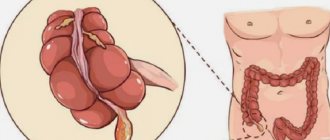
A disease that is caused by inflammation of the appendix is commonly called appendicitis. The appendix is the atrophied part of the large intestine. This process looks like a hollow worm-like tube and is located between the small and large intestines.
The causes of the disease are still poorly understood to date. But there are some factors that are more likely to contribute to the development of appendicitis. For example, some parasites and worm infestations. But, according to experts, it is quite difficult to predict the development of the disease and it is almost impossible to prevent it.
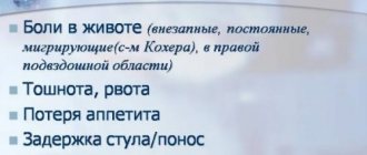
Doctors note that appendicitis most often develops in young people and children, explaining this by the high activity of their immune system. Symptoms of appendicitis:
- Acute pain in the abdominal area (pain is most often localized at the location of the appendix, namely in the right half of the abdomen, above the inguinal fold);
- High temperature (often the temperature rises to 38 degrees);
- Vomiting and nausea.
Taking medications that can relieve pain is not recommended for appendicitis. Since when taking analgesics, the picture of symptoms may change somewhat, which can mislead the attending physician when making a diagnosis.
A doctor can diagnose this disease by collecting the patient’s medical history, checking for specific syndromes and obtaining the results of an ultrasound examination. It is based on these indicators that a diagnosis can be reliably made. Ultrasound reveals blockage and swelling of this process. The process can only be removed through surgery.
Complication of appendicitis – peritonitis
Peritonitis is a process of inflammation of the peritoneum.
Appendicitis in itself is not dangerous. Much more dangerous are its complications. That’s why you shouldn’t think long about visiting a doctor if you start having symptoms that at least somehow raise doubts!
The most advanced forms of appendicitis can also lead to peritonitis. In this case, there are even fatal outcomes of such a disease.
What is peritonitis? This is an inflammation of the entire peritoneum (the peritoneum is a membrane that lines the abdominal cavity), which poses a danger to the patient’s life: choosing the right treatment is not always possible, unfortunately.
Purulent omentitis in childhood
UDC: 616.346.2 – 089.87 – 002.3 – 053.2
Acute appendicitis is a common disease in childhood. It may be complicated by the development of purulent omentitis. The article discusses the reasons for the development of this pathology, the complexity of diagnosis, and highlights the options for interventions performed and their effectiveness.
Key words: acute appendicitis, purulent omentitis, videolaparoscopy.
The acute of appendicitis is often disease in children's age. It can become complicated by development purulent omentitis. In the article the reasons of development of this pathology, complexity of diagnostics are considered, the variants carried out of operations, their efficiency are covered.
Despite the fact that in the twentieth century huge steps forward were made in surgery, the problem of early diagnosis and treatment of surgical infection in the abdominal cavity is still far from being solved.
In childhood, acute appendicitis is the most common inflammatory disease of the abdominal cavity. The often observed discrepancy between clinical manifestations and existing changes in the appendix indicates the difficulty of diagnosing acute appendicitis in childhood and the relevance of this issue at the present time. The active surgical tactics adopted in the past, when it was impossible to exclude inflammatory changes in the appendix, made it possible to significantly minimize the number of its complicated forms, as well as its mortality rate by almost 100 times. However, the structure of postoperative complications has also undergone changes towards a relative increase in the proportion of adhesive processes in the abdominal cavity and associated complications (Isakov Yu.F., Doletsky S.Ya.).
The abdominal cavity has its own local protective mechanisms, one of which is the greater omentum, to localize and relieve inflammatory processes. It carries out regulatory functions to maintain a constant environment in the abdominal cavity. Thanks to its high plastic ability and mobility, it quickly delimits foci of infection followed by their phagocytosis due to histiocytes and other macrophages (Bairov G.A.).
Prolonged interaction of the inflamed appendix with the greater omentum enveloping it can lead to decompensation of the protective immunological properties of the latter and its secondary involvement in the spread of infection. A decrease in its functional effectiveness can contribute to the activation of infectious processes in the abdominal cavity from a sluggish inflammatory-dystrophic course to severe purulent-fibrinous diffuse peritonitis.
The task of a pediatric surgeon during an operation for acute appendicitis is not only to perform an appendectomy, but also to assess the condition and changes in the organs adjacent to the appendix, and to sanitize the abdominal cavity. The surgeon’s desire to minimize surgical trauma mainly comes down to reducing the size of the access and the duration of the operation, which can lead to underestimation of inflammatory changes in the organs adjacent to the process and, in particular, the greater omentum. The “modified” omentum left in the abdominal cavity in the postoperative period can help maintain the activity of the inflammatory-adhesive process in it.
In the last century, many innovative technologies, characterized by high diagnosticity and minimally invasiveness, began to actively develop in surgery.
The purpose of the work was to present the relevance of the topic, to show the complexity of diagnosis and selection of adequate surgical tactics for purulent omentitis in children, the need to search for available methods of diagnosis and treatment of this pathology.
The results of 378 appendectomies and two cases of surgical treatment of postoperative intra-abdominal infiltrates were analyzed based on materials from the children's surgical department of the Gomel Regional Clinical Hospital for the period from 01/01/2001. to December 31, 2001. Incidental resection of the greater omentum during appendectomies was performed in 54 patients. In the removed processes of this group of patients, the biopsy showed pronounced destructive changes, complicated in 56% by perforation. These operations included simultaneous appendectomy due to torsion of the greater omentum (biopsy of the appendix revealed secondary phlegmonous appendicitis). The development of intra-abdominal postoperative infiltrates in both cases was preceded by an appendectomy for perforated appendicitis; one of the patients underwent an appendectomy in a district hospital. After unsuccessful conservative therapy, the children underwent resection of the altered omentum using video laparoscopic technology. The slight severity of pain in the area of the postoperative wound already in the first days after such operations made it possible to reduce the volume of rehabilitation measures and the rehabilitation period itself, in comparison with patients after operations with conventional approaches. No wound suppuration was noted. Biopsy shows purulent omentitis.
The statistical difference in incidence by month during the year was minimal, on average 4 - 5 cases monthly, an increase to 7 cases was noted in February, May and September. The age of patients ranged from 2 to 14 years, with a sharp increase in the number of patients aged 9 to 13 years; there were twice as many boys. Among the patients, urban residents accounted for about 70%. Two children were admitted with a diagnosis of chronic appendicitis for planned appendectomy. According to emergency indications, 46 patients were hospitalized by ambulance teams, the rest were referred by doctors from clinics and other hospitals or referred independently, and in those admitted, the time period from the onset of the disease was more than 24 hours.
When performing appendectomies with resection of the greater omentum area, the following approaches were used: Volkovich-Dyakonov - in 46 cases, median and lower-median - in 8. In two patients from this group, when it was difficult to diagnose, standard appendectomy was preceded by diagnostic videolaparoscopy.
Cultures of effusion were taken from 41 patients: in 14, no growth of flora was obtained; in the rest, the presence of opportunistic intestinal flora (E.coli, Str.epidermidis, etc.) was noted.
In the postoperative period, patients received antibiotic therapy and physical therapy (magnetic therapy, electrophoresis with antibiotics or CI, exercise therapy). The length of hospital days varied from 10 to 16 days, in rare cases longer.
The course of the postoperative period in 4 patients operated on for destructive appendicitis was complicated by the development of early adhesive intestinal obstruction. After dissection of adhesions using video laparoscopic technology, the condition of these patients dynamically returned to normal and were discharged with recovery.
The constant impact of unfavorable environmental and social environmental factors on children's bodies contributes to changes in the reactivity of their immune system. This is confirmed by the prevalence of urban residents, as well as the high proportion of children of secondary school age.
Active awareness of the population about the timeliness of medical examination for abdominal pain will help reduce the number of complicated forms of acute appendicitis.
The use of video laparoscopic technology in case of ineffectiveness of conservative treatment of postoperative complications in the abdominal cavity (intra-abdominal infiltrates and adhesive intestinal obstruction) contributes to the further progressive development of positive dynamics, leads to a rapid improvement in condition and recovery, and also improves the quality of life of such patients.
Peritonitis and its symptoms
The feeling of a gag reflex is characteristic of peritonitis.
The symptoms of peritonitis are similar to the symptoms of appendicitis, but they are much stronger and more pronounced.
If a patient has appendicitis complicated by peritonitis, he will note the following symptoms:
- Severe pain, which can intensify even with leisurely walking and when pressing on the sore spot. It is worth noting an interesting point: “imaginary well-being.” Over time, pain receptors begin to adapt to severe pain and the person at times feels a complete absence of pain. But these sensations are deceptive and further pain will manifest itself with renewed vigor.
- Vomit;
- Constipation;
- Loss of appetite;
- Dyspnea;
- Cardiopalmus;
- Scanty urination;
- High temperature, chills, fever;
- Tension of the muscles of the anterior abdominal wall;
- Bloating.
The most common symptom of peritonitis is vomiting. If at the beginning of the complication it may be single, then it intensifies: the vomit begins to turn green, and blood impurities appear.
Profuse vomiting during peritonitis does not bring relief to the patient.
Treatment of peritonitis
Peritonitis must be treated surgically.
It is worth noting that treatment of peritonitis is always emergency. Treatment can only be carried out by a competent and experienced specialist.
Emergency surgery is necessary, both in the case of acute appendicitis and in the case of peritonitis: therapeutic treatment will not bring any effect. If the appendix is inflamed, it must be urgently removed, and the abdominal cavity must be sanitized in case of peritonitis.
Doctors recommend that if you detect symptoms that are similar to peritonitis, call an ambulance immediately. Because quite often a person needs resuscitation assistance.
After surgery, pus may accumulate in the abdominal cavity. In such a situation, special drainage tubes are removed, through which pus is removed from the cavity and sanitation is carried out. After surgery, the doctor prescribes antibiotics, which reduce the risk of complications. You will also have to adhere to the necessary diet, the principles of which the doctor will definitely familiarize you with. Along with antibiotics, necessary vitamins are often prescribed - they help maintain tone and give vitality to the body.
Treatment protocol for patients with peritonitis
Classification.
According to the degree of prevalence of the inflammatory process in the abdominal cavity, there are:
1. Limited (local) peritonitis (occupies up to 2 of the nine anatomical areas of the abdominal cavity).
2. Generalized peritonitis (occupies three or more anatomical areas of the abdomen):
According to the nature of the exudate: serous, serous-fibrinous and purulent peritonitis.
Diagnostics.
The diagnosis of peritonitis should be made on the basis of an initial physical examination.
During the examination of the patient in the emergency department, on the basis of a clinical and instrumental examination, the possible causes of peritonitis should be established.
In the emergency department, a general clinical blood test, a biochemical blood test, a coagulogram, and a general urinalysis are urgently performed; determine blood type and Rh factor; ECG, chest x-ray in direct projection and plain x-ray of the abdominal cavity (in bedridden patients - in the later position), ultrasound of the abdominal cavity (assessment of the presence of gas and liquid in the abdominal cavity, dilatation of intestinal loops, peristalsis) are performed; According to indications, consultations are carried out by doctors of therapeutic specialties.
Therapeutic tactics.
The diagnosis of peritonitis is an indication for emergency surgery. The patient is subject to emergency surgery within 1 hour from the moment of diagnosis.
Patients with severe symptoms of intoxication, syndromic disorders and severe concomitant diseases are recommended to undergo short-term (within 1.5-2 hours) preoperative preparation in the intensive care unit. The question of the need and scope of preoperative preparation of the patient for surgery is decided jointly by the surgeon and anesthesiologist.
Any surgical intervention for peritonitis must be performed under endotracheal anesthesia.
Access.
Laparoscopy:
- with a clinical picture of limited peritonitis,
— if necessary, a differential diagnosis (acute pancreatitis, mesenteric thrombosis),
- in case of appendicitis, cholecystitis and perforated ulcer diagnosed before surgery.
Midline laparotomy:
- with a clinical picture of widespread peritonitis,
— in case of a laparoscopic finding that precludes the possibility of performing a surgical procedure using a laparoscopic approach, conversion to a midline laparotomy is indicated.
Surgical tactics.
The mandatory stages of surgical intervention for peritonitis are:
- Collection of material for microbiological research;
- Complete and systematic revision of the abdominal cavity;
- Evacuation of exudate;
- Elimination of the source of peritonitis;
- Sanitation of the abdominal cavity;
- Naso-intestinal intubation – according to indications;
- Drainage of the abdominal cavity;
- Formation of a laparostomy for subsequent programmatic sanitation - according to indications.
Radical elimination of the source of peritonitis involves:
— removal of the inflamed organ (appendectomy, cholecystectomy);
— hermetic suturing of perforations and fresh through wounds of hollow organs;
— organ resection (for ulcers, tumors, injuries or impaired mesenteric circulation); - formation of a colostomy (ileostomy) to exclude affected areas of the colon from passage.
In the absence of conditions for radical elimination of the source of infection, its localization (delimitation with tampons in the abdominal cavity) and extra-abdominization of the organ are possible.
The formation of interintestinal anastomoses on inflamed areas of intestinal loops during purulent peritonitis is contraindicated. In such cases, it is advisable to remove the terminal intestinal stomas, including, if necessary, the jejunum.
The application of an intestinal suture and the formation of interintestinal anastomoses is permissible in non-inflamed areas of the intestine with limited forms of purulent peritonitis. The interintestinal anastomosis is formed as a “side to side” type, its diameter should be greater than the diameter of the intestine. When suturing the colon and rectum, it is advisable to supplement the operation with the formation of a temporary unloading colostomy.
With right-sided hemicolectomy, the issue of forming a primary ileotransverse anastomosis or forming an ileostomy is decided individually depending on the condition of the ileal wall.
When resection of the left half of the colon, a single-barrel colostomy is formed.
In cases of severe paresis and dilatation of intestinal loops of more than 4-5 cm, decompression of the digestive tube is indicated by nasointestinal intubation throughout the entire length of the small intestine with a wide 2-channel (non-absorbent) silicone probe. Simultaneously with the installation of the naso-intestinal tube, a naso-gastric tube is installed.
Rinsing the abdominal cavity must be done to “clean water” with solutions of sodium chloride 0.9%, sodium hypochlorite solution, and an aqueous solution of chlorhexidine (0.05%).
Drainage of the abdominal cavity is carried out using drainage tubes installed through counter-apertures (preferably two-channel silicone drainages or drainages of the “Gallic cross” type) with their rational placement in the expected places of accumulation of exudate (in case of widespread peritonitis - in the subdiaphragmatic, subhepatic areas, the left lateral canal, the small pelvis).
The introduction of tampons into the abdominal cavity is permissible only if it is impossible to radically eliminate the source of peritonitis and for the purpose of hemostasis.
Options for completing the operation:
- Surgery with complete elimination of the source can be completed by drainage and suturing of the abdominal cavity tightly. When the first signs of progression of peritonitis or intra-abdominal complications requiring surgical correction appear, relaparotomy is performed “on demand”;
- The surgical intervention ends with the formation of a laparostomy with subsequent program sanitation for the following indications:
widespread fibrinous-purulent or fecal peritonitis;
· signs of anaerobic infection of the abdominal cavity;
· severe abdominal sepsis, septic shock;
· the underlying disease (its complication), which does not allow one to immediately eliminate or localize the source of peritonitis;
· concomitant disease or condition that does not allow an adequate amount of surgery to be performed;
· intra-abdominal hypertension syndrome;
· the condition of the laparotomy wound (purulent-necrotic process), which does not allow suturing of the abdominal cavity.
Indications for relaparotomy “on demand”:
· progression of peritonitis (unresolved primary source, emergence of a new source, undiagnosed source, tertiary peritonitis);
· complication of the underlying disease requiring surgical correction (abscess, phlegmon, bleeding into the abdominal cavity or gastrointestinal tract, early adhesive intestinal obstruction);
· occurrence or complication in the postoperative period of a competing disease requiring surgical intervention (bleeding from gastroduodenal ulcers, destructive pancreatitis, perforation of acute and stress ulcers, acute disorder of mesenteric bleeding);
· complications associated with violation of the technique of surgical intervention, manipulation (iatrogenic damage, gastrointestinal tract failure, stump failure or bleeding due to slipping of the ligature, clips, foreign bodies of the abdominal cavity).
Relaparotomy “according to the program” or staged sanitation of the abdominal cavity is performed 24-48 hours after the initial operation. Staged sanitation includes removal of temporary sutures from the laparotomy wound, revision of the abdominal cavity, evacuation of exudate, washing of the abdominal cavity with antiseptic solutions, and closure of the laparotomy wound. When forming a laparostomy and performing staged sanitation, it is recommended:
— isolation of the abdominal organs from the parietal peritoneum and laparotomy wound with perforated polyethylene film;
— bringing together the edges of the surgical wound using separate sparse skin sutures, leaving a diastasis of the wound edges of 5–10 cm; after each sanitation, stitches are placed in new places;
— after completion of the staged treatment, the edges of the wound are sutured in layers: alternating interrupted and 8-shaped sutures of the aponeurosis + rare sutures on the skin according to Donati.
Criteria for ending the programmable sanitization mode:
· guaranteed elimination or localization of the source of peritonitis;
· absence of irremovable foci of necrosis and purulent foci;
· transparent serous exudate;
· presence of peristalsis of the small intestine;
· absence of widespread purulent-necrotic lesions of the surgical wound or anterior abdominal wall, excluding the possibility of one-stage surgical correction.
Antibacterial therapy.
Antibacterial therapy begins immediately after the diagnosis of peritonitis is established. The first dose of an antibacterial drug is administered before surgery. Until the results of a microbiological study of abdominal exudate are obtained, an “empirical” prescription of antibiotics with a spectrum of action that includes gram-negative bacteria in combination with anaerobic microorganisms is used:
- cephalosporins (cefoperazone 4 g/day) + aminoglycosides (tobramycin, sisomycin, amikacin 150 mg - 2 g/day) + metronidazole (1500 mg/day);
— ofloxacins (ciprofloxacin 800 mg/day) + metronidazole (1500 mg/day);
- carbacenems (meropenem 3-6 g/day) as monotherapy.
After receiving the results of microbiological studies, antibiotic therapy must be adjusted according to the sensitivity of the microflora.
Pathogenetic posyndromic therapy.
1. Infusion-transfusion therapy is carried out with the aim of eliminating fluid and electrolyte deficiency, correcting acid-base balance, taking into account indicators of homeostasis, hemodynamics, the rate of diuresis in the mode of moderate hemodilution (Ht 27-32%), if necessary, forced diuresis. The daily infusion volume is 3000-3500 ml/day with a diuresis rate of 80-100 ml/hour. When the volume of blood is replenished in the absence of hypoproteinemia and a decrease in diuresis, stimulation with furosemide up to 2 mg/kg/day. Correction of anemia with blood products - when hemoglobin decreases below 100 g/l.
2. Correction of metabolic disorders:
— normalization of protein and carbohydrate metabolism;
- the use of energy materials in the form of solutions of dextrose, concentrated glucose (with the introduction of an adequate amount of insulin), potassium salts, coenzymes (vitamins);
- administration of antihistamines;
— antioxidant therapy;
- administration of protease inhibitors.
3. Correction of the aggregate state of the blood, prevention of DIC syndrome:
- improving blood rheology by creating hemodilution and using disaggregants (trental 30-50 ml/day, chimes 6-8 ml/day, acetylsalicylic acid 0.5 g/day);
- protease inhibitors;
— heparin therapy (the most rational is constantly dosed administration of heparin 20,000-30,000 units/day under the control of aPTT and thrombin time);
- blood products containing coagulation and fibrinolysis factors;
- prevention of cytotoxic effect, membrane stabilizing effect - dalargin (0.6-0.8 mg/day), dexazone 16-32 mg/day.
4. Correction of immunological status:
— passive immunization with hyperimmune plasmas and sera;
- use of immunomodulators (t-activin, thymalin, myelopid) and nonspecific immunocorrection drugs (dibazole, ascorbic acid);
- ultraviolet and laser blood treatment.
5. Constant decompression of the small intestine at low levels of vacuum (10-20 mm water column) with intestinal lavage with electrolyte solutions (SER - saline electrolyte solution) in a volume of 1000-1500 ml/day.
6. Correction of the state of the cardiovascular system:
7. Respiratory therapy, prevention of bronchopulmonary complications:
8. Active detoxification methods should be used for II and III degrees of endogenous intoxication:
— the method of choice is to consider plasmapheresis from the moment of stabilization of hemodynamics (it is advisable to collect 500-1000 ml of plasma with replacement with an adequate amount of donor plasma and albumin);
- in case of severe nephropathy, hemodialysis or hemodiafiltration is advisable;
- UV irradiation of blood (volume of irradiated blood 300-500 ml).
9. Pain relief in the postoperative period:
- epidural analgesia with solutions of anesthetics, narcotic analgesics;
- traditional pain relief: narcotic and non-narcotic analgesics.
10. Correction of energy potential by:
- regulation of oxygen and carbohydrate metabolism (adequate respiratory function, improved microcirculation, relief of insulin resistance);
— parenteral nutrition (with a total energy resource of 2500 kcal);
— enteral tube feeding from 2-3 days of the postoperative period.