There is an opinion that pain in the lumbar region, associated with nausea and general malaise, only affects athletes and those people who dressed inappropriately for the weather and “got a cold in their kidneys.” In fact, there may be many more reasons, and most of them are not associated with intense physical activity or hypothermia. If you feel nauseous, have a fever, lower back pain, and the discomfort does not go away on its own, it’s time to go to the doctor, who will determine the cause of the disease and prescribe an adequate treatment regimen.
Lower back pain and nausea: causes
Whatever the nature of the pain in the lower back, if at the same time you feel sick or have a fever, this means that there is an inflammatory process in the body. This condition cannot be corrected on your own: put the pills aside and make an appointment with a specialist. However, you can determine which pathology has manifested itself in your case even before visiting a therapist
,
gynecologist
,
urologist
or nephrologist. Depending on how much the lower back hurts, whether there is nausea, whether the attacks stop and whether there are additional symptoms in the form of fever, digestive disorders, dizziness and fainting conditions, you can find out more specific reasons that provoked the current condition.
Diseases and pathologies of the gastrointestinal tract
The combination of symptoms such as severe pain in the lumbar region, high fever, nausea and vomiting often signals the presence of pathological conditions of the gastrointestinal tract.
Intestinal infections
This broad group of diseases affects any person, regardless of gender, age, or social class. This includes such terrible and potentially fatal diseases as dysentery, cholera, intestinal tuberculosis, and typhoid fever. They can be recognized by the presence of fever and/or chills, mucus or pus in the stool, a general feeling of weakness, nausea, and also if the lower abdomen hurts.
Inflammation of the appendix
If you feel nauseous and have back pain in the lumbar region, along with a rise in temperature and an upset bowel movement, then the problem probably lies in inflammation of the vermiform appendix of the large intestine - the appendix. The problem can be solved exclusively through surgery; moreover, the sooner medical assistance is provided, the better. Otherwise, the appendix may rupture and cause peritonitis.
Colitis
The term “colitis” refers to inflammation of the wall of the large intestine that occurs as a result of infection entering the body. The disease affects both sexes, but in women it is most often diagnosed between the ages of 20 and 60 years, while for men the potentially dangerous period begins at 30–40 years.
Inguinal hernia
It is most often diagnosed in children, but can also occur in adults. A hernia is a protrusion of the intestines and pelvic organs through the anterior abdominal wall. Very often, patients mistake the disease for cancer, but there is no need to worry ahead of time, especially if there are no symptoms associated with cancer. But you need to visit a doctor.
Tumors
They are divided into benign and malignant. If we talk about cancer of the gastrointestinal tract, then in addition to lumbar pain, nausea or vomiting, you may be plagued by sudden weight loss not associated with changes in diet or stress, weakness (a consequence of a decrease in the level of hemoglobin in the blood), the presence of blood streaks during bowel movements, fever, which lasts steadily for several weeks.
Emergency care for acute pyelonephritis and hematuria
Acute pyelonephritis is a nonspecific infectious inflammation of the pyelocaliceal system and renal parenchyma. ARF is caused by Escherichia coli, Klebsiella, Proteus, Pseudomonas.
Routes of entry of an infectious agent into the kidney:
- ascending - foci of chronic inflammation are located in the female genital organs, lower urinary tract and less commonly in the colon;
- hematogenous - the source is an acute or subacute inflammatory process outside the urinary tract (mastitis, boil or carbuncle, etc.).
Predisposing factors for the development of acute pyelonephritis are hemodynamic or urodynamic disorders in the kidney or upper urinary tract. Acute pyelonephritis is more often observed in women.
The infection, once in the kidney, receives favorable conditions for its development in zones of hypoxia, where the inflammatory process occurs. An infected thrombus or embolus in the terminal vessels of the renal cortex causes an infarction followed by suppuration. The occurrence of multiple small suppurating infarctions in the cortex is classified as apostematous nephritis. The development of an extensive infarction with subsequent suppuration leads to the formation of a renal carbuncle.
With regard to the pathogenesis of the disease, primary and secondary pyelonephritis are distinguished (Fig. 1). Secondary pyelonephritis is based on organic or functional changes in the kidneys and urinary tract.
Depending on the passage of urine through the upper urinary tract, i.e. from the kidney to the pelvis and further along the ureter, acute pyelonephritis is distinguished between non-obstructive (if it is preserved) and obstructive (if it is disturbed).
Manifestations of acute pyelonephritis:
- chills;
- high fever (38–39°C and above);
- pain in the lower back (side);
- often nausea and vomiting;
- general intoxication.
Often acute pyelonephritis is preceded by frequent, painful urination at the end (clinical picture of acute cystitis).
Non-obstructive acute pyelonephritis can begin with dysuria and on the same day or 1-2 days later lead to high body temperature, chills and pain in the affected kidney. Chills may be replaced by heavy sweat with a short-term decrease in body temperature; pain in the lumbar region in some cases appears during urination and precedes chills and fever (vesicoureteral reflux!), and after them does not recur (rupture of the fornix of one or more calyces and resorption of urine - fornical reflux!).
Obstructive acute pyelonephritis (occlusion of the ureter by a stone, products of chronic inflammation of the kidney, external compression - retroperitoneal fibrosis, cancer of the internal genital organs, enlarged lymph nodes, etc.) begins with gradually increasing or acute pain in the lower back on the affected side, followed by chills and increased body temperature.
Complications of obstructive acute pyelonephritis:
- development of a purulent process;
- severe impairment of renal function;
- bacteriotoxic shock;
- urosepsis;
- toxic hepatitis;
- paranephritis;
- pyonephrosis.
Diagnostics
When collecting anamnesis, pay attention to:
- for recent hypothermia;
- chronic pyelonephritis;
- urolithiasis;
- diseases of the female genitalia, prostate gland;
- previous operations on the kidneys and urinary tract, etc.
They check that the pulse rate corresponds to body temperature, detect pain in the hypochondrium during palpation of the abdomen and a positive symptom of tapping on the lower back (Pasternatsky’s symptom) from the affected kidney.
Laboratory diagnostics. In a general urine test, leukocyturia is often noted, which may be absent in obstructive acute pyelonephritis, since urine from the affected kidney does not enter the bladder.
In a general blood test, leukocytosis is noted, often with a shift in the blood count to the left (the number of band neutrophils is 20% or higher).
In a biochemical blood test, an increase in urea and creatinine levels is possible, often in elderly and weakened patients, with chronic renal failure or damage to the only functioning kidney.
When culturing urine (carried out before antibacterial therapy), the causative agent of the disease is isolated and its sensitivity to antibacterial drugs is determined.
To clarify the diagnosis and form of acute pyelonephritis, the following is carried out:
- Ultrasound;
- X-ray examinations;
- computed tomography;
- magnetic resonance imaging.
Acute pyelonephritis is differentiated from the following conditions:
- infectious diseases that occur with fever and chills and are not accompanied by pain localized in the lumbar region;
- surgical pathology of the abdominal organs, in which severe pain is often observed, and a pronounced increase in temperature and dysuric phenomena are rare.
Main directions of therapy
The emergency care algorithm for acute pyelonephritis is presented in Figure 2.
- Normalization of urine passage from the kidney:
- installation of a ureteral catheter or stent;
- installation of a catheter in the bladder if vesicoureteral reflux is suspected (lower back pain during urination);
- nephrostomia.
- Antibacterial therapy for acute pyelonephritis is in most cases empirical and depends on the severity of the disease:
- mild - oral drugs from the fluoroquinolone group;
- moderate and severe - parenteral aminoglycosides in combination with or without ampicillin, fluoroquinolones, third and fourth generation cephalosporins in combination with or without aminoglycosides.
- Surgical treatment after further examination is prescribed:
- if antibacterial therapy is ineffective within 3 days;
- severe course of the disease;
- purulent pyelonephritis
Clinical pharmacology of individual drugs
Gentamicin is effective against infections caused by gram-positive and gram-negative microorganisms, bacteria of the intestinal group. The drug is rapidly absorbed when administered intramuscularly, the therapeutic concentration in the blood is reached after 1 hour and persists for 8–12 hours. A single dose is 80–160 mg, a daily dose is 160–320 mg. Side effects: nephro- and ototoxicity. Contraindications for use: decreased kidney function and decreased hearing.
Fluoroquinolones (ciprofloxacin, norfloxacin, ofloxacin) are active against a large number of gram-positive and gram-negative bacteria and are available in forms for oral and parenteral administration. The drugs are well absorbed in the intestine and widely distributed in body fluids and tissues, secreted mainly by the kidneys. The half-life is 3–7 hours. The most widely used drugs for the treatment of acute pyelonephritis are ciprofloxacin (medociprin, siflox, ciprovin) - 500 mg 2 times a day, norfloxacin (nolicin, norbactin) - 400 mg 2 times a day and ofloxacin (zonocin, oflo , ofloxacin) - 200 mg 2 times a day. The use of fluoroquinolones is contraindicated in children under 14 years of age, pregnant women, as well as in case of individual intolerance.
Common therapy errors:
- conservative management of obstructive pyelonephritis;
- continuation of intensive antibacterial therapy without effect for more than 3 days without further examination;
- prescription of antibacterial drugs from the group of semisynthetic penicillins (ampicillin, oxacillin);
- the use of drugs from the group of uroseptics (nitroxoline, palin, negram, etc.).
Indications for hospitalization are most cases of acute pyelonephritis, especially if there is a suspicion of an obstructive nature of the lesion.
Analysis of clinical cases
Patient S., 18 years old. Complaints of pain in the left lumbar region, increased body temperature to 38°C, accompanied by chills, nausea, increased frequency and painful urination. History: 3 days ago there was hypothermia, after which urination became more frequent and painful, the temperature increased and pain appeared. I took nitroxoline without effect. On examination: pain on palpation in the left lateral parts of the abdomen, Pasternatsky’s sign is sharply positive on the left. Diagnosis: acute non-obstructive pyelonephritis on the left. The patient was hospitalized in the urological hospital. Treatment: antibacterial therapy with gentamicin (80 mg 2 times a day IM).
Patient K., 29 years old. Complaints of sharp pain in the right lumbar region, radiating to the perineum, increased body temperature to 39°C with tremendous chills, nausea, and repeated vomiting. Medical history: he has been suffering from urolithiasis for a long time; previously, small stones passed on their own several times. Two days ago, sharp pain appeared in the right lumbar region, she took Baralgin and No-shpa with a temporary effect. A day after the onset of pain, she noted a rise in temperature with chills, nausea and vomiting. On examination: the patient groans in pain. The tongue is dry, heart rate 90 beats/min. The abdomen is soft, sharply painful on palpation in the right lateral regions, Pasternatsky's symptom is sharply positive on the right. Diagnosis: right-sided renal colic. Acute obstructive pyelonephritis on the right. The patient was hospitalized in the urological hospital. Treatment: catheterization of the right ureter; if this is impossible, nephrostomy on the right. Intensive detoxification and antibacterial therapy.
Patient Sh., 67 years old. Complaints of chills, increased body temperature to 39.5°C, lower back pain on the right. History: 3 months ago, a radical cystoprostatectomy was performed for bladder cancer, plastic surgery of the bladder with an intestinal flap according to Studer. Preoperative examination revealed aplasia of the left kidney in the patient. A month after the operation, constant nagging pain appeared in the right lumbar region; he did not consult a doctor. Over the past week, the amount of urine excreted has decreased and swelling of the legs has appeared. Three days ago he noticed an increase in temperature against the background of increased pain in the right lumbar region, the temperature progressively increased, reached 39.5°C and was accompanied by chills, the volume of urine per day did not exceed 200 ml. On examination: the patient is pale, asthenic in build, and there is swelling of the lower extremities. Palpation reveals severe pain in the right lateral parts of the abdomen, Pasternatsky's symptom is sharply positive on the right. Diagnosis: acute obstructive pyelonephritis on the right. Stricture of the right ureteroneocystanastomosis. Oliguria. The patient was hospitalized in the urological hospital. Treatment: emergency nephrostomy on the right.
Hematuria is a pathological symptom characterized by blood in the urine.
The main causes of renal bleeding are presented in Table 1.
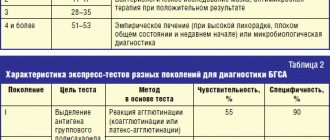
Essential hematuria (Table 2) combines a number of conditions in which the etiology and pathogenesis are unknown, and clinical, radiological and morphological studies do not allow us to accurately indicate the cause of bleeding.
Hematuria can be caused by taking non-narcotic analgesics, anticoagulants, cyclophosphamide, oral contraceptives, vincristine.
With kidney tumors, when the tumor grows in the pelvis or calyces, the integrity of the vascular wall is disrupted. If the tumor does not communicate with the pyelocaliceal system, the venous outflow from the area of the kidney with the tumor node is disrupted, the fornical veins dilate and rupture.
With prostate cancer, the tumor grows into the walls of the bladder or prostatic urethra, the veins of the bladder in its cervical section are compressed by the tumor, and venous stasis occurs. Free-floating villous tumors of the bladder, located near its neck, during urination are carried away by the flow of urine into the urethra, clog its lumen and, when pinched, swell, burst and begin to bleed.
With cystitis and prostatitis, the mucous neck of the bladder becomes inflamed, injured and bleeds easily when it comes into contact with other walls at the end of urination.
When renal hemo- and urodynamics are disturbed, the venous plexuses of the fornix, with impaired venous outflow or a significant increase in intrapelvic pressure, become overfilled with blood, the veins encircling the vaults of the calyces in a ring form expand and their integrity is disrupted, which leads to hematuria from the upper urinary tract.
With necrosis of the renal papillae, the blood supply to the papilla, and sometimes to the entire Malpighian pyramid, is disrupted, necrotic tissue is rejected, and bleeding occurs.
With benign prostatic hyperplasia, venous congestion occurs in the pelvic organs, and the integrity of the vessels is disrupted.
There are two types of hematuria:
- microscopic - the presence of red blood cells in the urine (more than three in the field of view) is determined only by microscopic examination of urine sediment;
- macroscopic - blood in the urine is determined visually; if only in the first portion of urine - initial (initial), in the last portion of urine - terminal (final), in all portions of urine - total.
Clinical picture
The appearance of red blood cells in the urine gives it a cloudy appearance and a pink, brown-red or reddish-black color, depending on the degree of hematuria. Hematuria can occur suddenly, be one-time or repeated. Its cessation is deceptive, since the cause of hematuria, as a rule, is not eliminated, and examination in case of repeated bleeding with a great delay often reveals an already advanced tumor.
With a kidney tumor, hematuria may be the first and for a long time the only symptom of the disease. In most cases, it is painless, but with profuse bleeding with the formation of clots in the renal pelvis and their passage through the ureters, dull, less often colicky pain occurs in the lumbar region. With papillary tumors of the bladder, hematuria is usually painless and profuse without the formation of clots. With villous tumors of the bladder located near its neck, the urine stream may be interrupted during urination.
With tuberculous kidney damage, total hematuria is observed in the very early stages of the process, which may be accompanied by persistent pyuria.
With benign prostatic hyperplasia, hematuria occurs for no apparent reason or during catheterization of the bladder due to a violation of the integrity of the loosened mucous membrane of the posterior urethra.
In acute cystitis and prostatitis, terminal hematuria is noted against the background of severe dysuria.
With urolithiasis, hematuria usually develops following an attack of renal colic; the urine stream may suddenly stop during urination.
Possible complications: with massive bleeding with the formation of shapeless clots, acute urinary retention is possible due to tamponade of the bladder with these clots. Rarely, shock may develop due to blood loss and pain.
Diagnostics
During the survey you need to find out:
- conditions for the occurrence of hematuria;
- degree, nature and duration of hematuria: - initial hematuria - a pathological process in the urethra (tumor, inflammation, foreign body, burn, etc.); – terminal hematuria - a pathological process in the neck of the bladder (acute cystitis, prostatitis, stones and bladder tumors); – total hematuria is a pathological process in the kidney, ureter or bladder (tumors, benign prostatic hyperplasia, renal tuberculosis, pyelonephritis, necrosis of the renal papillae, nephroptosis, etc.);
- the presence of blood clots in the urine, their shape: – worm-shaped – bleeding from the upper urinary tract and the formation of clots in the ureter; – shapeless clots – bleeding from the bladder;
- the presence of pain and its connection with changes in the color of urine: – pain in the lumbar region on the affected side – disruption of the passage of urine from the kidney due to blood clots, urolithiasis; – painless hematuria with subsequent development of renal colic (normal-colored urine) – tumor of the kidney or upper urinary tract;
- presence or absence of dysuric phenomena;
- whether there were injuries to the kidneys, bladder, urethra (in case of injury, blood is released from the urethra outside the act of urination).
Visual assessment of urine:
- scarlet blood - bleeding continues;
- brown urine - bleeding has stopped;
- brick shade - intense uraturia.
The color of urine may change when taking medications and foods:
- pink - when taking pyramidon;
- saffron yellow - nitroxoline;
- brown - rhubarb and hay;
- raspberry - purgena;
- red - phenolphthalein and beets;
- red-brown - madder dye.
With the syndrome of prolonged compression and crushing of tissues, myoglobin from the muscles enters the blood and penetrates into the urine, which gives it a red-brown color.
To determine the nature and source of bleeding in a hospital setting, the following is carried out:
- cystoscopy - in all cases of gross hematuria, with the exception of inflammatory diseases (acute urethritis, cystitis, prostatitis);
- digital rectal examination;
- Ultrasound of the bladder kidneys, prostate, transrectal ultrasound;
- excretory urography, retrograde reno- and urethrocystography, computed tomography, osteoscintigraphy according to indications in a hospital setting.
Laboratory diagnostics include:
- clinical analysis of blood, urine;
- blood chemistry;
- microscopic examination of urine sediment;
- urine culture for sterility;
- determination of the level of prostate-specific blood antigen.
With hemoglobinuria, the color of urine does not change even after standing for a long time, and with hematuria, red blood cells settle to the bottom of the vessel and the upper layers of urine become yellowish.
Hematuria in women should be differentiated from bleeding from the genitals. To do this, examine the average portion of urine during spontaneous urination or urine obtained from the bladder by catheterization.
Main directions of therapy
The emergency care algorithm for hematuria is presented in Figure 3.
| Figure 3. Emergency care algorithm for hematuria |
With the development of hypovolemia and arterial hypotension, restoration of bcc: crystalloid and colloid solutions intravenously.
If acute cystitis is suspected, after collecting urine for culture to ensure sterility, a broad-spectrum antibiotic is prescribed.
Therapy with hemostatic drugs should be carried out in a urological hospital, after diagnosis, under the control of blood clotting.
Clinical pharmacology of individual drugs
Etamsylate (dicinone) activates the formation of thromboplastin, the formation of blood coagulation factor III, and normalizes platelet adhesion. The drug does not affect the thrombotic time, does not have hypercoagulable properties, and does not contribute to the formation of blood clots. Etamsylate is administered in 2–4 ml (0.25–0.5 g) intravenously at once or drip, adding to conventional solutions for infusion.
The hemostatic effect develops:
- for intravenous administration - after 5–15 minutes; maximum effect - after 1–2 hours; the effect lasts 4–6 hours or more;
- when administered intramuscularly, the effect occurs somewhat more slowly;
- When taken orally, the maximum effect occurs after 3 hours.
Etamsylate should not be used for hemorrhages caused by anticoagulants.
Aminocaproic acid inhibits fibrinolysis by blocking plasminogen activators and partially inhibiting the effect of plasmin, and has a specific hemostatic effect in bleeding associated with increased fibrinolysis. To quickly achieve an effect, under the control of a coagulogram, a sterile 5% solution of the drug in saline is administered intravenously, up to 100 ml.
Protamine sulfate has an antihemorrhagic effect in bleeding caused by heparin during its overdose, after operations using extracorporeal circulation and the use of heparin. Apply 1 ml of a 1% solution intravenously in a slow stream over 2 minutes or drip; 1 mg of protamine sulfate neutralizes approximately 85 units of heparin. Contraindicated in hypotension, adrenal insufficiency, thrombocytopenia.
Common treatment errors: prescribing hemostatic drugs before identifying the source of hematuria.
Gross hematuria is an indication for emergency hospitalization in a urological hospital.
Analysis of clinical cases
Patient M., 30 years old. Complaints of cramping pain in the lumbar region on the left, aggravated by movement, after physical activity, blood in the urine, increased body temperature to 38.2°C. The disease began with dysuria in the form of frequent, painful urination, a day later pain appeared in the lumbar region, and blood in the urine. History: 7 years ago, a diagnosis was made of stage I nephroptosis on the left. The disease manifested itself as periodic attacks of pyelonephritis. On examination: the patient is restless. The kidney area is visually unchanged, painless on palpation, the effleurage symptom is weakly positive on the left, there is no pain along the ureters, the bladder is empty by percussion. Diagnosis: acute ascending left-sided pyelonephritis. Nephroptosis on the left. The patient was hospitalized in the urology hospital.
Patient J., 77 years old. Calling an emergency room due to intense blood in the urine and the release of blood clots during urination. History: no urological diseases. On examination: the patient is asthenic, the skin is pale. The kidney area is not visually changed; bimanual palpation of the right kidney reveals a mass formation in the lower segment; the effleurage symptom is negative on both sides. A digital rectal examination reveals that the prostate is enlarged 1.5–2 times, has a tight-elastic consistency, the median groove is smoothed, the rectal mucosa above the gland is mobile, palpation is painless. Diagnosis: tumor of the right kidney. The patient was hospitalized in the urology department.
Patient Ch., 24 years old. Complaints of constant bleeding through the urethra. History: About an hour ago I fell and hit my crotch on a pipe. On examination: bruising in the perineal area, discharge of scarlet unchanged blood from the urethra, outside the act of urination. A digital rectal examination reveals that the prostate gland is of normal size, has a tight-elastic consistency, the median groove is pronounced, the rectal mucosa above the gland is mobile, palpation is painless. Diagnosis: urethral injury. The patient is indicated for hospitalization in a hospital for examination and determination of treatment tactics.
Thus, the success of treatment of patients with pyelonephritis and hematuria is largely determined by the adoption of adequate measures in the provision of emergency medical care.
E. B. Mazo , Doctor of Medical Sciences, Professor, Corresponding Member of the Russian Academy of Medical Sciences, Russian State Medical University, Moscow
Table 1. Causes of renal bleeding
| Causes of hematuria | Pathological changes in the kidney, blood diseases, etc. |
| Congenital anomalies | Cystic diseases of the pyramids, papillary hypertrophy, nephroptosis, etc. |
| Mechanical | Trauma, stones, hydronephrosis |
| Hemodynamic | Disorders of the blood supply to the kidney (venous hypertension, infarction, thrombosis, phlebitis, aneurysms), nephroptosis |
| Hematological | Blood coagulation disorders, hemophilia, sickle cell anemia, etc. |
| Reflex | Vasoconstrictor disorders, shock |
| Allergic | Glomerulonephritis, arteritis, purpura |
| Toxic | Medicinal, infectious |
| Inflammatory | Glomerulonephritis (diffuse, focal), pyelonephritis |
| Tumor | Benign neoplasms, malignant neoplasms |
| Essential | (See Table 2) |
Table 2. Causes of essential renal hematuria
| Nature of the process | Immediate cause |
| Pathological processes | Pyelovenous reflux, calyceal venous channel, |
| in the papillary-fornical zone | venous varicose veins |
| kidneys (fornical bleeding) | |
| Diseases of the papillae and pelvis | Cystic processes in the papilla, “spongy kidney”, necrosis of the papillae, endometriosis, varicose veins |
| Pathological kidney mobility | Nephroptosis |
| Renal sinus diseases | Adhesions around the main renal vessels, sinus lipomatosis, aneurysms, varicose veins |
| Diseases of the renal interstitium | Microfibromatosis, interstitial hyperplasia with arterio- |
| Infectious and inflammatory | Pyelonephritis, necrotizing papillitis, hepatitis |
| processes | (hepatorenal syndrome) |
| Renal vascular damage | Infarction, venous thrombosis, compression of the main renal vein, varicose veins of the intrarenal veins, intraparenchymal aneurysms, angiomatous formations in Fornix |
| Blood diseases, dyscrasia | Sickle cell anemia, hemophilia, erythremia, |
| The harmful effects of drugs that disrupt the function of the blood coagulation system | Anticoagulant therapy, cytostatics |
Diseases of the urinary system
The second group of diseases, in which a person has lower back pain, nausea and dizziness, is associated with the kidneys, bladder and other organs of the genitourinary system. As a rule, in this case, the unpleasant symptoms are also accompanied by pain when urinating.
Infections and inflammations in the genitourinary system
As a rule, they are caused by the pathogenic activity of staphylococcus or streptococcus. This microorganism, entering the human body and beginning to actively multiply, can cause pyelonephritis
, pyelitis and other infectious and inflammatory diseases. Such conditions are usually treated with antibiotics.
Urolithiasis disease
It is associated with metabolic disorders and is often inherited. When kidney stones form, a person experiences periodic pain in the lower back, feels sick, and may have a fever. As a rule, people who lead a sedentary lifestyle and also have a history of diseases of the endocrine system are susceptible to the disease.
Lower back pain and woman nausea (gynecology)
Why does a woman have lower back pain and nausea? The answer to this question can most often be given by a gynecologist
. If the pain is regular, occurs at approximately the same time, and is successfully relieved with antispasmodics, then we can talk about the peculiarities of the menstrual cycle. Sometimes this condition occurs during pregnancy, regardless of the trimester. But in some cases, an inflammatory process may occur - for example, adnexitis, endometritis, colpitis, as well as oncology (ovarian or uterine cancer).
Lower back pain and nausea in a man (urology)
When a man experiences lower back pain and nausea, it can be assumed that he has urological diseases affecting the pelvic floor and organs of the genitourinary system. prostatitis, can manifest itself.
, as well as a number of other diseases: vesiculitis, inflammation of the bladder or ureters. Inflammatory processes often manifest themselves as a combination of the symptoms described above with fever and pain when urinating.
Signs of prostatitis
Manifestations of the disease can be either acute or almost invisible. If you notice the following symptoms, even minor ones, it is better to immediately consult a doctor to confirm or refute the diagnosis.
- Vomiting disorders: frequent urge, especially during night sleep, weak or intermittent stream, pain accompanied by difficulty urinating.
- Pain in the lower abdomen, which radiates to the anus, scrotum, perineum.
- Decreased libido, decreased potency or sexual dysfunction.
- Changes in consistency, color, amount of ejaculate, difficulty with ejaculation.
If you identify signs and symptoms of prostatitis in men, do not self-medicate and do not use recommendations from the Internet. At best, they will not help you, at worst, they can harm you. In addition, failure to see a doctor in a timely manner can worsen the condition.
Diagnostics
It doesn’t matter what disease is the basis of your ailment - if your lower back begins to tighten, a headache and fever appear, or you feel sick, then it is advisable to go to a qualified medical institution on the same day, or at most in the next 24 hours. At the Energo clinic you will be seen at the most convenient time. We employ experienced specialists who have extensive experience in diagnosing and treating pathological conditions of various types. All examinations are carried out using modern imported equipment, which allows you to quickly and accurately make a diagnosis and prescribe further therapy.
The influence of osteochondrosis on the functioning of the gastrointestinal tract
back pain caused by osteochondrosis indicates vertebral degeneration. This means that the nerve endings in this area are pinched and inflamed, which negatively affects the innervation of the digestive system. Thus, the deterioration of the stomach is a pattern.
What is the connection between back diseases and gastrointestinal tract?
The affected areas can be located in different parts of the spine. According to statistics, the thoracic vertebrae, from which nerves begin to emerge that affect the functioning of the gastrointestinal tract, are destroyed less often than others. The problem is that stomach pain that occurs in such a situation may be misdiagnosed. This leads to the prescription of erroneous treatment.
To trace the connection between stomach pain and osteochondrosis, one should take into account the characteristics of destructive processes. The list is as follows:
- Acute pain and a feeling of heaviness in the abdominal cavity - pinched nerves in the 5-8th thoracic vertebra;
- Gastralgia – right-sided pinching of nerve endings in the 9th thoracic vertebra;
- Pain in the duodenum - damage to the nerve endings in the 8th and/or 9th thoracic vertebra;
- Pain in the right hypochondrium is a right-sided pinching of the nerve endings in the 7-9th thoracic vertebra.
What causes stomach pain?
Many patients believe that the development of pathology is indicated by pain in the back and spinal column, as well as a decrease in the level of mobility. If the spinal nerves are affected, the blood supply to the organs of certain systems is disrupted. This can lead to vascular spasms, paralysis of nerve endings, or inflammation. The body gives signals about this. One of them is stomach pain caused by osteochondrosis.
In the described situation, it is necessary to prescribe appropriate treatment and its subsequent completion. If this is neglected, the patient’s condition will only worsen. Gastritis, colitis, or irritable bowel syndrome may occur.
Learn more about determining the cause of abdominal pain
Many people who have been diagnosed with a disease such as spinal osteochondrosis simply do not know that this disease can negatively affect the functioning of the gastrointestinal tract. They do not see any connection here and turn to a gastroenterologist, who may make an erroneous diagnosis.
In terms of intensity, pain in the stomach caused by osteochondrosis and those that indicate a disease of the gastrointestinal tract are identical. To find out the nature of their appearance, you should pay attention to the conditions under which they made themselves felt. In particular, it is worth remembering that stomach pain with osteochondrosis:
- They occur not after eating, but at the moment of movement, for example, during sharp turns;
- They are distinguished by their static nature if the patient remains in one position for a long time.
Diagnostics and preventive measures
As a rule, in order to make a correct diagnosis, the doctor sends a person suffering from osteochondrosis to undergo an X-ray or tomography of the spine. Based on the results of the procedure, you can learn about the condition of the affected areas, which facilitates the process of making a diagnosis and prescribing treatment.
In most cases, osteochondrosis develops in people accustomed to leading a sedentary lifestyle. Lack of movement leads to vertebral degeneration and subsequent stomach pain. To avoid all this, you should lead a healthy lifestyle, including proper nutrition and regular exercise.
It will be useful to know that sometimes an incorrect diagnosis made it possible to identify a really existing disease of the gastrointestinal tract.
Meanwhile, in modern conditions, the probability of such an event remains low, which guarantees timely relief from stomach pain due to osteochondrosis. Author: K.M.N., Academician of the Russian Academy of Medical Sciences M.A. Bobyr