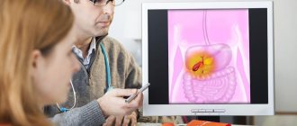
Gastroscopy and colonoscopy on the same day
The efficiency of same-day gastroscopy and colonoscopy has many potential benefits, as it can reduce costs, shorten hospital stays and speed up patient care. General indications for these endoscopy methods are hidden or obvious gastrointestinal bleeding, a history of relevant pathology (polyps, ulcers), unspecified abdominal pain and anemia, diarrhea or other symptoms, radiological studies. The practice of gastroscopy and colonoscopy on the same day is diagnostically more informative; the procedures can be performed even in elderly patients and does not carry increased risks, therefore it is offered when indicated.
Types of gastroscopy
Modern endoscopes are equipped with an instrument channel at the bottom of the handle, through which, if necessary, an instrument can be passed at any time to perform a biopsy, treatment or other manipulation. These replacement tools include:
- flexible forceps that are used to grasp a tissue sample;
- biopsy forceps to remove a tissue sample or suspicious lesion;
- a cytology brush, which is used to collect cell samples;
- pliers for removing sutures.
Endoscopy can be not only a diagnostic procedure, but also a therapeutic one; it can be used to:
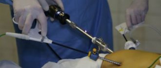
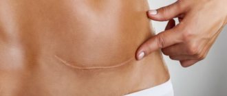
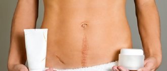
- laparoscopic surgery, making only small incisions in the skin;
- laser endoscopic therapy using laser radiation delivered from a laser device to the site of treatment via a light guide, which is passed through the working channel of the device [1];
- microwave ablation, which uses hyperthermic technologies for interstitial thermal ablation of tumors for their destruction and subsequent necrosis;
- surgical endoscopic resection of the mucosa;
- photodynamic therapy, in which, after injection of a light-sensitive drug into the tissue, tumor destruction is performed using a laser;
- administration of medications.
Transnasal gastroscopy
Transnasal gastroscopy is performed through the nose and is much easier to tolerate than the traditional method because the intranasal route of administration does not cause a gag reflex. This method is mainly used in elderly patients with a complex of concomitant diseases. Patients can speak if necessary during the procedure. This means that fewer patients require sedation and can continue their daily activities after the procedure.
Capsule gastroscopy
Capsule gastroscopy is a procedure that uses a tiny wireless video camera embedded inside a small capsule that the patient swallows. As the capsule passes through the digestive tract, the video camera captures an image at a frequency of 2 frames per second and transmits it to an external storage device, after which the doctor uses a special program to view and analyze the resulting images. This method is performed without swallowing the probe, as with traditional gastroscopy. The procedure is considered complete after the capsule leaves the body naturally. This method does not completely replace endoscopic gastroscopy, since many of its functions are not available to the capsule, for example, it is impossible to control the movement of the capsule, therefore, once in the stomach, it can take a picture of only part of one of the walls of the stomach, from which it is impossible to judge its condition . With capsule endoscopy, the large intestine is not straightened, and the capsule moves uncontrollably through the large intestine, which is large enough to be viewed; the images it transmits from this part of the intestine are absolutely uninformative and cannot in any way be analyzed by a doctor. Capsule endoscopy is intended only for examining the small intestine.
The most typical questions about preparing for endoscopy
Is smoking allowed before the procedure?
Nicotine increases the production of gastric juice, complicating the study. Smoking is strictly prohibited 3 hours before endoscopy.
Can I brush my teeth before the procedure?
Brushing your teeth is prohibited. During an endoscopy, the doctor measures the acidity of gastric juice. Brushing your teeth and drinking water can change the acidity and the measurements will be erroneous.
What can you drink before an endoscopy?
On the day of the study, you should not drink coffee, tea, juices, or carbonated drinks. If you are very thirsty (no later than 3 hours before the test), you can drink no more than 100 ml of pure water without gas.
How many hours should you not eat before the test?
If the procedure is scheduled for the morning, then the last meal should be the night before no later than 18:00. Only easily digestible foods are allowed. If an endoscopy is scheduled for the afternoon, then a light snack is allowed early in the morning. During the examination, the doctor carefully examines the gastric mucosa, so it should be free of food.
What food and when can you take before endoscopy?
The last meal of the day before the study should be no later than 6 pm. Food can be taken in small portions, warm, preferably with a puree-like consistency.
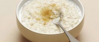
Allowed are oatmeal and buckwheat porridge, potato soup without fried ingredients, mashed potatoes, soft-boiled eggs, boiled lean fish, steamed white poultry cutlets, baked vegetables and fruits, and dried white bread.
How to get ready for research psychologically?
A calm psychological state and the right attitude on the day of the procedure are an important component of a successful study. Modern equipment allows you to perform EGDS as comfortably and painlessly as possible. Taking tissue samples (biopsy) will also not cause pain.
If during the procedure the patient experiences nervous tension, panic increases, and there is a feeling of lack of air, it is important for him to calm down, take a deep breath, and concentrate on breathing evenly. The gastroscope cannot prevent the flow of air; it is too thin for this. The examination will go faster if the patient tries to relax as much as possible during the procedure.
If the patient cannot cope with anxiety and this interferes with the study, at the discretion of the doctor, he can be put into medicated sleep. For 24 hours after sedation, the patient will not be able to drive a vehicle and will need an accompanying person to get home.
How long does the study last?
If endoscopy is performed using local anesthesia, the patient is relaxed, does not feel anxious and follows all the doctor’s recommendations, the endoscopic examination will take 3-5 minutes. Along with the preparation, the patient will stay in the endoscopy room for about 15 minutes.
When using medicated sleep, the procedure will take approximately 30 minutes.
The main criterion for the effectiveness of esophagogastroduodenoscopy is not the time spent on the procedure, but the thoroughness of the examination!
What anesthesia is usually used for endoscopy?
The insertion of an endoscope is often accompanied by a gag reflex. If the patient does not have an allergic reaction to anesthetics (lidocaine), then the endoscopist uses it to irrigate the upper part of the pharynx and the root of the tongue a few minutes before the procedure. The drug has a bitter taste and does not alter consciousness. The patient is awake, he sees and hears everything that happens during the examination, he may have a feeling of discomfort when swallowing.
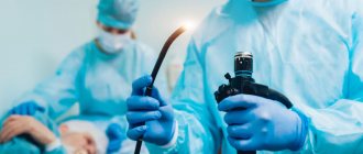
How is endoscopic examination performed?
EGD is performed by a certified endoscopist.
Before the examination, the doctor treats the upper part of the pharynx and the root of the patient’s tongue with an anesthetic to reduce gagging during insertion of the gastroscope.
The patient is then asked to clamp the mouthpiece between his teeth, which facilitates insertion of the sensor and protects the teeth from possible injury.
The patient is asked to breathe through the mouth and, if possible, not to swallow - this helps reduce the urge to vomit.
If the patient cannot cope with anxiety and interferes with the procedure, he may undergo endoscopy “in his sleep.”
After inserting the gastroscope, the endoscopist gradually moves the device through the mouth and pharynx towards the esophagus and stomach. When the gastroscope reaches the area required for diagnosis, the doctor straightens and cleans the mucous membrane, supplying air and water through a special channel of the device. This allows you to better visualize possible changes in the mucosa. The supply of air and water may be accompanied by a feeling of tension in the stomach.
If necessary, the doctor takes a biopsy. To do this, special forceps are used to take tissue samples from suspicious areas on the mucous membranes of the esophagus and stomach, after which they are submitted for histological examination. Taking samples is painless.
At the end of the study, the doctor removes the gastroscope, and his assistant frees the patient from the mouthpiece.
When can you drink and eat after endoscopy?
The patient may experience a feeling of numbness after using the anesthetic, so it is advisable to postpone drinking water and food for a while to avoid choking. 1-2 hours after the procedure, you can drink a few sips of water; if swallowing is restored and the process does not cause discomfort, you can start eating and drinking as usual.
If a biopsy was performed, then during the day after the procedure you can only eat chilled food.
Performing a gastroscopy procedure
Before gastroscopy, the mouth and throat are anesthetized using an aerosol. All dentures must be removed before the procedure. The patient may require a sedative. The time for gastroscopy is about 5-10 minutes, it may be longer, depending on the purpose of the procedure. Usually the procedure is performed in a lying position on the left side; in some exceptional cases, gastroscopy is performed while standing. A small tube or protective ring is placed between the teeth, leaving the mouth open, the patient is asked to take a sip so that the gastroscope can enter the esophagus, and it is then slowly advanced into the stomach and down to the entrance of the duodenum.
Using video images, the doctor controls the advancement of the probe and visualizes the area being examined. During gastroscopy, the monitor reveals bleeding, varicose veins, unusually narrow passages and stomach ulcers. If necessary, a tissue sample is taken for subsequent histological examination, which is not painful for the patient.