Gastroenterologists put forward 4 general theories for the appearance of formations in the gastrointestinal tract:
- Infectious - based on research in the field of gastroenterology, during which a relationship was identified between benign tumors of the gastrointestinal tract (as well as ulcers, gastritis) and the bacterium Helicobacter pylori.
- Genetic – for benign tumors, inheritance has been observed.
- Radiation – polyps appear under the influence of radiation.
- Chemical - the mucous membrane of organs is injured by aggressive substances entering the gastrointestinal tract with food and water.
Provoking factors are uncontrolled intake of drugs (medicines), the presence of a general inflammatory process in the gastrointestinal tract, unhealthy diet with the consumption of fatty, spicy foods, smoked foods, coffee, lemonade, and the presence of bad habits.
Gastrointestinal polyps differ in several ways:
- shape – mushroom or pear-shaped;
- size – average parameter 4mm;
- surface type – smooth, villous, lobulated, ulcerated;
- quantity – single, multiple.
The most common location is in the stomach, less often they appear in the rectum and colon, and even less often in the esophagus. Inflammatory growths develop asymptomatically and initially do not pose a threat. Over time, they can develop into cancerous tumors, so it is important to get diagnosed regularly.
Polyps in the stomach
Gastric polyps are epithelial or glandular growths of a benign nature, which are growths on the mucous membrane. They account for approximately 90% of all tumors of this kind. The number of males who face this disease is 2-4 times higher than the number of women. Formations begin to be detected during studies at 40-50 years of age.
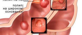
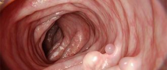
Externally, the outgrowths look like a mushroom cap or the top of a cauliflower and have one of two shapes - on a stalk or with a wide base. Large growths without a stalk require special attention from the point of view of transformation into cancer.
In 80% of patients, polyps are found in the antrum, but they are also found in others:
- pyloric polyp;
- body polyp.
Formations can appear singly (47% of cases), in groups (51% of diagnoses) or in the form of diffuse polyposis, which account for only 2% of the total number of diseases.
Histology divides them into 2 groups:
- Adenomatous - diagnosed in 5% of cases, consist of glandular tissue. They are the ones who transform into malignant tumors, especially when they grow beyond 2 cm in size.
- Hyperplasiogenic - diagnosed in other cases, consisting of epithelial cells. They always remain benign and do not develop into cancer.
Large adenomatous growths develop into malignant ones in 40% of cases, and tumors of any size of this type develop into cancer in 10-15% of patients. They are divided into three groups depending on which tissue cells they were formed from:
- tubular - tubular cells;
- papillary – papillary structures;
- papillotubular - two types of cells.
Symptoms of the disease are nonspecific, and only from the clinical picture can a large number of gastrointestinal pathologies be suspected. The intensity of symptoms depends on age and the number of formations. Newly formed polyps may not cause any pain or other symptoms. Sometimes a patient first develops gastritis and feels its symptoms, after which growths in the stomach are discovered during the examination.
If a large number of growths have already accumulated in the organ, tangible problems begin:
- feeling of bloating, frequent gases, including belching;
- excessive secretion of acid, and as a result, heartburn, excessive salivation, unpleasant taste and odor in the mouth;
- loss of appetite, change in diet, weight loss;
- discomfort in the part of the stomach where polyps have accumulated, pain that radiates to the back (under the shoulder blades most intensely);
- problems with stool - alternating diarrhea and constipation;
- general symptoms of a weakened body, possibly an increase in temperature.
The most acute symptoms appear during complications (they may be characteristic of ulcers and other gastrointestinal diseases). These include almost black stool of unusual thick consistency (indicating bleeding), severe dizziness (indicating anemia), vomiting blood (also a sign of bleeding). Require immediate medical attention.
Diagnosis and treatment
The task of diagnosis and treatment lies with the gastroenterologist. He gets acquainted with the patient's medical history, identifies possible heredity, carefully examines the patient, asks about the nature of the sensations and the period of their onset.
The next stage is laboratory and instrumental research:
- blood tests to detect complications;
- coprogram for searching blood;
- serological tests - detect Helicobacter pylori;
- gastroscopy;
- FGDS;
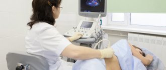
- Ultrasound;
- endoscopy with simultaneous biopsy;
- ultrasonography;
- MRI;
- CT.
Treatment begins with conservative methods - eliminating the disease that caused the polyps to develop (using medications and diet). The polyps themselves can only be cured by surgery. For these purposes, polypectomy, open surgery or resection are recommended.
Polypectomy is applicable if the diameter of the formation is no more than 5-30 mm. It is performed endoscopically using a metal loop through which an electric current is applied. Open surgery is necessary when the tumor has grown beyond 3 cm. Resection is used in extreme cases - when it becomes malignant or diffuse. The patient faces a large number of complications with this choice, but in order to save lives, this operation may be necessary.
After the operation, conservative treatment is reintroduced - the patient’s task is to recover and not provoke complications. From this point on, only steamed, boiled, baked and stewed foods are shown that do not contain large amounts of fat and sugar. You will also have to give up fruits and berries, baked goods, sweets, spices and sauces, sausages, smoked meats, and a lot of fiber. The timing and content of the diet is regulated by the doctor.
Surgical methods of treatment
Esophagogastroduodenoscopy (EGDS)
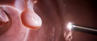
It is a diagnostic examination that allows the doctor to assess the condition of the inner lining of the stomach. It is carried out using an endoscope. During the procedure, any gastric tumors, including polyps, are effectively removed, wounds from them are cauterized, and bleeding can be clipped.
Endoscopic laser polypectomy
Soft tissues are evaporated layer by layer under the influence of a directed laser beam. The surrounding tissues are minimally injured. During the removal of tumors, the blood vessels are sealed, healing occurs in a very short time.
After removal of gastric polyps through any operation, the resulting material is sent for histological examination. It allows us to reveal the nature of education.
Polyps in the intestines
Polyps in the intestines, like stomach polyps, can interfere with the movement of the food bolus and blood circulation in the intestinal walls, and also develop into malignant tumors. The disease can manifest itself in both children and adults. In the early stages it develops asymptomatically, and the younger the patient, the higher the likelihood of asymptomatic development.
Common symptoms of the disease include:
- the presence of bloody spots in the stool;
- regular constipation, not justified by any external signs or food taken;
- pain in the lower abdomen, occurring with increased regularity;
- “tightness” in the intestines means that inflammation has begun.
It is important for a doctor who has received such complaints from a patient to differentiate the disease from angioma, uterine fibroids, actinomycosis, Crohn's disease and other gastrointestinal pathologies. To do this, you need to undergo a full diagnosis.
Diagnosis and treatment
In order to promptly identify growths in the gastrointestinal tract, you should make it a practice to donate stool every year to detect blood in it. This not only indicates the presence of polyps, but is also a good test for identifying the general condition of the gastrointestinal tract in asymptomatic diseases. However, the absence of blood does not mean the absence of polyps, therefore, if suspected, the following is carried out:
- finger examination;
- irrigoscopy (makes polyps visible on x-ray);
- colonoscopy.
Such studies are recommended to be carried out every 5 years. They allow you to identify a wide range of gastrointestinal diseases.
Treatment is only possible through surgical removal. Transanal excision is the most popular surgical option. It can be carried out if specific conditions regarding the location of the polyp are met. If they are located deeper than 10 cm from the anal sphincter, the use of this method is not possible. The second option is electrocoagulation. A colonoscope is used to grasp the polyp and separate it.
Other methods:
- transanal resection of the intestine - only for malignant tumors, requires removal of part of the intestine along with the formation;
- transanal endomicrosurgical excision - for large growths that previous methods cannot cope with;
- colotomy – requires an incision in the abdominal cavity, used when for some reason other methods are not available.
Polyps are always examined after separation to determine their histological nature and quality. If the biopsy sample turns out to be malignant, a repeat operation is performed to remove part or all of the intestine.
Possible complications, prevention
The long course of the pathology is associated with an increased risk of ulcerations and erosions due to the constant maceration of the formations by the food consumed. It should be taken into account that possible massive bleeding from the esophageal vessels poses a threat to the patient’s life.
With repeated low-intensity blood loss, iron deficiency hyporegenerative anemia occurs, accompanied by dizziness, general weakness, paleness of the skin and mucous membranes, and flickering of spots before the eyes.
The most dangerous complication of esophageal tumors is their malignancy with the formation of adenocarcinoma or esophageal cancer. Most often, malignancy is observed in adenomatous growths, in 0.25–7.1% of cases – hyperplastic ones.
In the later stages of the disease, due to the impossibility of adequate enteral nutrition, many patients may develop cachexia.
Specific measures to prevent the disease have not been developed. In order to prevent polyposis, it is recommended to promptly diagnose and treat esophageal pathologies, chew food well, avoid eating rough foods and limit alcohol consumption.
In the esophagus
The starting material for esophageal polyposis is epithelial cells. Most often, the disease affects middle-aged males. There are several places in the esophagus where outgrowths form especially often - in the area of the gastroesophageal sphincter, in the area of narrowings, on the mucous membrane of the lower part of the esophagus (this area is affected by more damaging factors). They are also characterized by two types of structure, like the others. A pedunculated polyp is dangerous because it can fall into the lumen of the pharynx (if it is close to it) or become pinched in the cardiac region.
Large growths rarely form in the esophagus. As a rule, they are discovered accidentally during research not related to the search for polyps, for example, during fibrogastroscopy. Heredity, concomitant diseases (esophagitis, gastritis, ulcers), and chronic stress predispose to the development of the disease. Injuries from hot and hard foods, alcohol, and cigarette smoke can also affect the formation of outgrowths. The injured area needs healing, and at some point the epithelial cells grow too much.
Symptoms depend on the number of formations, location and size. With large formations, a person feels a lump in the throat, compression in the chest, and heartburn. If the polyp is pinched in any way, pain may occur, especially while eating. As mentioned above, polyps are often discovered by chance - they are usually small in size and grow slowly.
If the formations still go unnoticed and manage to grow, the patient will face complications and serious symptoms, in particular:
- constant painful sensations;
- urge to vomit;
- severe pain when eating;
- loss of appetite;
- weight loss;
- irritability.
Diagnosis and treatment
The disease is diagnosed by one of three methods - examination and history taking, endoscopic examination, x-ray.
The results of the examination and survey allow one to suspect the presence of growths and prescribe appropriate diagnostic measures. Endoscopic examination successfully assesses the condition of the mucosa and is highly likely to detect formations. It allows not only to determine the condition of the mucosa, but also to take a biopsy sample for further examination for signs of malignancy.
If endoscopic examination is impossible for some reason, a decision is made to use radiography with contrast. Thanks to the contrast agent, the walls of the organ and all the protrusions and outgrowths on it are visible in detail.
Treatment, as in other cases, is surgical. It is necessary to start it as quickly as possible in order to avoid ulceration, internal bleeding, and pinching of the polyp, which causes pain. Removal is performed endoscopically using a loop, which cuts off the polyp and immediately “seals” the vessels to prevent bleeding. This method is also suitable for removing multiple polyps. It only takes 1-2 weeks to heal.
During the recovery period, you should consume warm, soft foods, steamed or boiled. You should not take sour, peppery, salty foods, hot or cold foods. Foods with fiber, fatty meat, and some cereals are excluded from the diet. A more detailed consultation on diet is provided by a gastroenterologist.


