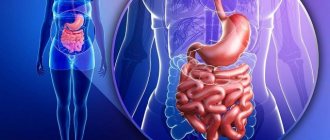
The digestive system is a complex of organs that are united by common functions. Thanks to it, a person has the opportunity to take food and assimilate it.
Snoring, apnea, insomnia or other problems? Contact the Sleep Medicine Center at the Rehabilitation Clinic in Khamovniki. We will definitely help you! Ask questions and schedule a consultation by phone.
This organ system includes the gastrointestinal tract and auxiliary structures: salivary glands, liver, gallbladder, pancreas.
Food is a source of energy and building material
To maintain his life, a person must eat food.
Food products contain all the substances necessary for life: water, mineral salts and organic compounds. Proteins, fats and carbohydrates are synthesized by plants from inorganic substances using solar energy. Animals build their bodies from nutrients of plant or animal origin. Nutrients that enter the body with food are building materials and at the same time a source of energy. During the breakdown and oxidation of proteins, fats and carbohydrates, a different but constant amount of energy is released for each substance, characterizing their energy value.
Digestion
Once in the body, food products undergo mechanical changes - they are crushed, moistened, split into simpler compounds, dissolved in water and absorbed. The set of processes by which nutrients from the environment pass into the blood is called digestion.
Enzymes play a huge role in the digestion process - biologically active protein substances that catalyze (accelerate) chemical reactions. During digestion processes, they catalyze reactions of hydrolytic breakdown of nutrients, but do not themselves change.
Main properties of enzymes:
- specificity of action - each enzyme breaks down nutrients only of a certain group (proteins, fats or carbohydrates) and does not break down others;
- act only in a certain chemical environment - some in alkaline, others in acidic;
- enzymes are most active at body temperature, and at a temperature of 70–100ºС they are destroyed;
- a small amount of enzyme can break down a large mass of organic matter.
Digestive organs
The alimentary canal is a tube that runs throughout the body. The canal wall consists of three layers: outer, middle and inner.
The outer layer (serous membrane) is formed by connective tissue that separates the digestive tube from surrounding tissues and organs.
The middle layer (muscular layer) in the upper parts of the digestive tube (oral cavity, pharynx, upper part of the esophagus) is represented by striated muscle tissue, and in the lower parts - smooth muscle tissue. Most often, the muscles are located in two layers - circular and longitudinal. Thanks to the contraction of the muscular membrane, food moves through the digestive canal.
The inner layer (mucosa) is lined with epithelium. It contains numerous glands that secrete mucus and digestive juices. In addition to small glands, there are large glands (salivary, liver, pancreas) lying outside the digestive canal and communicating with them through their ducts. The following sections are distinguished in the digestive canal: oral cavity, pharynx, esophagus, stomach, small and large intestines.
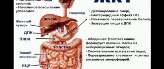
Diagram of the digestive tract as part of the digestive system:
|
Organs and functions of the human digestive system
Digestion is a combination of mechanical, chemical and enzymatic processing of food products included in the daily diet. The initial stages of this long process are represented by mechanical grinding, which greatly facilitates the subsequent digestion of nutrients. It is achieved mainly through the physical impact of the teeth, gums and oral cavity on each bite consumed. Chemical breakdown, in turn, acts more subtly and scrupulously: under the action of enzymes secreted by the glands of the digestive system, finely chewed food is broken down into its constituent ingredients, gradually breaking down into the initial nutrients - lipids, proteins and carbohydrates.
Each section of the digestive system has its own internal environment, which serves as the basis for the functions assigned to it. The organs of the gastrointestinal tract, together with the auxiliary glands, gradually break down each component of food, releasing what the body needs and sending the rest of the absorbed food to waste. If a failure occurs at any of these stages, organs and systems do not receive enough energy resources and, therefore, cannot fully perform their functions, causing an imbalance in the entire body.
The digestive system itself is conventionally divided into 3 key sections: anterior, middle and posterior. The processes of food digestion begin in the anterior section, represented by the oral cavity, pharynx and esophagus - here large pieces are crushed, softened by the incoming salivary fluid and pushed to the stomach. Chemical processing of food occurs in the middle section, which includes the stomach, intestines (large and small), as well as enzymatic organs - the liver and pancreas. It is in this area of the gastrointestinal tract that an optimal balance of microflora and pH is provided, due to which the main nutritional components are absorbed and residual masses are formed, the so-called ballast, which is subsequently released through the caudal part of the rectum. It is here, in the posterior gastrointestinal tract, that the digestive chain ends.
Digestion in the mouth
The oral cavity is the initial section of the digestive tract. It is bounded above by the hard and soft palate, below by the diaphragm of the mouth, and in front and on the sides by the teeth and gums.
The ducts of three pairs of salivary glands open into the oral cavity: parotid, sublingual and submandibular. In addition to these, there is a mass of small mucous salivary glands scattered throughout the oral cavity. The secretion of the salivary glands - saliva - moistens food and participates in its chemical changes. Saliva contains only two enzymes - amylase (ptialin) and maltase, which digest carbohydrates. But since food does not remain in the oral cavity for long, the breakdown of carbohydrates does not have time to complete. Saliva also contains mucin (a mucous substance) and lysozyme, which has bactericidal properties. The composition and quantity of saliva may vary depending on the physical properties of the food. During the day, a person secretes from 600 to 150 ml of saliva.
In the oral cavity, an adult has 32 teeth, 16 in each jaw. They grab food, bite it off and chew it.
Teeth consist of a special substance called dentin, which is a modification of bone tissue and has greater strength. The outside of the teeth is covered with enamel. Inside the tooth there is a cavity filled with loose connective tissue containing nerves and blood vessels.
Most of the oral cavity is occupied by the tongue, which is a muscular organ covered with mucous membrane. It is distinguished by the top, root, body and back, on which taste buds are located. The tongue is the organ of taste and speech. With its help, food is mixed during chewing and pushed through when swallowing.
Food prepared in the oral cavity is swallowed. Swallowing is a complex movement that involves the muscles of the tongue and pharynx. During swallowing, the soft palate rises and blocks the food from entering the nasal cavity. At this time, the epiglottis closes the entrance to the larynx. The bolus of food enters the pharynx, the upper part of the digestive canal. It is a tube, the inner surface of which is lined with mucous membrane. Through the pharynx, food enters the esophagus.
The esophagus is a tube about 25 cm long, which is a direct continuation of the pharynx. No food changes occur in the esophagus, since digestive juices are not secreted in it. It serves to carry food into the stomach. The movement of the food bolus through the pharynx and esophagus occurs as a result of contraction of the muscles of these sections.
The structure of the human digestive system
All organs related to the digestive system are most often classified based on their location, distinguishing the anterior, middle and posterior sections, which are described above. However, from a functional point of view, it is much easier to consider the digestive system as a complex of organs of the gastrointestinal tract, through which food passes the main path from a familiar dish to complete breakdown, and an enzymatic system responsible for the release of certain substances that greatly facilitate the movement and breakdown of food masses. Let's study each organ in this chain in more detail in order to clearly assess its importance in the complex mechanism of food digestion.
Main organs of the gastrointestinal tract
1. Oral cavity
The oral cavity is an opening through which food enters directly into the body in the familiar form of ready-made dishes on the daily menu. This includes the lips, teeth, tongue and salivary glands, which greatly facilitate the mechanical grinding process. The lips are the closing link and hold food in the oral cavity, the teeth cope with grinding larger and harder pieces, the tongue and gums grind small soft pieces, forming a food bolus, which is moistened with saliva and thanks to this easily passes into distant parts of the digestive tract.
The main function of mechanical grinding is performed by the dentition. 99.8% of newborn babies have no teeth, so they can only eat special homogenized food. However, by six months, as a rule, babies have one or even several milk teeth, which is a signal for the introduction of complementary foods - the child can already accept other products in addition to breast milk or adapted infant formula. As the number of teeth increases, the menu becomes more varied, and by the age of 10–12, when all milk teeth are replaced by permanent ones, the child can grind and digest food on the same basis as an adult.
However, not only the mechanical process of grinding food takes place in the oral cavity: other, much more significant functions are also performed here. The papillae located on the tongue allow you to evaluate the temperature, taste and quality of food, preventing possible poisoning from spoiled foods, thermal burns and damage to the mucous membrane. And the salivary glands secrete not only the liquid part of saliva, which softens the food bolus, but also enzymes, under the influence of which the primary breakdown of products occurs and their preparation for further digestion.
2. Throat
The pharynx is a funnel-shaped digestive tube that connects the oral cavity and the esophagus itself. Its only function is the swallowing process, which occurs reflexively. Its length is about 10 cm, which are divided approximately equally between the oropharynx, nasopharynx and laryngeal part. This is where the respiratory and digestive systems intersect, separated by the epiglottis, which normally prevents food from entering the lungs. However, if it is insufficient or spontaneous swallowing, this protective process is disrupted, which may result in asphyxia.
3. Esophagus
The anterior section of the gastrointestinal tract ends in a hollow tube about 25 cm long, the upper part of which is formed predominantly by striated muscle fibers, and the lower part by smooth ones. Thanks to this alternation, a wave-like contraction and relaxation occurs in the esophagus, which gradually moves the crushed food prepared for digestion into the stomach cavity. This process is the only significant function of the esophagus; no other physical, chemical or metabolic processes occur here.
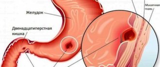
4. Stomach
The stomach looks like a hollow muscular organ located in the left hypochondrium. It is an extension of the esophagus with highly developed muscular walls that contract well, facilitating the digestion of food. Thanks to the coordinated work of muscle fibers, the shape and size of the stomach can change depending on eating habits and a certain phase of the digestive chain. For example, the empty stomach of an average adult has a volume of no more than one and a half liters, but after eating it can easily increase to 3 or even 4 liters, that is, more than 2 times.
The same applies to people prone to frequent overeating: regular consumption of large portions leads to overstretching of muscle fibers, which is why the walls of the stomach become flabby and the overall volume increases. This, in turn, causes disruption of eating habits and contributes to the accumulation of excess weight. Therefore, all nutritionists without exception recommend eating often, but in fractional portions: such a diet is more physiological.
During swallowing, the muscles that form the walls of the stomach relax, allowing the bolus of food, or chyme, as it is called in nutrition, to pass through. This happens until the meal is over (or the stomach is full), after which the walls contract again - this is how the metabolic process begins. Under the pressure of peristalsis, the chyme is mixed, ground and loosened, exposed to gastric juice. The acidic component of the internal environment of the stomach is produced in the folds of the mucous membrane, where special secretory glands are located. The food is gradually saturated with this secretion, crushed, becoming softer and looser, which contributes to its rapid decomposition into molecules.
Then special enzymes in the gastric juice - proteases - begin the process of breaking down protein structures. However, the process does not end with this; in the stomach, proteins are only prepared for complete decomposition, breaking down into complex multicomponent substances. In addition, the breakdown of emulsified lipids into glycerols and fatty acids occurs here, and the metabolism of starches is completed.
The composition and concentration of gastric juice directly depends on a person’s eating habits. Thus, the largest amount of it is synthesized in response to protein foods, and the smallest – to fatty foods. This is why lipids are much more difficult to break down and more often lead to excess weight than other substances included in the diet.
5. Small intestine
The small intestine is the longest part of the human digestive system. Its total length can reach 5–6 meters, which fit into the abdominal cavity only thanks to a thoughtful loop-like arrangement. The following areas are distinguished in the small intestine:
- duodenum (about 30 cm),
- jejunum (about 2.5 meters),
- iliac (2.5–3.5 m).
Starting from the pylorus of the stomach, up to the colon, the lumen of the small intestine is constantly narrowing. Peristaltic contraction gradually propels the chyme, continuing to break it down into nutrient molecules. Here the food bolus is mixed several more times, softened and gradually absorbed by the cells of the mucosa.
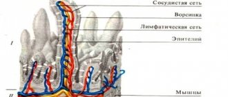
The inner side of the small intestine has many circular folds, within which numerous villi are hidden. Thanks to this, the total area of the mucous membrane increases several times, which means that the absorption capacity of the intestine also increases. Each villi has its own network of lymphatic and blood capillaries, through the thin walls of which molecules of proteins, fats and lipids leak into the blood, spreading throughout the body and forming an energy depot. This allows you to get the maximum amount of nutrients from the food you consume.
6. Large intestine
The large intestine completes the digestive chain. The total length of this intestine is about one and a half meters, from which at the very beginning a small blind appendix extends - the appendix. A very small organ is a kind of pouch, which in some cases can become inflamed and cause an acute condition requiring immediate surgical intervention.
Under the influence of the mucus of the large intestine, the absorption of certain vitamins, glucose, and amino acids synthesized by flora microorganisms occurs. In addition, most of the fluid and electrolytes necessary to maintain water balance in the body's cells are absorbed here.
The final section of the intestine is the rectum, ending in the anus, through which the body leaves unnecessary substances formed into feces. If the entire digestive process is not disturbed, in total it takes about 3 days, of which 3–3.5 hours are spent delivering chyme to the colon, another 24 hours for filling it and a maximum of 48 for emptying.
Accessory organs of the digestive system
1. Salivary glands
The salivary glands are located in the oral cavity and are responsible for the synthesis of an enzymatic fluid that moistens food and prepares it for breakdown. This organ is represented by several pairs of larger glands (parotid, sublingual, submandibular), as well as numerous small glands. Human saliva normally contains a watery and mucous secretion, as well as enzymes that provide the initial chemical breakdown of the products included in the dishes consumed.
Normally, the following enzymes are present in salivary fluid:
- amylase breaks down starches into disaccharides,
- maltase completes this process by converting disaccharides into glucose molecules.
The concentration of these enzymes is usually very high, since food remains in the mouth for an average of 18–23 seconds before being swallowed. However, this time is not always enough, so gastroenterologists recommend chewing each piece thoroughly and for a long time, then the starches will have time to completely break down, and the food itself will become softer and more homogeneous.
2. Pancreas
The pancreas is another auxiliary enzymatic organ that synthesizes substances necessary for the complete digestion of nutrients. Its cells produce pancreatic juice, which contains all the necessary chemical compounds for the preparation and subsequent breakdown of lipids, proteins and carbohydrates. In addition, the pancreatic juice contains pancreatic substance, which is produced by duct cells. Due to bicarbonate ions, this liquid neutralizes the acidic component of residual digestive products, thereby preventing irritation and damage to the mucous membranes.
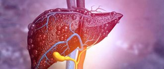
3. Liver
Due to its versatility, the liver belongs to several systems of the body, one of which is the digestive system. In liver cells, amino acids, free fatty acids, lactic acid and glycerol are transformed into glucose, which serves as an energy reserve for the human body. In addition, the liver plays a key role in neutralizing toxic compounds that enter the digestive system. This protective reaction prevents the serious consequences of food poisoning and cleanses the gastrointestinal tract of harmful components that have entered the body.
4. Gallbladder
Anatomically, the gallbladder is an appendage of the liver, in which a supply of bile accumulates in case of urgent need for the body. When a large amount of food is received, especially harmful food (fatty, fried, smoked, etc.), the accumulated bile is released into the lumen of the small intestine to support and speed up metabolic processes. However, such a mechanism is not always necessary, so the flow of bile is clearly dosed using valves and bile ducts and increases only if food that is difficult to break down enters the gastrointestinal tract.
Summary
Human digestion is a complex and delicate mechanism, the quality of which directly depends on the proper functioning of each organ, each cell that forms this system. Such a balance is possible only if you treat your own digestive tract with care and sensitivity. You should not overload it with exorbitant portions, fatty, heavy and fried foods, meat products, which pollute the body and do nothing but harm, and then you will not be bothered by metabolic problems, and the body will always be provided with a sufficient amount of energy without the risk of deficiency or, conversely , excess accumulation of body fat and excess weight. Take care of the correct diet today, and tomorrow you won’t have to go to a gastroenterologist and waste time on expensive and sometimes ineffective treatment of the digestive system!
Digestion in the stomach
The stomach is the most expanded section of the digestive tube with a capacity of up to three liters. The size and shape of the stomach changes depending on the amount of food taken and the degree of contraction of its walls. At the point where the esophagus flows into the stomach and where the stomach passes into the small intestine, there are sphincters (squeezers) that regulate the movement of food.
The mucous membrane of the stomach forms longitudinal folds and contains a large number of glands (up to 30 million). The glands consist of three types of cells: main (producing enzymes of gastric juice), parietal (secreting hydrochloric acid) and accessory (secreting mucus).
Contractions of the stomach walls mix food with juice, which promotes better digestion. Several enzymes are involved in the digestion of food in the stomach. The main one is pepsin. It breaks down complex proteins into simpler ones, which are further processed in the intestines. Pepsin acts only in an acidic environment, which is created by hydrochloric acid in gastric juice. Hydrochloric acid plays a major role in the disinfection of stomach contents. Other gastric juice enzymes (chymosin and lipase) are able to digest milk protein and fats. Chymosin curdles milk, so it stays in the stomach longer and undergoes digestion. Lipase, present in small quantities in the stomach, breaks down only the emulsified milk fat. The action of this enzyme in the stomach of an adult is weakly expressed. There are no enzymes that act on carbohydrates in gastric juice. however, a significant portion of the food's starch continues to be digested in the stomach by salivary amylase. The mucus secreted by the glands of the stomach plays an important role in protecting the mucous membrane from mechanical and chemical damage and from the digestive action of pepsin. The glands of the stomach secrete juice only during digestion. In this case, the nature of juice secretion depends on the chemical composition of the food consumed. After 3-4 hours of processing in the stomach, the food gruel enters the small intestine in small portions.
What work does the digestive system do?
Conventionally, all functions assigned to the human digestive system can be divided into 4 key categories:
- Mechanical. This stage involves grinding the incoming food products for further breakdown and processing.
- Secretory. This function is rather complex and consists in the production of enzymes necessary for digestive processes - gastric and intestinal juices, bile, saliva.
- Suction. After products are broken down into nutrient molecules, the food chain does not end; it is still necessary for them to be absorbed in the gastrointestinal tract and be able to perform the functions assigned to them - energy supply, metabolism, various physiological processes, etc.
- Excretory. Not everything that comes with food is equally beneficial to the body. In the digestive tract, the necessary beneficial substances are filtered out, and the remaining part is formed into feces and excreted from the body.
All these functions are performed in stages: first, the food is crushed and softened by the liquid part of saliva, then it is split into various substances, the useful part of which is absorbed by the body, and the ballast part is excreted. At the slightest failure at any of these stages, this chain is interrupted, and in this case several outcomes are possible, each of which is associated with certain complications. Either the body does not receive enough nutritional components, suffering from a lack of energy resources, or the unfulfilled functions are compensated by other parts of the digestive system, which sooner or later causes even more serious problems. Therefore, it is very important to know how well each organ that is part of the digestive system performs its assigned function; not only complete digestion, but also the health of the body as a whole depends on this.
Small intestine
The small intestine is the longest part of the digestive tube, reaching 6–7 meters in an adult. It consists of the duodenum, jejunum and ileum.
The excretory ducts of two large digestive glands - the pancreas and liver - open into the initial section of the small intestine - the duodenum. Here the most intensive digestion of food gruel occurs, which is exposed to the action of three digestive juices: pancreatic, bile and intestinal.
The pancreas is located behind the stomach. It distinguishes between the apex, body and tail. The apex of the gland is surrounded in a horseshoe shape by the duodenum, and the tail is adjacent to the spleen.
Gland cells produce pancreatic juice (pancreatic). It contains enzymes that act on proteins, fats and carbohydrates. The enzyme trypsin breaks down proteins into amino acids, but is active only in the presence of the intestinal enzyme enterokinase. Lipase breaks down fats into glycerol and fatty acids. Its activity increases sharply under the influence of bile produced in the liver and entering the duodenum. Under the influence of amylase and maltose in pancreatic juice, most food carbohydrates are broken down into glucose. All pancreatic juice enzymes are active only in an alkaline environment.
In the small intestine, food gruel undergoes not only chemical, but also mechanical processing. Thanks to the pendulum-like movements of the intestine (alternate lengthening and shortening), it mixes with digestive juices and liquefies. Peristaltic movements of the intestines cause contents to move towards the large intestine.
The liver is the largest digestive gland in our body (up to 1.5 kg). It lies under the diaphragm, occupying the right hypochondrium. The gallbladder is located on the lower surface of the liver. The liver consists of glandular cells that form lobules. Between the lobules there are layers of connective tissue in which nerves, lymphatic and blood vessels and small bile ducts pass.
Bile, produced by the liver, plays a large role in the digestion process. It does not break down nutrients, but prepares fats for digestion and absorption. Under its action, fats break up into small drops suspended in liquid, i.e. turn into an emulsion. In this form they are easier to digest. In addition, bile actively influences absorption processes in the small intestine, enhances intestinal motility and the secretion of pancreatic juice. Despite the fact that bile is produced continuously in the liver, it enters the intestines only when eating. Between periods of digestion, bile is collected in the gallbladder. Through the portal vein, venous blood flows into the liver from the entire digestive canal, pancreas and spleen. Toxic substances that enter the blood from the gastrointestinal tract are neutralized here and then excreted in the urine. In this way, the liver carries out its protective (barrier) function. The liver is involved in the synthesis of a number of important substances for the body, such as glycogen, vitamin A, and influences the process of hematopoiesis, the metabolism of proteins, fats, and carbohydrates.
Test your knowledge
If you want to test your knowledge on the topic of this lesson, you can take a short test consisting of several questions. For each question, only 1 option can be correct. After you select one of the options, the system automatically moves on to the next question. The points you receive are affected by the correctness of your answers and the time spent on completion. Please note that the questions are different each time and the options are mixed.
Statistics Full screen
Kirill Nogales
← Proper nutrition Proteins, fats, carbohydrates →
Nutrient Absorption
In order for the amino acids, simple sugars, fatty acids and glycerol resulting from the breakdown to be used by the body, they must be absorbed. These substances are practically not absorbed in the oral cavity and esophagus. Water, glucose and salts are absorbed in the stomach in small quantities; in the large intestines - water and some salts. The main processes of nutrient absorption occur in the small intestine, which is quite well adapted to carry out this function. The mucous membrane of the small intestine plays an active role in the absorption process. It has a large number of villi and microvilli, which increase the absorption surface of the intestine. The walls of the villi contain smooth muscle fibers, and inside them there are blood and lymphatic vessels.
Villi take part in the absorption of nutrients. By contracting, they promote the outflow of blood and lymph, rich in nutrients. When the villi relax, fluid from the intestinal cavity again enters their vessels. The products of the breakdown of proteins and carbohydrates are absorbed directly into the blood, and the bulk of digested fats are absorbed into the lymph.