Gastric adenocarcinoma is one of the most common types of cancer in this location. It accounts for up to 95% of all malignant neoplasms of the stomach. The disease is insidious in that in the initial stages it does not manifest itself in any way, then pain, nausea, loss of appetite occur, and against the background of these symptoms, weakness and weight loss increase. The only radical treatment for adenocarcinoma today is surgery. Radiation therapy and chemotherapy are prescribed as additional treatment or when surgery is not possible.
- Reasons for development
- Types of gastric adenocarcinoma
- Symptoms
- Diagnosis of gastric adenocarcinoma
- Stages of adenocarcinoma
- Diagnostics
- Metastasis of gastric adenocarcinoma
- Treatment of gastric adenocarcinoma
- Prognosis of gastric adenocarcinoma
- Prevention
Reasons for development
The cause of malignant transformation of mucosal cells has not been fully identified, but scientists have identified several factors that reliably increase this likelihood:
- Chronic atrophic gastritis. Against this background, up to 60% of all cases of stomach cancer develop. When the atrophic process is localized in the body of the stomach, the risk increases by 3-5 times, in its antral part - by 18 times, and if the entire stomach is affected - by 90 times.
- Malnutrition - consumption of large amounts of spicy and salty foods, marinades, smoked meats, dried meat, as well as fats, especially heat-treated ones. The nature of food intake matters. Overeating and insufficiently thorough chewing of food can lead to chronic trauma to the stomach wall and, against this background, to malignant degeneration of cells.
- Infection with the bacterium Helicobacter pylori increases the risk by 3-4 times.
- Smoking and alcohol abuse.
- A history of gastric surgery. The risk of developing cancer increases 4 times.
- Pernicious anemia is a pernicious anemia associated with the inability to absorb vitamin B12. Against this background, a decrease in immunity occurs, which in 10% of cases leads to the development of cancer.
- Immunodeficiency conditions, including AIDS.
- Hereditary predisposition, according to various authors, increases the risk by 5-20%. The scientific literature describes cases of inheritance of gastric adenocarcinoma.
- Working with harmful production factors (nickel, asbestos).
- Stomach ulcer. When it is located in the body of the stomach, the risks of malignancy increase by 2 times, but when the antrum is affected, this pattern is not observed.
Possible complications
Complications can occur with any type of highly differentiated adenocarcinoma lesion. The appearance of complications is primarily due to the impact of the lesion as it grows. Large tumors can damage organs. Thus, a rupture of the intestinal wall is possible, leading, in turn, to intestinal bleeding. In addition, the specific stage of the oncological process also has a negative impact. Everything is simple here: the further the tumor has gone in its development, the higher the likelihood of damage to nearby organs. The growth of oncological components also plays a detrimental role. For example, increased pathogenic cells in colorectal cancer lead to intestinal obstruction. Also, it is worth remembering concomitant pathologies that can complicate the course of cancer.
Types of gastric adenocarcinoma
According to the modern classification, the following types of adenocarcinomas affecting the stomach are distinguished:
- Papillary - visually resembles a polyp.
- Tubular. Grows from cubic or columnar epithelial cells.
- Mucinous. Grows from mucus-producing cells.
According to the degree of differentiation, the following forms are distinguished:
- Well-differentiated adenocarcinoma of the stomach. Tumor cells have a high level of differentiation and “resemble” healthy tissue. It is characterized by slow growth and, with timely diagnosis, is highly treatable. The main problem is that at the very beginning this tumor is difficult to detect, even with endoscopic examination, and its detection at later stages does not allow achieving the desired result.
- Moderately differentiated gastric adenocarcinoma. The cells of this tumor are difficult to identify with the tissue from which it grows. It is characterized by moderate malignancy and growth rate.
- Poorly differentiated gastric adenocarcinoma. The cells of a given tumor cannot be identified with a specific tissue. This is the most malignant type of stomach cancer. Characterized by rapid growth and metastasis.
What's happened
Adenocarcinoma is a malignant tumor developing from secretory glandular epithelial cells. It is localized on various internal organs, on the internal membranes of hollow organs, as well as human skin.
A distinctive feature of adenocarcinoma is the ability to produce secretions.
Such tumors can have a variety of sizes and shapes, which are directly dependent on the cellular and tissue functions, cellular and tissue structure of the organ that was affected.
Symptoms
- Loss of appetite.
- The symptoms of dyspepsia are bloating, nausea, belching, a feeling of heaviness. If the tumor is large, it can block the lumen of the stomach, which causes vomiting when eating, but this happens in the last stages.
- Weakness and apathy.
- Pain. As a rule, it does not occur immediately, but mainly at advanced stages. At this time, it is permanent and does not depend on food intake. It occurs for no apparent reason and often worsens after eating. In the final stages, it is so strong that it cannot be stopped with the usual analgesics.
As we can see, these symptoms are nonspecific, and a person can try to stop them on their own for a long time.
Diagnosis of gastric adenocarcinoma
Timely diagnosis of stomach cancer is of utmost importance, since the prognosis in the early stages of the disease is much more favorable than with a widespread tumor process. But adenocarcinoma in the initial stages is characterized by either an asymptomatic course, or its symptoms are perceived as signs of another disease, for example, gastritis, ulcers, cholelithiasis, pancreatitis. As a result, patients do not go to the doctor and relieve symptoms on their own using medications purchased at the pharmacy.
What is the danger
In 90% of cases, it metastasizes to nearby and distant organs. Often the disease is diagnosed at the 4th stage of progression, when surgery and chemotherapy are no longer effective. General indicators of a progressive disease are:
- pain in the so-called epigastric region, - a change in taste preferences (for example, there is an aversion to any dishes containing protein), - pathologically active salivation, - frequent vomiting (particular attention should be paid to vomiting with blood, because it is an important indicator stomach cancer), blood impurities in the stool, change in its consistency.
However, even with active progression, cancer can proceed silently. Doctors call such oncological lesions “silent”; they are fraught with the greatest danger. Among the indirect symptoms of the disease in question are a mild manifestation of anemia, general weakness, and an increase in body temperature to 37.5 degrees. Patients are often tormented by prolonged depression.
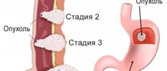
Stages of adenocarcinoma
- Stage 0 or cancer in situ. Malignant cells do not spread beyond the epithelial layer.
- Stage 1. The tumor grows into the epithelium and spreads to other layers of the stomach wall, but does not go beyond it.
- Stage 2. The tumor increases in size and metastasizes to regional lymph nodes.
- Stage 3. Adenocarcinoma grows into neighboring organs (pancreas, liver) and/or metastasizes to several groups of lymph nodes.
- At the fourth stage, adenocarcinoma metastasizes to distant organs and lymph nodes.
Classification
Adenocarcinoma is usually classified according to the location (organ) of the lesion, according to its morphological characteristics, and according to its histological characteristics.
By location of the lesion (organ)
Adenocarcinoma very often affects organs such as:
- Lungs,
- Mammary gland,
- pancreas,
- prostate gland,
- Esophagus,
- Intestines.
According to histological characteristics
There are three types of adenocarcinoma:
G1 (Highly differentiated)
This type of adenocarcinoma is characterized by low-grade malignancy. The morphology of such adenocarcinoma: normal and not quite normal cells that can multiply quickly.
G2 (Moderately differentiated)
The composition of this adenocarcinoma is malignant cells that are very different from healthy ones and can multiply quickly. The nature of these cancers is more aggressive.
G3 (Low differentiated)
Cells of this type of adenocarcinoma are characterized by a high degree of malignancy. They are characterized by complete loss of signs of normal cells. They reproduce actively, unpredictably and haphazardly.
According to morphological characteristics
Based on this criterion, adenocarcinoma is divided into the following subtypes:
Acinar
This adenocarcinoma is a tumor of the prostate gland, is diagnosed more often than others and has a long asymptomatic course. During the study, tissue differentiation is clearly visible: cancer cells of various sizes with nuclei and cytoplasm.
Small-acinar
This is a type of cancer found in the peripheral (most often) prostate gland or transition zone of the prostate. It consists of small acini with structural disturbances in several places.
Mucinous
Adenocarcinoma with different types of cells, characterized by the presence of a cavity containing mucin. In this type of tumor, mucus is produced in large quantities and comes to the surface.
Dark cell
Cancer, inside the cells of which there are polymorphic nuclei with a predominant dark color. The structure is formed from these cells and cytoplasm. It has a glandular appearance. Typically, this subtype of adenocarcinoma progresses quickly, but it all depends on the differentiation of the neoplasm.
Serous
Cancer is formed from differentiated polymorphic and cellular structures. The nature of this tumor is quite aggressive.
Clear cell
Such adenocarcinoma has a cystic-tubular structure and may have a glandular structure. This cancer also develops rapidly, and metastases can often occur.
Tubular
The structure of such adenocarcinoma is cylinders, tubes and cubes (tubular structures). They are localized in fibrous connective tissue.
Follicular
The structure of such adenocarcinoma is colloidal particles. The size of the tumor is inversely proportional to the progression of the lesion: the larger the tumor, the slower the progression and vice versa. Metastases in this subtype of cancer spread through the bloodstream to various parts of the body.
Papillary (papillary)
The structure of this subtype of cancer is similar to papillae or papillomas. This type of adenocarcinoma has a good prognosis in most cases if treated on time.
Invasive
Characterized by the penetration of cancer cells into nearby organs and tissues. If suddenly such a carcinoma enters the blood and lymph, then the spread to parts of the body becomes hematogenous.
Diagnostics
Diagnosis of gastric adenocarcinoma is complex and includes a number of examinations that not only help determine the type of tumor, but also the stage of the disease:
- FGDS is an examination that is carried out using a special device - a flexible endoscope. With its help, a visual examination of the gastric mucosa is carried out, and the device transmits an enlarged image of the area under study to the monitor, which makes it possible to detect minor changes in the mucosa. Secondly, the endoscope is equipped with a special manipulation system, with which you can take a piece of tissue for histological analysis and accurately determine the type of tumor and the degree of differentiation of its cells.
- Ultrasound. This research method makes it possible to clarify the size of the tumor, its relationship with neighboring organs, and the presence of metastases in regional lymph nodes.
- CT and MRI also help verify the size of the malignant tumor and its ingrowth into surrounding tissues, but the main goal is to look for metastases in the lymph nodes and distant organs (for example, the lungs).
- PET-CT (positron emission computed tomography) can detect distant metastases up to 1 mm in size.
- Determination of tumor markers CEA, CA72-4, CA19-9. In gastric cancer, these markers do not have diagnostic value as such, but if the norm is initially elevated, they can be used to monitor treatment and relapse. After surgery, the level of tumor markers should decrease, it may reach normal. If a relapse or progression of the disease occurs, it will increase again.
Dr. M.S. Burdyukov conducts a diagnostic examination of the stomach - FGDS
Diagnosis
The process of identifying highly differentiated carcinoma includes collecting anamnesis and carrying out standard diagnostic measures: detailed blood and urinary analysis, stool analysis, ultrasound examination of the area of possible localization of the lesion, removal of biomaterial for subsequent laboratory study. If there is a suspicion of intestinal cancer, a digital examination of the rectum is performed. In addition, a colonoscopy is performed to assess the condition of the mucous membrane of the inner border of the large intestine. Another important procedure is irrigoscopy (x-ray of the large intestine).
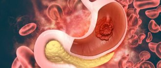
Metastasis of gastric adenocarcinoma
Adenocarcinoma is characterized by lymphogenous, hematogenous and implantation metastasis.
Implantation metastasis involves the spread of cells through contact of a tumor with a host surface. In the case of stomach cancer, such a surface may be the peritoneum, pleura, pericardium, or diaphragm. At the fourth stage, carcinomatosis (massive metastasis to various tissues) is often detected.
Lymphogenous metastasis involves the spread of the tumor through the lymphatic vessels. First, nearby lymph nodes are affected, and then more distant ones. For stomach cancer, there are specific forms of lymphogenous metastases:
- Damage to the nodes of the left supraclavicular region is Virchow's metastases.
- Damage to pararectal lymph nodes—Schnitzler metastases.
- Damage to axillary lymph nodes—Irish metastases.
Hematogenous metastasis involves the transfer of cancer cells through blood vessels. The liver is most often affected as a result of cell migration through the portal vein. Damage to the lungs, kidneys, brain, and bone marrow is also possible.
Symptoms
At the latent stage of the disease, there are no symptoms.
The first symptoms appear due to tumor growth:
- Pain in the area where the tumor is located;
- Presence of blood clots;
- The appearance of constipation;
- Growth of lymph nodes;
- Weight loss;
- Decreased hemoglobin;
- Fatigue;
- Low ability to work;
- Bad dream.
At the stage of intensive growth of cancer cells and the appearance of metastases, the severity of symptoms increases.
Let's look at the symptoms of the formation of adenocarcinoma in various organs.
Intestines
- Painful stomach;
- Unpleasant sensations after eating;
- Poor intestinal permeability;
- Loose stools alternating with constipation;
- Blood and mucus in the stool.
Esophagus
- Pain when swallowing;
- Dysgaphia;
- Active salivation.
Nasal cavity
- Swelling of the tonsils;
- Persistent inflammation of the tonsils;
- Pain in the larynx, pharynx, nose;
- Unpleasant sensations when swallowing;
- Ear pain;
- Speech impairment;
- Enlarged lymph nodes.
Liver
- Pain in the area of the right rib and hypochondrium;
- The eyes and skin are yellowish.
Treatment of gastric adenocarcinoma
Treatment of adenocarcinoma depends on the stage of the disease and the histological type of the tumor. As a rule, it is complex and involves a combination of surgery with chemotherapy or radiation therapy. The surgical component here is the key factor.
Treatment may involve removing the entire stomach (gastrectomy) or part of it (gastrectomy). At the same time, tissues affected by malignant cells are removed - regional lymph nodes, parts of organs where the tumor has grown (liver, small intestine, peritoneum, etc.).
Chemotherapy and radiation therapy can be used preoperatively (neoadjuvant regimen) and postoperatively (adjuvant regimen). In the first case, their goal is to reduce the size of the tumor so that it can be removed with the least amount of tissue, and in the second, they aim to destroy the remaining cancer cells. In addition, the use of chemotherapy and radiation therapy can reduce the severity of pain.
If radical removal of the formation is not possible, palliative treatment is carried out. In this case, it is aimed at eliminating complications caused by adenocarcinoma and improving the patient’s quality of life. For example, if a tumor has blocked the lumen of the stomach, bypass anastomoses are performed or a gastrostomy tube is removed, so that the patient can eat.
Prognosis of gastric adenocarcinoma
The prognosis for adenocarcinoma depends on the stage of the disease. The sooner treatment begins, the more effective it will be:
- In the first stage, the five-year survival rate reaches 80%. Moreover, the chances of a full recovery are high. Unfortunately, at this stage, stomach cancer is detected very rarely, usually by chance.
- In the second stage, the five-year survival rate approaches 55%. Half of these people have a chance of complete recovery. According to the literature, less than 10% of malignant gastric tumors are detected at the second stage.
- In the third stage, the five-year survival rate is less than 40%, and in the fourth stage it does not exceed 5%. Unfortunately, up to 75% of adenocarcinomas are detected at the fourth stage.
Prevention
Prevention of stomach cancer is aimed at preventing or reducing exposure to risk factors leading to the development of this disease:
- Normalization of nutrition. Eating enough dietary fiber (vegetables, fruits, cereals), limiting the consumption of salt, spices, marinades and smoked meats.
- Quitting smoking and alcohol abuse.
- Treatment of infections and precancerous diseases - chronic gastritis, stomach polyps.
- Maintaining adequate levels of physical activity
| More information about the treatment of stomach cancer at Euroonco: | |
| Treatment of stomach cancer | |
| Oncologist-gastroenterologist | RUB 5,100 |
| Chemotherapy appointment | RUB 6,900 |
| Emergency oncology care | from 12,100 rub. |
| Radiologist consultation | RUB 11,500 |
Book a consultation 24 hours a day
+7+7+78